. 2024 May 13;20(5):e1011749.
doi: 10.1371/journal.ppat.1011749. eCollection 2024 May.

Protective role of RIPK1 scaffolding against HDV-induced hepatocyte cell death and the significance of cytokines in mice

Affiliations

Protective role of RIPK1 scaffolding against HDV-induced hepatocyte cell death and the significance of cytokines in mice

Gracián Camps et al. PLoS Pathog. .

Abstract

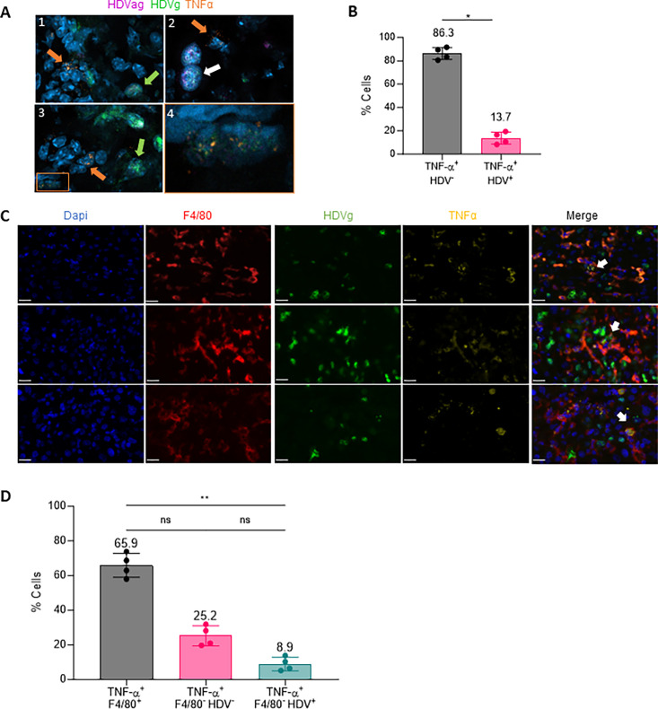
Hepatitis delta virus (HDV) infection represents the most severe form of human viral hepatitis; however, the mechanisms underlying its pathology remain incompletely understood. We recently developed an HDV mouse model by injecting adeno-associated viral vectors (AAV) containing replication-competent HBV and HDV genomes. This model replicates many features of human infection, including liver injury. Notably, the extent of liver damage can be diminished with anti-TNF-α treatment. Here, we found that TNF-α is mainly produced by macrophages. Downstream of the TNF-α receptor (TNFR), the receptor-interacting serine/threonine-protein kinase 1 (RIPK1) serves as a cell fate regulator, playing roles in both cell survival and death pathways. In this study, we explored the function of RIPK1 and other host factors in HDV-induced cell death. We determined that the scaffolding function of RIPK1, and not its kinase activity, offers partial protection against HDV-induced apoptosis. A reduction in RIPK1 expression in hepatocytes through CRISPR-Cas9-mediated gene editing significantly intensifies HDV-induced damage. Contrary to our expectations, the protective effect of RIPK1 was not linked to TNF-α or macrophage activation, as their absence did not alter the extent of damage. Intriguingly, in the absence of RIPK1, macrophages confer a protective role. However, in animals unresponsive to type-I IFNs, RIPK1 downregulation did not exacerbate the damage, suggesting RIPK1's role in shielding hepatocytes from type-I IFN-induced cell death. Interestingly, while the damage extent is similar between IFNα/βR KO and wild type mice in terms of transaminase elevation, their cell death mechanisms differ. In conclusion, our findings reveal that HDV-induced type-I IFN production is central to inducing hepatocyte death, and RIPK1's scaffolding function offers protective benefits. Thus, type-I IFN together with TNF-α, contribute to HDV-induced liver damage. These insights may guide the development of novel therapeutic strategies to mitigate HDV-induced liver damage and halt disease progression.


Conflict of interest statement

The authors have declared that no competing interests exist.

Figures

Fig 1
Fig 1
Macrophages are the main source of TNF-α production in AAV-HBV/HDV mouse livers. (A) TNF-α mRNA (orange) and HDV RNA genome (HDVg in green) and antigenome (HDVag in pink) distribution was analyzed by in situ hybridization (ISH) in the liver of C57BL/6 mice 21 days after receiving adenoassociated viral (AAV) vectors delivering both HBV and HDV genomes (HBV/HDV) at a dose of 5x10¹⁰ vg/mouse. Images show cells expressing TNF-α (orange arrows), cells positive for HDVg (green arrows) and cells positive for both HDVg and HDVag (white arrows). Panel 4 shows a magnification of a HDV positive cell expressing TNF-α. (B) The percentage of TNF-α+ HDV+ and TNF-α+ HDV- was quantified using image J (mean ± SD, n = 4). Statistical differences were determined by Mann-Whitney test p <0.05 (*). (C) Liver sections were analyzed for TNF-α mRNA (yellow) and HDVg (green) expression by in situ hybridization (ISH) followed by immunofluorescence (IF) to detect F4/80 protein (red). Representative images of hybridized liver sections taken with the Vectra Polaris Automated Imaging System are depicted in; white arrows pointed to macrophages expressing TNF-α and that contain HDV RNA. The white bar in each image is 20μm long. (D) The percentage of TNF-α+ F4/80+, TNF-α+ F4/80+ HDV-, TNF-α+ F4/80- HDV+ and TNF-α+ F4/80+ HDV+ cells was quantified as described in methods. Individual data points and mean values ± standard deviations are plotted. Statistical differences were determined by Kruskal-Wallis test following Dunn’s multiple comparison test p <0.05 (*).
Fig 2
Fig 2
RIPK1 scaffolding function partially prevents hepatocellular apoptosis in HBV/HDV mice. (A) Schematic representation of the experimental procedure, 6/8-week-old C57BL/6 wt mice were intravenously injected with 1011 vg/mouse of AAV-SaCas9-RIPK1g2 (RIPK1edit) or an AAV expressing SaCas9 without guide (control) and after 30 hours they were administered with 5x10¹⁰ vg/mouse AAV-HBV (HBV) or AAV-HBV/HDV (HBV/HDV). Peripheral blood was extracted at 7, 14 and 21 days post-injection (dpi). Liver damage was analyzed by (B) quantification of serum ALT levels (international units (IU)/L) and (C) quantification of a-Casp3+ hepatocytes/area after Immunohistochemistry (IHC). (D) HDV viremia in circulation by RT-PCR was determined in control and RIPK1edit mice 7, 14 and 21 dpi (E). IHC analysis against HDAgs was performed at 21 dpi in the liver sections of both groups and (F) quantified using Image J. (G) The presence of HDV genomes and antigenomes in mouse liver samples was quantified by RT-PCR. (H) Analysis of serum ALT levels in wt mice daily treated with Nec1 or saline was performed 7, 14 and 21 days after administration of HBV or HBV/HDV. (I) The presence of HDV genomes and antigenomes in mouse liver samples was quantified by RT-PCR. Individual data points and mean values ± standard deviations are shown. Statistical analysis was performed by one-way ANOVA followed by Bonferroni multiple-comparison test. p <0.05 (*), p <0.01 (**), p <0.001 (***), p <0.0001 (****) and ns = non-significant. IU: International units. (A) Created with BioRender.
Fig 3
Fig 3
The exacerbation of liver damage in RIPK1-edited mice upon HBV/HDV co-injection is not mediated by macrophages or TNF-α. (A) The presence of macrophages in the livers of control and RIPK1edit wt mice was analyzed 21 dpi of HBV or AAV- HBV/HDV by F4/80 immunohistochemistry (IHC). Representative IHC images of F4/80-stained livers are shown (200x). (B) Quantification of F4/80 IHC stained area (mean ± SD). Significant differences were determined by one-way ANOVA followed by Bonferroni multiple-comparison test. (C) Schematic representation of the experimental procedure, 6/8-week-old C57BL/6 wt mice were treated as described in Fig 2A and for macrophage depletion, animals were iv injected with clodronate-loaded liposomes (CLL) every 4 days starting the day before HBV/HDV injection up to day 20. Liver damage was analyzed by (D) determination of serum ALT levels (IU/L) (E) quantification of a-Casp3+ hepatocytes/area (Individual data points and mean values ± SD are shown) and (F) liver histology analysis on H&E-stained sections, representative images are shown. Statistical differences were determined by two-way (D) or one-way (E) ANOVA followed by Bonferroni multiple-comparison test. (G-H) 6-8-week-old C57BL/6 wt and TNF-α KO mice were treated as described in Fig 2A. Liver damage was analyzed by (G) determination of serum ALT levels (IU/L) and (H) quantification a-Casp3+ hepatocytes. Individual data points and mean values ± standard deviations are shown. Statistical differences were determined by two-way (F) or one-way (G) ANOVA followed by Bonferroni multiple-comparison test. p <0.05 (*), p <0.01 (**), p<0.001 (***), p <0.0001 (****) and ns = non-significant.
Fig 4
Fig 4
RIPK1 downregulation has no effect over HDAg-induced liver damage. 6/8-week-old C57BL/6 wt mice were injected with Cas9 control or RIPK1edit and 30 hours later both groups were injected with an AAV expressing HDV-Small Antigen (S-HDAg). Liver damage was analyzed by (A) determination of serum ALT levels (IU/L) at 7 and 14 dpi (dpi) and (B) quantification of a-Casp3+ hepatocytes/area at 14 dpi. (C) Western blot analysis to determine S-HDAg expression was performed at 14 dpi in the liver extracts. Individual data points and mean values ± standard deviations are shown in A and B. Statistical differences were determined by two-way ANOVA. p <0.05 (*), p <0.01 (**) and ns = non-significant.
Fig 5
Fig 5
RIPK1 downregulation has no effect over HDV-induced liver damage in the absence of type-I IFN response. 6/8-week-old C57BL/6 wt and IFN-α/βR KO mice were treated as described in Fig 2A. Liver damage was analyzed by (A) determination of serum ALT levels (IU/L) at 7, 14, and 21 dpi and (B) quantification of a-Casp3+ hepatocytes/area at 21 dpi. (C) IHC against HDAgs was performed at 21 dpi in the liver sections of control and RIPK1edit mice injected with the HBV/HDV vectors. (D) Quantification of HDAg expression in the aforementioned IHC images for each mouse. (E) The presence of HDV genomes and antigenomes in mouse liver samples was quantified by RT-PCR. Individual data points and mean values ± standard deviations are shown. Statistical differences were determined by two-way ANOVA (A and E) or one-way ANOVA (B and D) followed by Bonferroni multiple-comparison test. p <0.01 (**), p <0.001 (***), p <0.0001 (****) and ns = non-significant.

References

    1. Lempp FA, Ni Y, Urban S. Hepatitis delta virus: insights into a peculiar pathogen and novel treatment options. Nat Rev Gastroenterol Hepatol. 2016; 13:580–589. doi: 10.1038/nrgastro.2016.126 - DOI - PubMed
    1. Palom A, Rodríguez-Tajes S, Navascués CA, García-Samaniego J, Riveiro-Barciela M, Lens S, et al.. Long-term clinical outcomes in patients with chronic hepatitis delta: the role of persistent viraemia. Aliment Pharmacol Ther. 2020; 51:158–166. doi: 10.1111/apt.15521 - DOI - PubMed
    1. Wedemeyer H. The burden of hepatitis D—defogging the epidemiological horizon. J Hepatol. 2020; 73:493–495. doi: 10.1016/j.jhep.2020.06.037 - DOI - PubMed
    1. Stockdale AJ, Kreuels B, Henrion MYR, Giorgi E, Kyomuhangi I, de Martel C, et al.. The global prevalence of hepatitis D virus infection: Systematic review and meta-analysis. J Hepatol. 2020; 73:523–532. doi: 10.1016/j.jhep.2020.04.008 - DOI - PMC - PubMed
    1. Koh C, Heller T, Glenn JS. Pathogenesis of and New Therapies for Hepatitis D. Gastroenterology. 2019; 156:461–476.e1. doi: 10.1053/j.gastro.2018.09.058 - DOI - PMC - PubMed

Substances