. 2024 Jun 3;147(6):2053-2068.
doi: 10.1093/brain/awae078.

Mitigation of TDP-43 toxic phenotype by an RGNEF fragment in amyotrophic lateral sclerosis models

Affiliations

Mitigation of TDP-43 toxic phenotype by an RGNEF fragment in amyotrophic lateral sclerosis models

Cristian A Droppelmann et al. Brain. .

Abstract

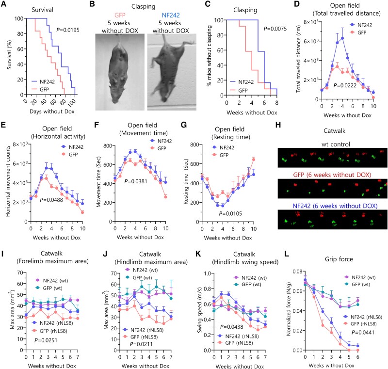
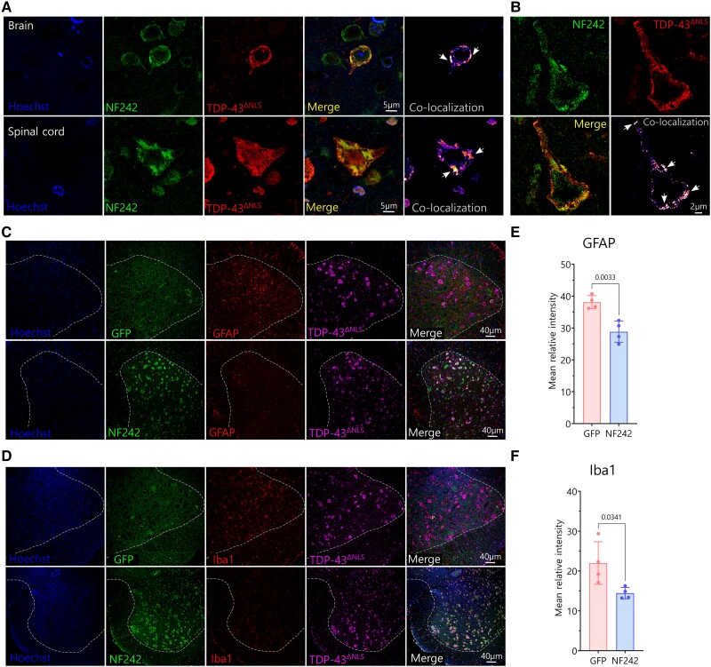
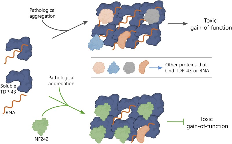
Aggregation of the RNA-binding protein TAR DNA binding protein (TDP-43) is a hallmark of TDP-proteinopathies including amyotrophic lateral sclerosis (ALS) and frontotemporal dementia (FTD). As TDP-43 aggregation and dysregulation are causative of neuronal death, there is a special interest in targeting this protein as a therapeutic approach. Previously, we found that TDP-43 extensively co-aggregated with the dual function protein GEF (guanine exchange factor) and RNA-binding protein rho guanine nucleotide exchange factor (RGNEF) in ALS patients. Here, we show that an N-terminal fragment of RGNEF (NF242) interacts directly with the RNA recognition motifs of TDP-43 competing with RNA and that the IPT/TIG domain of NF242 is essential for this interaction. Genetic expression of NF242 in a fruit fly ALS model overexpressing TDP-43 suppressed the neuropathological phenotype increasing lifespan, abolishing motor defects and preventing neurodegeneration. Intracerebroventricular injections of AAV9/NF242 in a severe TDP-43 murine model (rNLS8) improved lifespan and motor phenotype, and decreased neuroinflammation markers. Our results demonstrate an innovative way to target TDP-43 proteinopathies using a protein fragment with a strong affinity for TDP-43 aggregates and a mechanism that includes competition with RNA sequestration, suggesting a promising therapeutic strategy for TDP-43 proteinopathies such as ALS and FTD.

Keywords: RNA binding proteins; RNA metabolism; amyotrophic lateral sclerosis; motor neuron disease; neuronal cytoplasmic inclusions; therapeutic.


Conflict of interest statement

The authors report no competing interests.

Figures

Figure 1
Figure 1
Interaction between RGNEF/NF242 and TDP-43. (A) Representative SPR sensorgrams showing the interaction between His-GST-TDP-43wt (analyte) and His-MBP-RGNEF1–275 (ligand) at different concentrations of His-GST-TDP-43wt. KD = 1.78 ± 0.49 μM (n = 4). (B) NanoBiT experiment showing interaction between TDP-43wt (structure detailed) and NF242 (NF) (n = 3; NC = negative control). (C) NanoBiT experiment showing interaction between TDP-431–366 (structure detailed) and NF242 (NF) (n = 3; NC = negative control). (D) NanoBiT experiment showing interaction between TDP-431–274 (structure detailed) and NF242 (NF) (n = 3; NC = negative control). (E) NanoBiT experiment showing an absence of interaction between TDP-43ΔRRM1–2 (structure detailed) and NF242 (NF) (n = 3; NC = negative control). (F) Representative SPR sensorgrams showing the interaction between His-TDP-431–269 (analyte) and His-MBP-RGNEF1–275 (ligand) at different concentrations of His-TDP-431–269. KD = 4.11 ± 1.33 μM (n = 3). (G) Representative SPR sensorgrams demonstrating the absence of interaction between His-TDP-431–102 (analyte) and His-MBP-RGNEF1–275 (ligand) at different concentrations of His-TDP-431–102 (n = 3). (H) NanoBiT experiment showing interaction between TDP-43ΔRRM1 (structure detailed) and NF242 (NF) (n = 3; NC = negative control). (I) NanoBiT experiment showing the lack of interaction between TDP-43ΔRRM2 (structure detailed) and NF242 (NF) (n = 3; NC = negative control). (J) Representative SPR sensorgrams demonstrating the interaction between His-RRM1 (analyte) and His-MBP-RGNEF1–275 (ligand) at different concentrations of His-RRM1(n = 4). (K) Representative SPR sensorgrams demonstrating weak interaction (low signal intensity) between His-RRM2 (analyte) and His-MBP-RGNEF1–275 (ligand) at different concentrations of His-RRM2 (n = 4). (L) NanoBiT experiment showing interaction between TDP-431–192 (structure detailed) and NF242 (NF) (n = 3; NC = negative control). RRM = RNA recognition motif; SPR = surface plasmon resonance spectroscopy.
Figure 2
Figure 2
Inhibition of TDP-43 and RNA binding by NF242. (A) Luciferase assay measuring TDP-43 stabilizing activity over NEFL 3′ UTR (fixed amount of TDP-43) in presence of increasing amounts of NF242 (blue dots, n = 3; dotted line with associated dots). NF242 decreases TDP-43 stabilizing activity in a dose-dependent manner. The red dot (lower right corner outside of dotted line) shows the control in the absence of TDP-43 (n = 3). (B) Luciferase assay measuring TDP-43 stabilizing activity over NEFL 3′ UTR in presence (red; lower curve) of absence (blue; upper curve) of 120 ng of NF242 at increasing amounts of TDP-43. Displacement of the dose-response curve suggests competition of NF242 and RNA for TDP-43 (n = 3). (C) Competition experiment between NF242 and RNA for TDP-43 binding using surface plasmon resonance spectroscopy (SPR). A biotinylated RNA oligo was attached to an SPR neutravidin chip (ligand) and then 100 nM of His-TDP-431–269 was used as analyte. In the accompanying schematic of the binding of His-TDP-431–269 (TDP-43) to the chip (curve i; upper curve in C), the capability of this protein to bind RNA as observed in the sensorgram is illustrated. When His-TDP-431–269 (TDP-43) and His-NF242 (NF242) were pre-incubated together at two NF242 concentrations (1 μM or 10 μM; curves ii and iii, middle and lower curves, respectively) to ensure an effect of NF242 over TDP-43, and this was injected into the SPR machine, a 10% reduction of the signal in the sensorgram (inhibition) was observed at 1 μM NF242 and 50% at 10 μM NF242 indicating that an important fraction of TDP-43 was bound to NF242 and not interacting with RNA. This confirms that NF242 blocks the access of TDP-43 to the RNA on the chip through its binding to the same site that binds RNA in TDP-43. Panel C(iiii) created with BioRender.com.
Figure 3
Figure 3
Modelling of TDP-43-NF242 interaction. (A) NF242 structure based in the atomic coordinates of RGNEF residues 1–242 (NF242) extracted from the AlphaFold Protein structure database (accession Q8N1W1). (B) Minimized structure of TDP-43 in complex with AUG12 RNA from experimental NMR coordinates (PDB accession: 4BS2). (C) Region of high inter-molecular contacts occurring between TDP-43 and RNA. (D) Minimized structure of TDP-43 in the complex with NF242. (E) Region of high intermolecular contacts occurring between TDP-43 and NF242 (yellow square in D) showing the most important amino acid interactions from the loop 76–81 of NF242 and the interface between RRM1 and RRM2 of TDP-43. (F) Summary of all intermolecular contacts. (G) Schematic showing the mutants used to study the importance of the loop 76–81 of the TIG domain of NF242 in the interaction with TDP-43. (H) NanoBiT experiment showing that the mutants NF242-mut 77–79 and NF242-Δ77–79_P81G, both fused to smBiT in the C-terminal end, do not interact with TDP-43 (P = 0.9636 and P = 0.9644, respectively). The interaction with NF242 is shown as positive control (P < 0.0001). RRM = RNA recognition motif.
Figure 4
Figure 4
Co-expression of RGNEF or NF242 with TDP-43 in fruit flies. (A) Kaplan-Meier graph showing the survival of elav>RGNEF (elav>R; n = 156), elav>NF242 (elav>NF; n = 101) RGNEF no driver control (R n.e.; n = 117), NF242 no driver control (NF n.e.; n = 93) and elav>w− (control of driver crossed with parental line; n = 123). The elav>RGNEF line shows an increased lifespan compared to RGNEF no driver control (P = 0.0035) and elav>w− (P < 0.0001) lines. The elav>NF242 line shows an increased lifespan compared to NF242 no driver control (P < 0.0001) and elav>w− (P < 0.0001) lines. (B) Kaplan-Meier graph showing the survival of elav>GFP;TDP-43wt (elav>G; T; n = 178), elav>RGNEF;TDP-43wt (elav>R; T, n = 132) and elav>NF242;TDP-43wt (elav>NF; T, n = 224). The elav>GFP;TDP-43wt line shows a reduced lifespan, an effect that is suppressed in the elav>RGNEF; TDP-43wt (P < 0.0001) and elav>NF242;TDP-43wt (P < 0.0001) lines. (C) Kaplan-Meier graph showing the survival of D42>GFP; TDP-43wt (D42>G; T; n = 143), D42>NF242;TDP-43wt (D42>NF; T, n = 181) and D42>w (control of driver crossed with parental line, n = 98). The D42>GFP;TDP-43wt line shows a reduced lifespan, an effect that is suppressed in the D42>NF242 line (P < 0.0001). The latter also show an increase in lifespan compared to the control D42>w line (P < 0.0001). (D and E) Negative geotaxis assay showing the climbing score at Days 3 and 6 for elav>RGNEF;TDP-43wt (elav>R; T, n = 11; 110 flies), elav>NF242;TDP-43wt (elav>NF; T, n = 12, 120 flies), elav>GFP;TDP-43wt (elav>G; T; with n = 12; 120 flies at Day 1) and elav>w (n = 8; 80 flies) lines. The elav>GFP;TDP-43wt line shows a severe motor phenotype that is suppressed when RGNEF or NF242 is co-expressed with TDP-43wt in neurons (P < 0.0001). (F and G) Negative geotaxis assay showing the climbing score at Days 6 and 9 for D42>RGNEF;TDP-43wt (D42>R; T, n = 12; 120 flies) and D42>NF242;TDP-43wt (D42>NF; T, n = 16, 160 flies), D42>GFP;TDP-43wt (D42>G; T; n = 14; 140 flies) and D42>w (n = 9; 90 flies). The D42>GFP; TDP-43wt line shows a significant motor phenotype that is suppressed when RGNEF or NF242 is co-expressed with TDP-43wt in motor neurons (P < 0.0001). (H) Representative images showing the eye phenotype of GMR>w (negative control), GMR>36R (positive control), GMR>GFP;TDP-43wt and GMR>NF242;TDP-43wt lines. NF242 co-expression with TDP-43wt suppresses the eye degeneration observed in the GMR>NF242;TDP-43wt line. (I) Immunofluorescence of adult elav>NF242;TDP-43wt fly brain tissue showing the co-localization between NF242 and TDP-43wt in neurons. (J and K) Confocal images at higher magnification of adult elav>NF242;TDP-43wt fly brain tissue showing the co-aggregation between NF242 and TDP-43wt in neurons. Nuclei are indicated with dashed lines. Arrows show nuclear co-localization and arrowheads cytoplasmic co-localization.
Figure 5
Figure 5
Ectopic expression of NF242 in rNLS8 mice. (A) Kaplan-Meier graph showing the increased lifespan after doxycycline (Dox) retrieval of rNLS8 mice injected with AAV9/GFP (n = 12) compared to mice injected with AAV9/NF242 (n = 11) (P = 0.0195). (B) Representative pictures showing a rNLS8 mouse injected with AAV9/GFP with clasping and a rNLS8 mouse injected with AAV9/NF242 after 5 weeks without Dox. (C) Kaplan-Meier graph showing clasping quantification of rNLS8 mice injected with AAV9/GFP (n = 12) or AAV9/NF242 (n = 12). AAV9/NF242 injected rNLS8 mice show a significant delay in clasping occurrence (P = 0.0075). (DG) Open field test comparing rNLS8 mice injected with AAV9/GFP (n = 12) or AAV9/NF242 (n = 12). AAV9/NF242 injected rNLS8 mice show an increase in (D) total distance travelled (P = 0.0222), (E) horizontal activity (P = 0.0488), (F) movement time (P = 0.0105) and (G) a decrease in resting time (P = 0.0105). (H) Representative visualizations of gait assessment (Catwalk) that compares the improved gait pattern of an AAV9/NF242 injected rNLS8 mouse with an AAV9/GFP injected rNLS8 mouse at 6 weeks without Dox. Wild-type mouse shows normal gait. (IK) Catwalk quantification comparing rNLS8 and wild-type (wt) mice injected with AAV9/GFP or AAV9/NF242 (n = 12 for each group of rNLS8 mice; n = 6 for each group of wild-type mice). The AAV9/NF242 injected rNLS8 mice show an improvement in (I) the forelimb maximum area (P = 0.0251), (J) the hindlimb maximum area (P = 0.0211) and (K) the hindlimb swing speed (P = 0.0438). (L) Grip force experiment showing that the AAV9/NF242 injected rNLS8 mice (n = 12) have a slight increase in the force compared to the AAV9/GFP injected mice (n = 12) (P = 0.0441). Wild-type mice injected with AAV9/GFP (n = 6) or AAV9/NF242 (n = 6) are shown as healthy grip force controls. GFP = green fluorescent protein.
Figure 6
Figure 6
Pathology of rNLS8 mice expressing ectopic NF242 at Week 3. (A) High magnification confocal images showing the co-localization and co-aggregation (indicated by white arrows) between NF242 and TDP-43ΔNLS in the brain cortex (cortical layer II–III) and spinal cord of a rNLS8 mouse injected with AAV9/NF242 after 3 weeks without doxycycline (Dox). (B) Super-resolution stimulated emission depletion (STED) microscopy images showing in detail the co-aggregation (indicated by white arrows) between NF242 and TDP-43ΔNLS in the brain cortex (cortical layer II–III) of a rNLS8 mouse injected with AAV9/NF242 after 3 weeks without Dox. (C and D) Representative immunofluorescences of rNLS8 mice injected with AAV9/GFP and AAV9/NF242 showing the decrease in the amount of glial fibrillary acidic protein (GFAP) (C) and Iba1 (D) in the spinal cord. The anterior grey horn is separated from the white matter by a dashed white line. (E and F) Quantification showing the reduction of the levels of GFAP (E, P = 0.0033) and Iba1 (F, P = 0.0341) in the ventral horns of the lumbar spinal cord of rNLS8 mice injected with AAV9/GFP and AAV9/NF242, after 3 weeks without Dox (n = 4). GFP = green fluorescent protein.
Figure 7
Figure 7
Schematic of the proposed mechanism of action for NF242. We hypothesize that under pathological conditions, TDP-43 aggregates sequester RNAs, other RNA-binding proteins or other proteins that can interact with TDP-43, leading to a toxic gain-of-function. When NF242 is expressed genetically or using adeno-associated viruses, it binds to TDP-43 aggregates and blocks sequestration of RNAs and proteins, thus inhibiting the toxic gain-of-function. Created with BioRender.com.

References

    1. Strong MJ, Kesavapany S, Pant HC. The pathobiology of amyotrophic lateral sclerosis: A proteinopathy? J Neuropathol Exp Neurol. 2005;64:649–664. - PubMed
    1. Corcia P, Beltran S, Bakkouche SE, Couratier P. Therapeutic news in ALS. Rev Neurol (Paris). 2021;177:544–549. - PubMed
    1. Droppelmann CA, Campos-Melo D, Ishtiaq M, Volkening K, Strong MJ. RNA metabolism in ALS: When normal processes become pathological. Amyotroph Lateral Scler Frontotemporal Degener. 2014;15(5–6):321–336. - PubMed
    1. Arai T, Hasegawa M, Akiyama H, et al. TDP-43 is a component of ubiquitin-positive tau-negative inclusions in frontotemporal lobar degeneration and amyotrophic lateral sclerosis. Biochem Biophys Res Commun. 2006;351:602–611. - PubMed
    1. Neumann M, Sampathu DM, Kwong LK, et al. Ubiquitinated TDP-43 in frontotemporal lobar degeneration and amyotrophic lateral sclerosis. Science. 2006;314:130–133. - PubMed