. 2024 Jun 1;165(6):1336-1347.
doi: 10.1097/j.pain.0000000000003139. Epub 2023 Dec 27.

Contribution of mechanoreceptors to spinal cord injury-induced mechanical allodynia

Affiliations

Contribution of mechanoreceptors to spinal cord injury-induced mechanical allodynia

Christopher Sliwinski et al. Pain. .

Abstract

Evidence from previous studies supports the concept that spinal cord injury (SCI)-induced neuropathic pain (NP) has its neural roots in the peripheral nervous system. There is uncertainty about how and to which degree mechanoreceptors contribute. Sensorimotor activation-based interventions (eg, treadmill training) have been shown to reduce NP after experimental SCI, suggesting transmission of pain-alleviating signals through mechanoreceptors. The aim of the present study was to understand the contribution of mechanoreceptors with respect to mechanical allodynia in a moderate mouse contusion SCI model. After genetic ablation of tropomyosin receptor kinase B expressing mechanoreceptors before SCI, mechanical allodynia was reduced. The identical genetic ablation after SCI did not yield any change in pain behavior. Peptidergic nociceptor sprouting into lamina III/IV below injury level as a consequence of SCI was not altered by either mechanoreceptor ablation. However, skin-nerve preparations of contusion SCI mice 7 days after injury yielded hyperexcitability in nociceptors, not in mechanoreceptors, which makes a substantial direct contribution of mechanoreceptors to NP maintenance unlikely. Complementing animal data, quantitative sensory testing in human SCI subjects indicated reduced mechanical pain thresholds, whereas the mechanical detection threshold was not altered. Taken together, early mechanoreceptor ablation modulates pain behavior, most likely through indirect mechanisms. Hyperexcitable nociceptors seem to be the main drivers of SCI-induced NP. Future studies need to focus on injury-derived factors triggering early-onset nociceptor hyperexcitability, which could serve as targets for more effective therapeutic interventions.

Trial registration: ClinicalTrials.gov NCT01571531.


Conflict of interest statement

The authors have no conflicts of interest to declare.

Figures

Figure 1.
Figure 1.
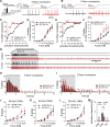
Post-SCI ablation of TrkB-expressing mechanoreceptors. (A) Schematic of experimental design. (B) Representative lumbar (L4-L6) DRG neurons of saline-treated control animals stained for HB-EGF shows robust expression of DTR in a subset of sensory neurons. (C) In ablated TrkBDTR animals, DTR expression is almost completely absent (scale bar, 100 μm). (D) Quantification (t test, P < 0.0001; DTX, n = 7; saline n = 6, L4-L6 bilaterally). (E–G) SCI-induced mechanical hypersensitivity to small-diameter light touch eliciting von Frey hair filaments (0.04, 0.07, and 0.16 g, 2-way RM ANOVA, time differences P < 0.0001, baseline vs 7 dpi FLSD #P < 0.05, ##P < 0.01, ###P < 0.001, ####P < 0.0001), but DTX-mediated ablation of TrkB+ sensory neurons (28-49 dpi) did not change SCI-induced mechanical hypersensitivity. (H) The Place Escape Avoidance Paradigm (PEAP, 0.16 g) performed at the end of the study shows that both groups spent more time on the light side of the chamber (2-way RM ANOVA P = 0.23 for group differences). (I) Injured TrkBDTR animals of both groups develop thermal hypersensitivity assessed by the Hargreaves test (2-way RM ANOVA, baseline vs 7 dpi P < 0.0001, FLSD ##P < 0.01, group differences P = 0.02, no significant Sidak pairwise comparisons). (J) Except for the SCI-induced motor deficits scored by the Basso Mouse Scale (BMS), no further motor deficits are observed after post-SCI ablation of TrkB+ sensory neurons. Mean ± SD for all graphs. 2-way RM ANOVA, 2-way eepeated-measures analysis of variance; DTR, diphtheria toxin receptor; DTX, diphtheria toxin; FLSD, Fisher least significant difference; FLSD, Fisher least significant difference; HB-EGF, heparin binding EGF-like growth factor ; SCI, spinal cord injury; TrkB, tropomyosin receptor kinase B.
Figure 2.
Figure 2.
Pre-SCI ablation of TrkB-expressing mechanoreceptors. (A) Schematic of experimental design. (B) Saline-injected control animals show robust DTR expression in lumbar (L4-L6) DRG neurons. Compared with control animals, DTR+ neurons are almost completely ablated in DTX-injected animals (C) (scale bar, 100 μm). (D) Quantification (t test P< 0.0001; DTX, n = 9; saline n = 7, L4-L6 bilaterally). (E–G) SCI-induced mechanical hypersensitivity to small-diameter filaments (0.04, 0.07, and 0.16 g, 2-way RM ANOVA, time differences P < 0.0001, post-DTX vs 7 dpi FLSD #P < 0.05, ##P < 0.01, no significant (ns) difference between baseline and post-DTX) is significantly altered in ablated animals, but inconsistently (2-way RM ANOVA group differences: 0.04 g, P = 0.004; 0.07 g, P = 0.008; 0.16 g, P = 0013, Sidak pairwise comparisons *P < 0.05). The response rate towards the 0.16-g von Frey hair filament (G) of ablated animals is not significantly different from control animals anymore at the end of the experiment 28 dpi. (H) This is confirmed by supraspinal processing of nociception using the PEAP (0.16 g) where both groups equally spent more time on the light side of the box. (I) DTX treatment does not affect pre-injury or post-injury thermal sensitivity, but SCI did induce thermal sensitivity in both groups (2-way RM ANOVA, time differences P < 0.0001, post-DTX vs 7 dpi FLSD ##P < 0.01, ####P < 0.0001). (J) SCI induces significant motor deficits in all animals scored by the Basso Mouse Scale (BMS), and ablation of TrkB+ sensory neurons does not induce further motor deficits. Mean ± SD for all graphs. DTX, diphtheria toxin; FLSD, Fisher least significant difference; PEAP, place escape/avoidance paradigm; SCI, spinal cord injury; TrkB, tropomyosin receptor kinase B.
Figure 3.
Figure 3.
Peptidergic nociceptive fiber changes in the dorsal horn after TrkB-expressing mechanoreceptor ablation. Lumbar (L4-L6) spinal cord sections of injured saline control (A and D) and DTX-treated injured (B and E) TrkBDTR mice stained for CGRP (scale bar, 100 μm). Representative images indicate the SCI-induced increase in CGRP-labeling density in deeper laminae (III-IV) of the spinal dorsal horn in all groups. (C and F) Quantification of the CGRP-labeling density in laminae III-IV of the lumbar (L4-L6) dorsal horn shows no difference comparing the injured saline-injected control group with injured DTX-injected animals but a significant increase compared to previous sham animals (dotted line) (mean ± SD) (post-SCI; saline n = 6; DTX n = 7) (pre-SCI; saline n = 7; DTX n = 9). CGRP, Calcitonin gene-related peptide; DTX, diphtheria toxin; SCI, spinal cord injury; TrkB, tropomyosin receptor kinase B.
Figure 4.
Figure 4.
Mechanoreceptor and nociceptor activity after SCI. Single-unit teased fiber recordings from hind paw glabrous skin-nerve preparations show that A-fiber (A) and C-fiber nociceptors (B) have lowered activation thresholds 7 dpi T11 (50 kDyn) SCI in mice. This is quantitatively shown in (C) for A-fiber and (E) for C-fiber nociceptors responding to forces smaller than 125 mN (multiple χ2 tests). While following SCI A-fibers (D) do not fire more frequently, C-fibers (F) significantly fire more frequently in response to mechanical ramp-and-hold stimuli. Example traces exhibit continuous firing after removal of stimulus (150 mN denoted in gray area) in the SCI group (G). This is quantitatively shown for A-fiber (H) and C-fiber (I) nociceptors (multiple Mann–Whitney tests). The firing of RA-Aβ-LTMRs (J), SA-Aβ-LTMRs (K), and DH-Aδ-LTMRs (L) was not significantly altered by SCI. Additionally, LTMR activation thresholds were not altered by SCI (M). Mean ± SEM. * P < 0.05, ** P < 0.01. DH, D hairs; LTMRs, low-threshold mechanoreceptors; RA, rapidly adapting; SA, slowly adapting; SCI, spinal cord injury.
Figure 5.
Figure 5.
Prolonged nocifensive behavior after experimental SCI. (A) T11 SCI mice showed prolonged nocifensive behavior (licking, shaking, and holding) compared with Sham mice when responding to 0.16-g von Frey hair filaments 7 dpi (slow-motion video analysis, unpaired 2-tailed t test, *P < 0.05). (B) The response rate of SCI rate was significantly higher than Sham mice at 7 dpi with 0.16-g von Frey hair filament (Sham n = 4, SCI n = 4, 2-way RM ANOVA, group difference P = 0.03, Sidak pairwise comparison ***P < 0.001). 2-way RM ANOVA, 2-way repeated-measures analysis of variance SCI, spinal cord injury.
Figure 6.
Figure 6.
Quantitative sensory testing in SCI subjects. Fourteen sensory incomplete SCI individuals (and 1 initial complete SCI subject) were examined 1, 3, and 12 months post-SCI for mechanical detection threshold (MDT), reflecting Aβ mechanoreceptors, and mechanical pain threshold (MPT), reflecting Aδ nociceptors. SCI individuals were measured at the L4 (right shin) and L5 (dorsum of right foot) dermatomes and found to be consistently altered compared with healthy database matched controls in MPT but not MDT (represented by Z-scores). Red dashed lines visually mark the Z-scores at 1.96 and −1.96, the cutoff for hypersensitivity or hyposensitivity, respectively. In the 12-month timepoint, open symbols represent at-level NP, lower filled symbols represent below-level NP, and fully filled symbols represent no NP. Mean ± SD. NP, neuropathic pain; SCI, spinal cord injury.

References

    1. Arcourt A, Gorham L, Dhandapani R, Prato V, Taberner FJ, Wende H, Gangadharan V, Birchmeier C, Heppenstall PA, Lechner SG. Touch receptor-derived sensory information alleviates acute pain signaling and fine-tunes nociceptive reflex coordination. Neuron 2017;93:179–93. - PubMed
    1. Attwell CL, van Zwieten M, Verhaagen J, Mason MRJ. The dorsal column lesion model of spinal cord injury and its use in deciphering the neuron-intrinsic injury response. Dev Neurobiol 2018;78:926–51. - PMC - PubMed
    1. Baastrup C, Jensen TS, Finnerup NB. Pregabalin attenuates place escape/avoidance behavior in a rat model of spinal cord injury. Brain Res 2011;1370:129–35. - PubMed
    1. Baumgärtner U, Magerl W, Klein T, Hopf HC, Treede RD. Neurogenic hyperalgesia versus painful hypoalgesia: two distinct mechanisms of neuropathic pain. PAIN 2002;96:141–51. - PubMed
    1. Bavencoffe A, Li Y, Wu Z, Yang Q, Herrera J, Kennedy EJ, Walters ET, Dessauer CW. Persistent electrical activity in primary nociceptors after spinal cord injury is maintained by scaffolded adenylyl cyclase and protein kinase A and is associated with altered adenylyl cyclase regulation. J Neurosci 2016;36:1660–8. - PMC - PubMed

Publication types

MeSH terms

Associated data