Review
. 2024;16(1):295-323.
doi: 10.1159/000539278. Epub 2024 May 13.

Pattern-Recognition Receptors and Immunometabolic Reprogramming: What We Know and What to Explore

Affiliations
Review

Pattern-Recognition Receptors and Immunometabolic Reprogramming: What We Know and What to Explore

Vijay Kumar et al. J Innate Immun. 2024.

Abstract

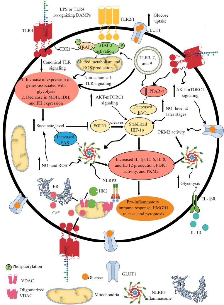
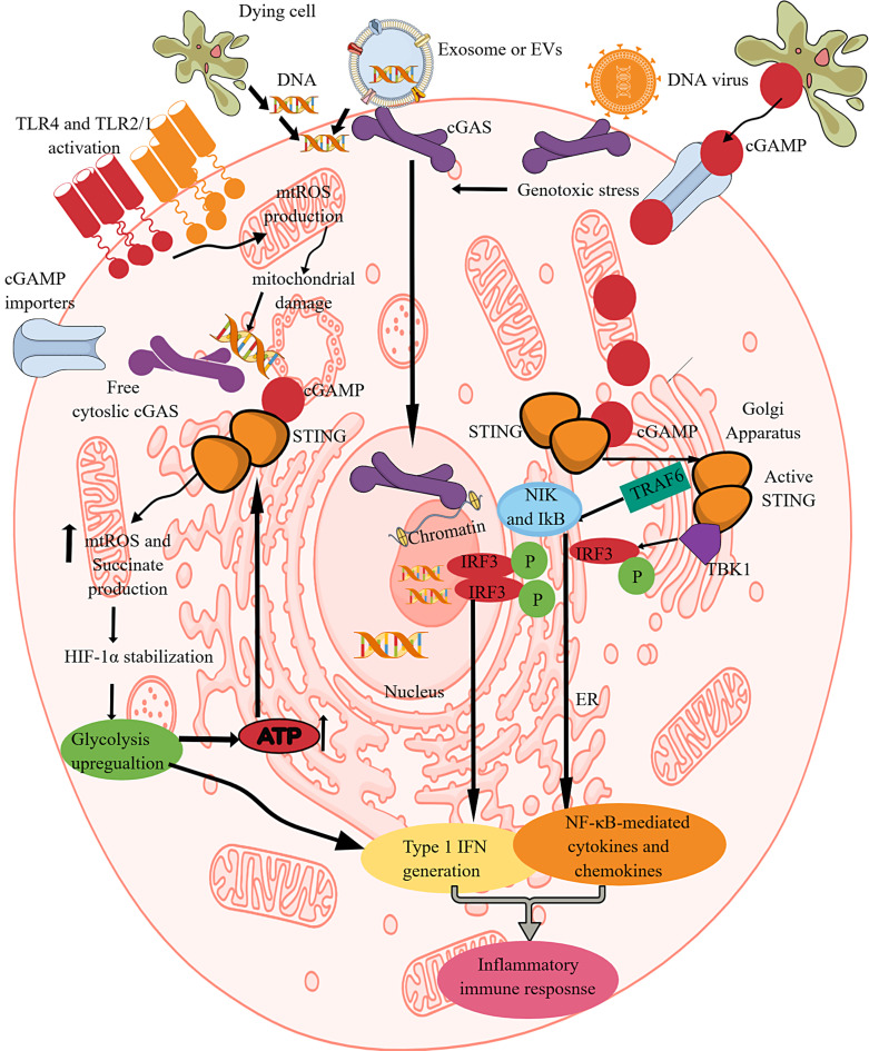
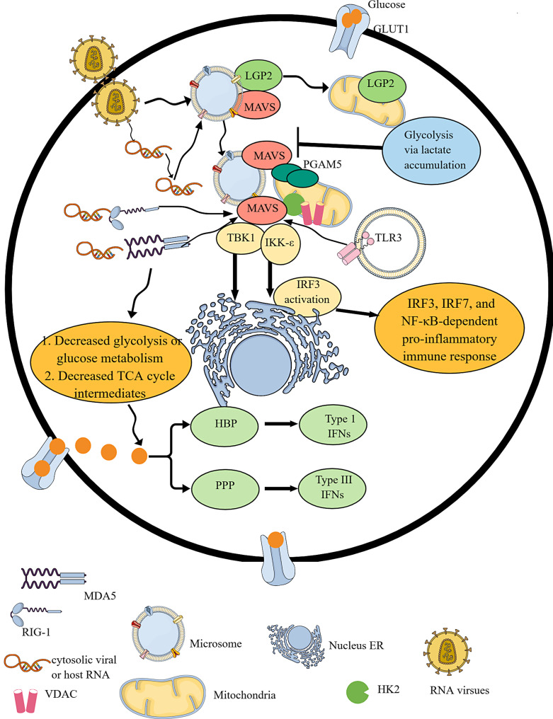
Background: Evolutionarily, immune response is a complex mechanism that protects the host from internal and external threats. Pattern-recognition receptors (PRRs) recognize MAMPs, PAMPs, and DAMPs to initiate a protective pro-inflammatory immune response. PRRs are expressed on the cell membranes by TLR1, 2, 4, and 6 and in the cytosolic organelles by TLR3, 7, 8, and 9, NLRs, ALRs, and cGLRs. We know their downstream signaling pathways controlling immunoregulatory and pro-inflammatory immune response. However, the impact of PRRs on metabolic control of immune cells to control their pro- and anti-inflammatory activity has not been discussed extensively.

Summary: Immune cell metabolism or immunometabolism critically determines immune cells' pro-inflammatory phenotype and function. The current article discusses immunometabolic reprogramming (IR) upon activation of different PRRs, such as TLRs, NLRs, cGLRs, and RLRs. The duration and type of PRR activated, species studied, and location of immune cells to specific organ are critical factors to determine the IR-induced immune response.

Key message: The work herein describes IR upon TLR, NLR, cGLR, and RLR activation. Understanding IR upon activating different PRRs is critical for designing better immune cell-specific immunotherapeutics and immunomodulators targeting inflammation and inflammatory diseases.

Keywords: Glycolysis; Immunometabolism; Infection; Inflammation; NOD-like receptors; Oxidative phosphorylation; Retinoic acid-inducible gene-1-like receptors; Toll-like receptors; cGLRs.


Conflict of interest statement

Authors declare no competing interest.

Figures

Fig. 1.
Fig. 1.
TLR and NLR signaling pathway activation-induced IR. Activation of different TLRs, such as TLR4, TLR2/1, TLR3, TLR7, and TLR9, shifts OXPHOS to glycolysis as indicated by the upregulation of glycolysis genes and downregulation of mitochondrial genes involved in OXPHOS and FAO. This IR occurs downstream of canonical and non-canonical (involves TBK1-TRAF6-STAT3 axis) TLR signaling pathways. The GLUT1 overexpression upon TLR activation further supports glycolysis by increasing glucose uptake. The TLR signaling decreases the PPAR-γ expression, which further decreases FAO to support the pro-inflammatory immune cell phenotype and function. TLR activation increases glucose uptake via increased mTOR-AKT signaling that also supports HIF-1α stabilization. The succinate accumulation upon pro-inflammatory TLR signaling activation further supports HIF-1α stabilization by inhibiting EGLN1. The NO. generation at later stages activates NLRP3 inflammasome activity and succinate accumulation. The TLR signaling-induced glycolysis, increased succinate level, HIF-1α stabilization and accumulation, PKM2, mammalian target of rapamycin complex 1, and AKT overactivity support NLRP3 inflammasome activation and IL-1β release. The HK2 dissociation from VDAC at the outer mitochondrial membrane during TLR signaling-induced glycolysis activates IP3 receptors in the ER to release Ca2+ in the cytosol – mitochondria uptake cytosolic Ca2+ molecules for VDAC oligomerization. The oligomerized VDACs aggregate with NLRP3 during its initial assembly to form the NLRP3 inflammasome complex. Furthermore, IL-1β released due to the NLRP3 inflammasome activity supports glycolysis through binding to IL-1βR. Thus, TLRs and NLRs (NLRP3) support each other’s pro-inflammatory function through IR.
Fig. 2.
Fig. 2.
cGAS/STING (cGLR) signaling-dependent IR. cGLRs or cGAS/STING signaling is critical for recognizing the cytosolic dsDNA and generating type 1 IFNs and NF-κB-dependent cytokines. cGAS-mediated cytosolic dsDNA recognition by cGAS generates cGAMP. STING recognizes cGAMP and undergoes dimerization to become active. The activated STING activates TBK1 and TRAF6, which activate IRF3 and NF-κB-dependent type 1 IFNs and cytokines. This process also activates glycolysis by increasing mtROS production, succinate accumulation, and HIF-α stabilization. The increased glycolysis overproduces ATP molecules, which further increases STING activation. Furthermore, TLR activation induced mtROS production and mitochondrial damage, releasing the mitochondrial DNA into the cytosol that the cGAS recognizes to initiate the cGAS/STING signaling. Hence, TLR and cGAS/STING signaling support each other through IR or glycolysis.
Fig. 3.
Fig. 3.
RLR signaling activation-mediated IR. RLR signaling activation involves the recognition of cytosolic RNA via RIG-1 and MDA5. During homeostasis, LGP2 is bound to the MAVS in the microsome. LGP2 moves to mitochondria upon viral infection, leaving MAVS free in the microsome. Thus, upon recognizing cytosolic RNA, RIG-1 and MDA5 interact with MAVS, which directly interacts with the oligomerized mitochondrial PGAM5. The RIG-1 and MDA5 interaction with MAVS interacting with oligomerized PGAM5 is critical for the downstream TBK1 and IRF3 phosphorylation-mediated type 1 IFN release. IRF3 activation occurs at the ER. The RIG-1 and MDA5 activation suppress glycolysis and the TCA cycle. Instead of glycolysis, cellular glucose undergoes PPP and HBP to generate type III and IFNs. Furthermore, glycolysis via increased lactate accumulation suppresses MAVS activity through binding to its TM domains. TLR3 activation also activates MAVS.

References

    1. Fullerton JN, Gilroy DW. Resolution of inflammation: a new therapeutic frontier. Nat Rev Drug Discov. 2016;15(8):551–67. - PubMed
    1. Furman D, Campisi J, Verdin E, Carrera-Bastos P, Targ S, Franceschi C, et al. . Chronic inflammation in the etiology of disease across the life span. Nat Med. 2019;25(12):1822–32. - PMC - PubMed
    1. Greten FR, Grivennikov SI. Inflammation and cancer: triggers, mechanisms, and consequences. Immunity. 2019;51(1):27–41. - PMC - PubMed
    1. Michels N, van Aart C, Morisse J, Mullee A, Huybrechts I. Chronic inflammation towards cancer incidence: a systematic review and meta-analysis of epidemiological studies. Crit Rev Oncol Hematol. 2021;157:103177. - PubMed
    1. Pisetsky DS. Pathogenesis of autoimmune disease. Nat Rev Nephrol. 2023;19(8):509–24. - PMC - PubMed

Substances

LinkOut - more resources