. 2024 Jun;630(8018):961-967.
doi: 10.1038/s41586-024-07515-9. Epub 2024 May 13.

Plasmid targeting and destruction by the DdmDE bacterial defence system

Affiliations

Plasmid targeting and destruction by the DdmDE bacterial defence system

Jack P K Bravo et al. Nature. 2024 Jun.

Abstract

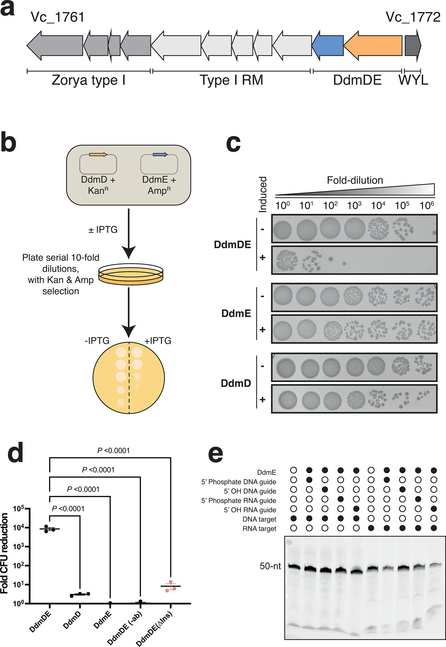
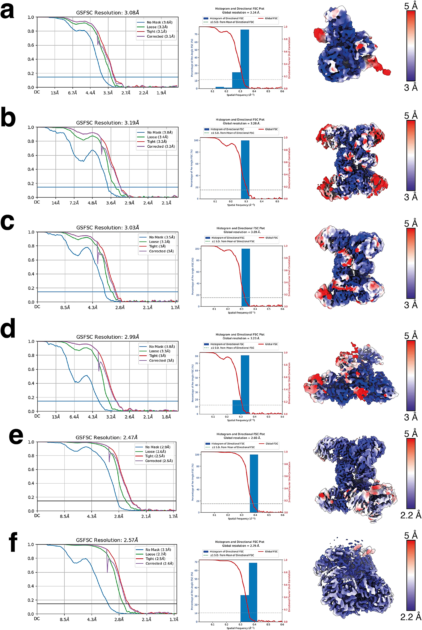
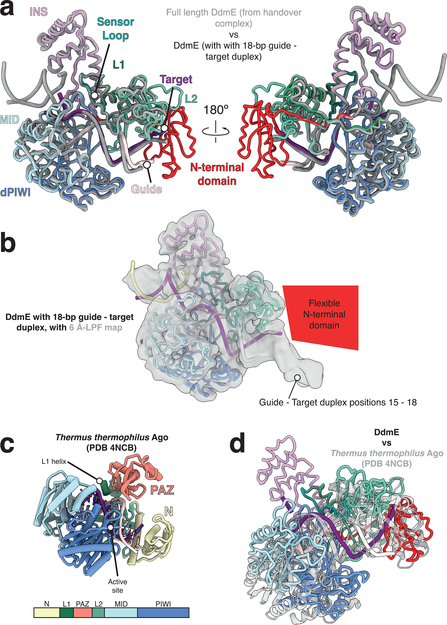
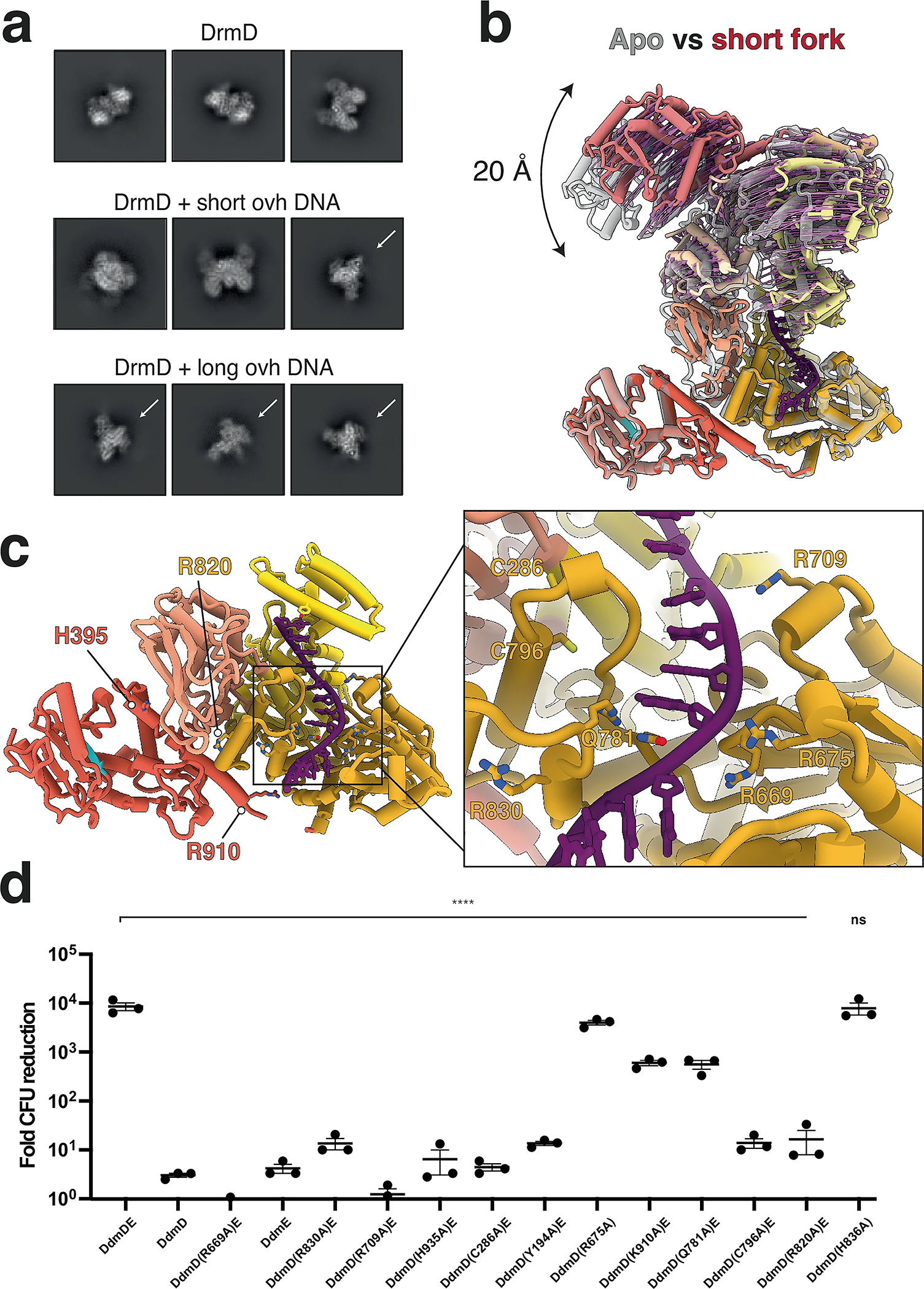
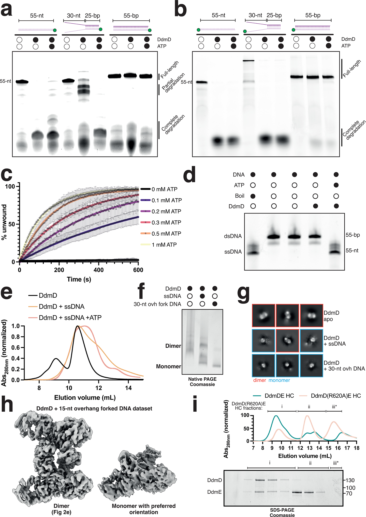
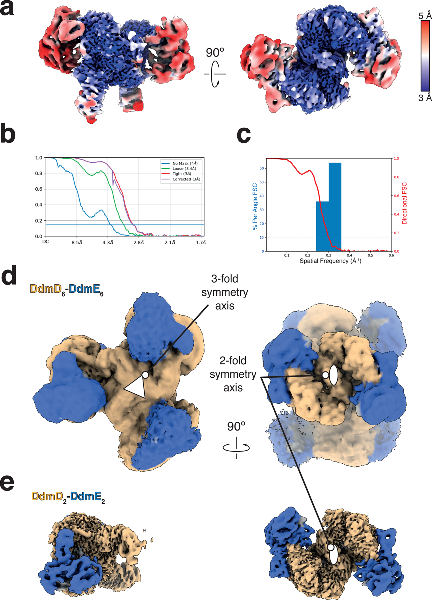
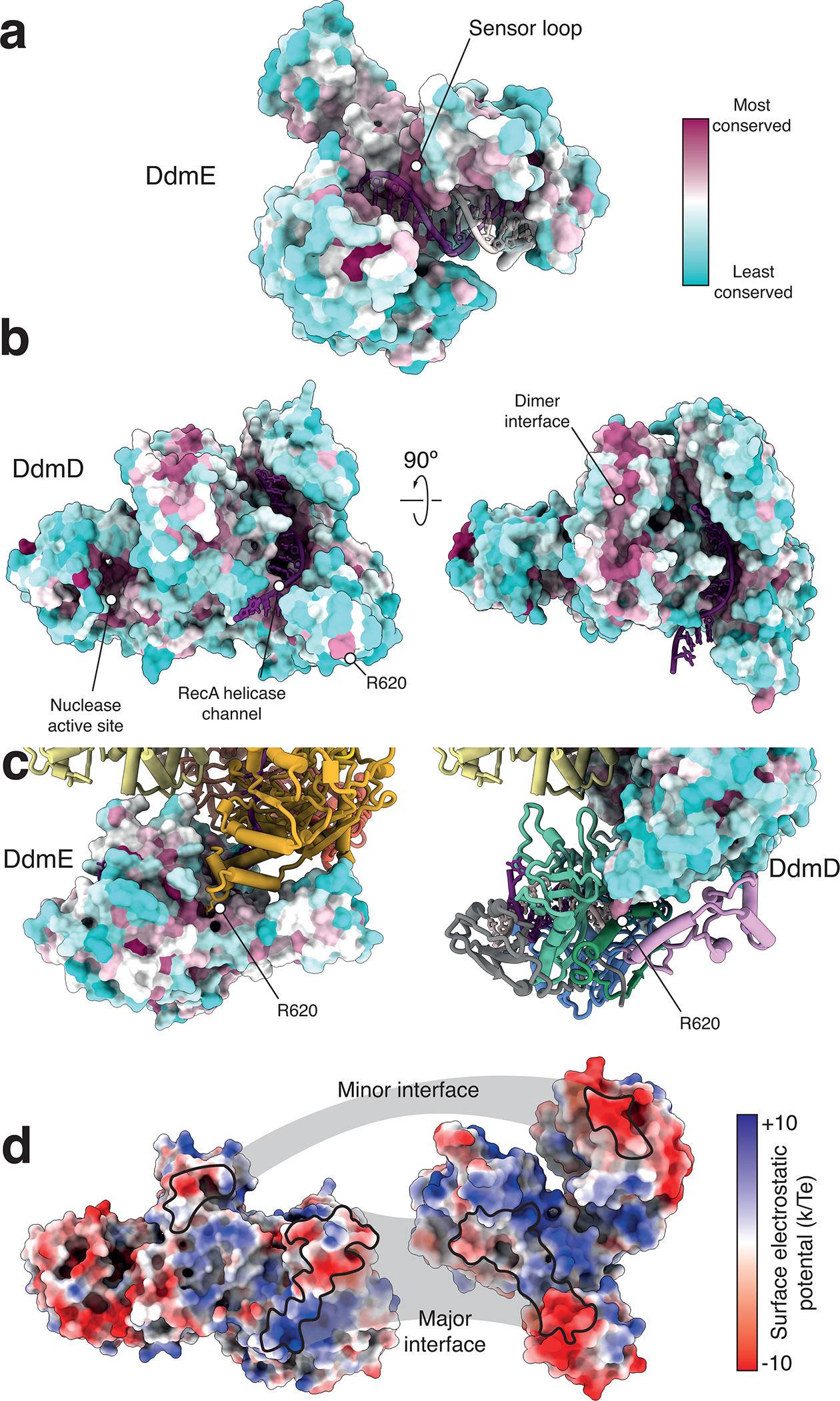
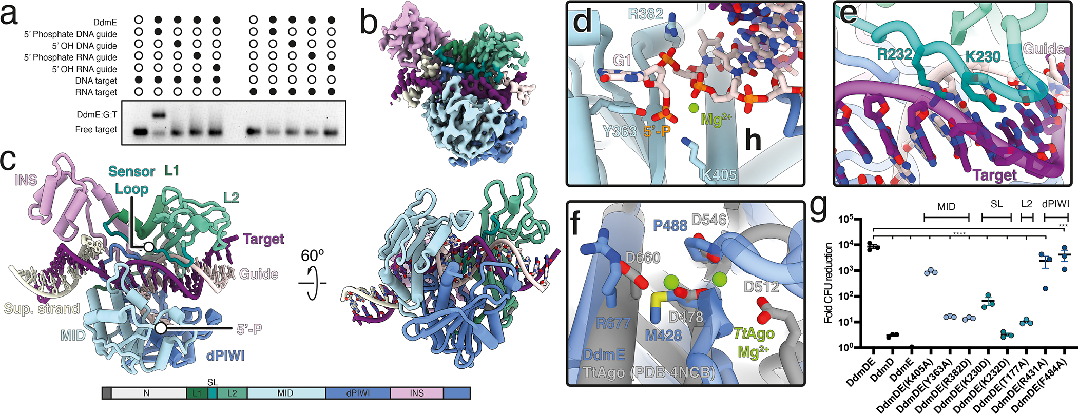
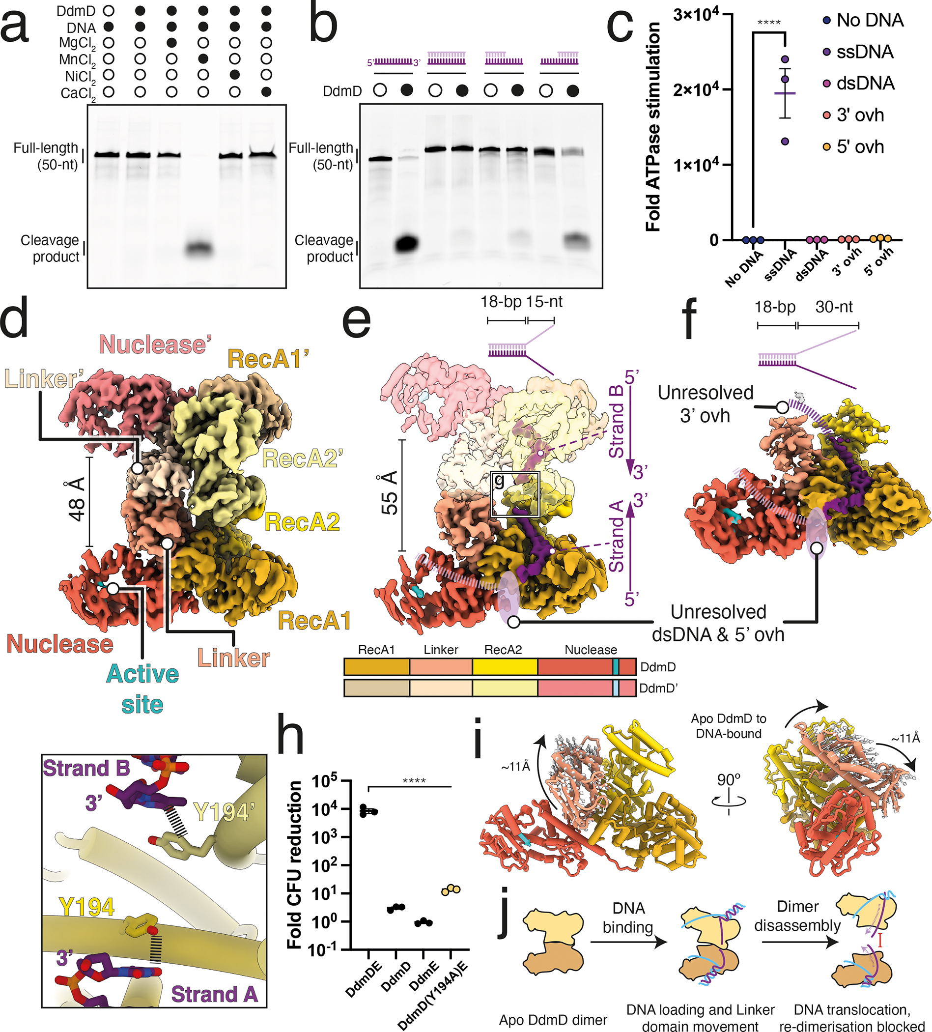
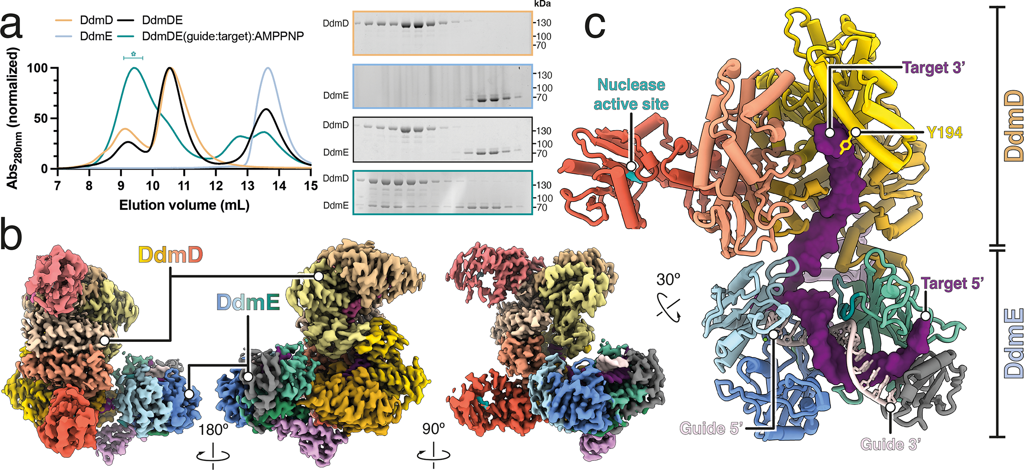
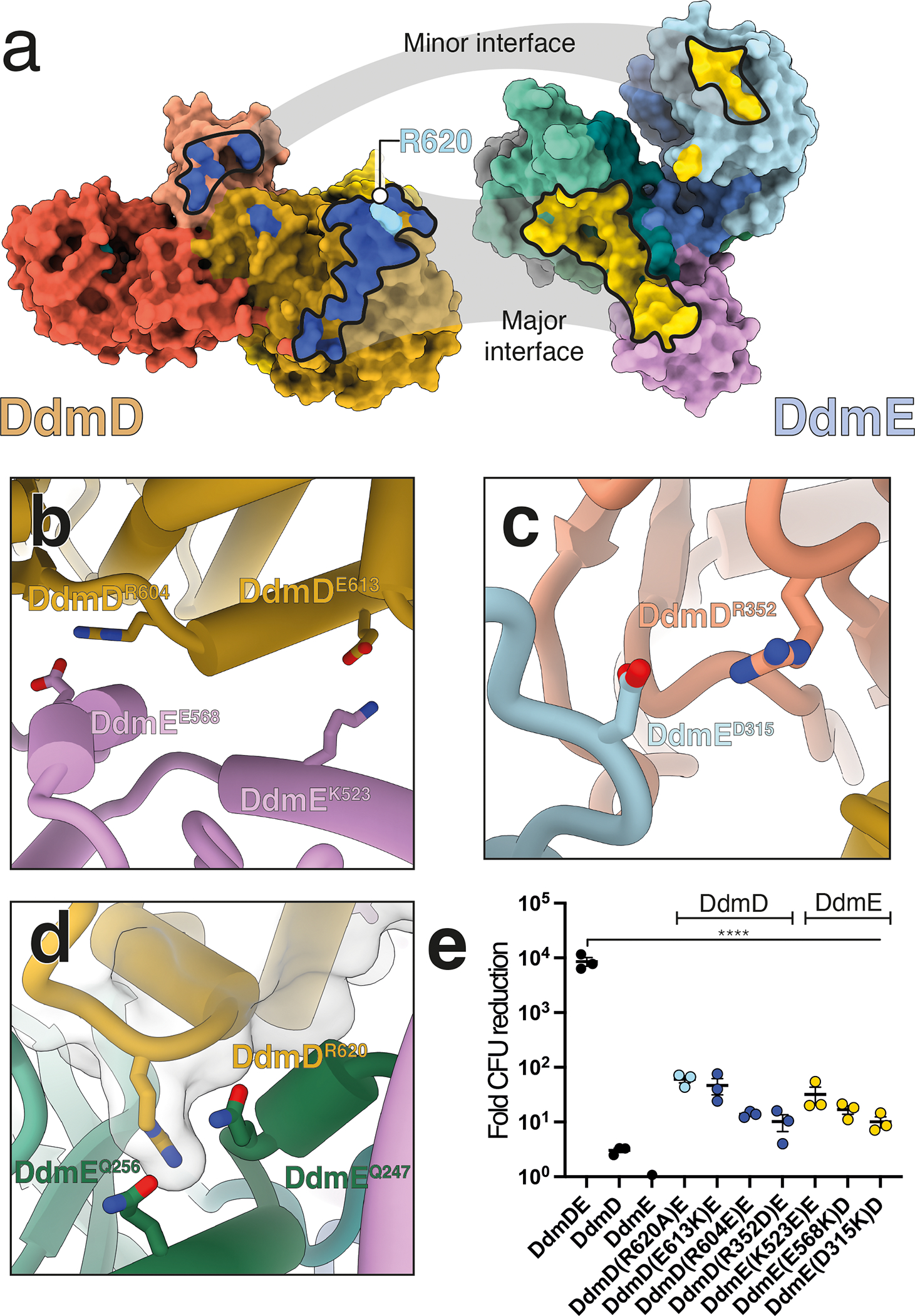
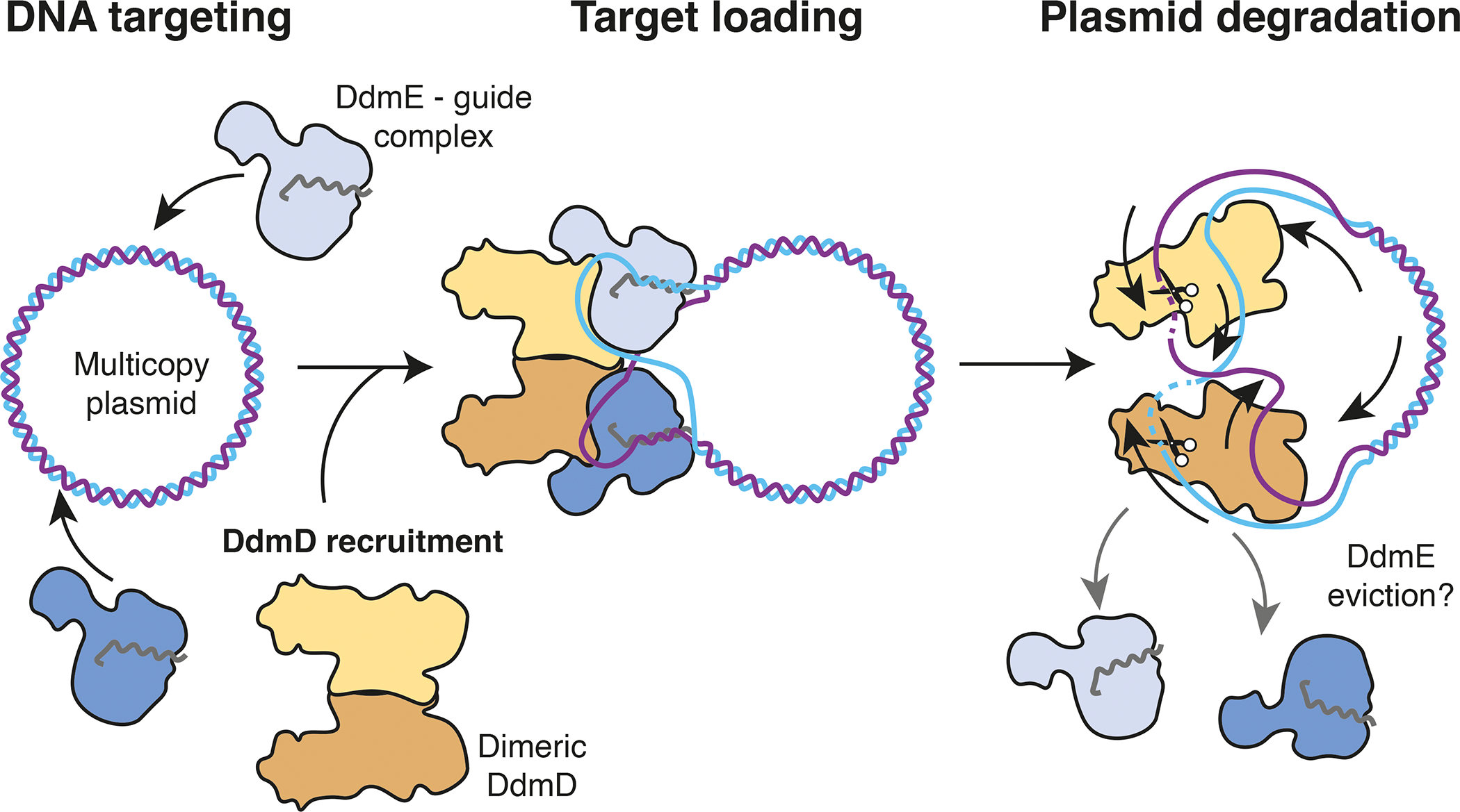
Although eukaryotic Argonautes have a pivotal role in post-transcriptional gene regulation through nucleic acid cleavage, some short prokaryotic Argonaute variants (pAgos) rely on auxiliary nuclease factors for efficient foreign DNA degradation1. Here we reveal the activation pathway of the DNA defence module DdmDE system, which rapidly eliminates small, multicopy plasmids from the Vibrio cholerae seventh pandemic strain (7PET)2. Through a combination of cryo-electron microscopy, biochemistry and in vivo plasmid clearance assays, we demonstrate that DdmE is a catalytically inactive, DNA-guided, DNA-targeting pAgo with a distinctive insertion domain. We observe that the helicase-nuclease DdmD transitions from an autoinhibited, dimeric complex to a monomeric state upon loading of single-stranded DNA targets. Furthermore, the complete structure of the DdmDE-guide-target handover complex provides a comprehensive view into how DNA recognition triggers processive plasmid destruction. Our work establishes a mechanistic foundation for how pAgos utilize ancillary factors to achieve plasmid clearance, and provides insights into anti-plasmid immunity in bacteria.


Conflict of interest statement

Competing interests

The authors declare no competing interests.

Figures

Extended Data Fig 1.
Extended Data Fig 1.. DdmDE denfence system.
a, Representation of VPI-2 defense island. Annotation was performed by PADLOC. b, Schematic representation of in vivo plasmid clearance assay. In brief, DdmD and/or DdmE were transformed into E. coli BL21 DE3 cells. c, representative dilution series of cultures either induced or uninduced. d, Quantification of transformation-fold reduction for DdmDE co-transformed, DdmD and DdmE alone, DdmDE co-transformed in the absence of antibiotic selection, and DdmD – DdmEΔINS domain. Significance between DdmDE and other variants was determined by ordinary one-way ANOVA test. Data are mean ± s.d. of at three independent experiments started from separate colonies. e, Denaturing urea-PAGE gel of DNA and RNA targets bound by DdmE (same samples as in figure 1a). No cleavage was observed. Representative of three independent experiments. For gel source data, see Supplementary Information Figure 1.
Extended Data Fig 2.
Extended Data Fig 2.. Resolutions of cryo-EM structures.
Gold-standard Fourier shell correlation (FSC) curves, Three-dimensional FSC curves and maps colored by local resolution for DdmE; FSC 3.1 Å (a), DdmD apoprotein; FSC 3.2 Å (b), DdmD + short overhang DNA; FSC 3.0 Å (c), DdmD monomer with long overhang DNA; FSC 3.0 Å (d), DdmDE consensus reconstruction; FSC 2.5 Å (e), DdmDE local refinement of DdmE; FSC 2.6 Å (f).
Extended Data Fig 3.
Extended Data Fig 3.. Conformational changes during substrate handover.
a, Comparison of DdmE structure (colored) with DdmE in the context of the DdmDE handover complex. In the handover complex, the N-terminal domain is ordered and present (red). The structures are otherwise identical (RMSD <2 Å). b, 6 Å-low pass filtered map of DdmE with model fitted, showing the flexible density for the extended guide – target duplex. The position of the N-terminal domain is shown as a red box. c & d, comparison of TtAgo (PDB ID 4NCB) with DdmE.
Extended Data Fig 4.
Extended Data Fig 4.. Binding of DNA by DdmD.
a, Cryo-EM 2D class averages of DdmD in the absence and presence of DNA substrates with different overhang lengths. White arrow denotes monomeric DdmD. Structural changes between DdmD apo, short fork DNA and long for DNA. b, Conformational changes in DdmD dimer upon short fork DNA loading. c, Interactions between DdmD and DNA. Residues highlighted are tested in panel c. d, Plasmid interference assay to analyse DdmD-DdmE interactions. Significance between DdmDE and other variants was determined by ordinary one-way ANOVA test. ****P < 0.0001 Data are mean ± s.d. of at three independent experiments started from separate colonies. DdmDE, DdmD and DdmE are the same data as shown in Fig 1h and ED Fig 1 c & d.
Extended Data Fig 5.
Extended Data Fig 5.. DdmD is helicase-nuclease.
a & b, TBE-Urea PAGE gel analysis of DNA cleavage by DdmD in the presence and absence of ATP. A 3’-FAM-labeled DNA substrate was incubated with DdmD as ssDNA (left), annealed to a partially complementary strand (creating a forked duplex with a 30-nt overhang and a 25-bp duplex), and a 5’ overhang substrate (through annealing to a 25-nt complementary strand). Within the structures of DdmD bound to DNA, the 3’ end occupies the RecA helicase channel. ssDNA and forked DNA substrates are degraded, while a larger, incomplete degradation product for the 5’ overhang substrate is observed, since only the ssDNA overhang itself can be cleaved by DdmD. This indicates that unwinding and translocation is essential for full duplex degradation by DdmD. Representative of three independent experiments. b, Nuclease assay as in a, but the FAM is on the 5’ single-stranded end of the forked substrate. Since ssDNA is readily cleaved by DdmD, complete degradation is observed in the absence of ATP. Representative of three independent experiments. c, DdmD DNA unwinding assay, where a fluorophore-quencher pair (FAM and BlackHole Quencher) are on each strand of the forked substrate. DdmD unwinding is ATP-dependent. Data are mean ± s.d. of at three technical replicates d, Gel-based unwinding assay. ATP and DdmD are required for duplex unwinding, as monitored using native TBE 10% PAGE gel. e, Visualization of DNA-bound dimeric (left) and monomeric (right) complex by cryoEM. The monomeric DNA-bound DdmD suffers from severe preferred orientation. Representative of three independent experiments. f, Native PAGE gel analysis of DdmD oligomeric state in the absence and presence of DNA substrates. Apo DdmD runs as a dimeric species, which shifts to a mixture of monomer and dimer with ssDNA, and predominantly monomer with the 30-nt overhang forked DNA substrate used for structural analysis in Fig 2. Representative of three independent experiments. g, Negative stain EM 2D classes of DdmD in complex with DNA substates used for panel f. h, Visualization of DNA-bound dimeric (left) and monomeric (right) complex by cryoEM. The monomeric DNA-bound DdmD suffers from severe preferred orientation. i, SEC chromatogram of DdmDE handover complex (as shown in Fig 3A, green), and handover complex reconstituted with DdmD(R620A) point mutant (pink). A280nm absorbance has been normalized to the size of the largest peak. The DdmD(R620A)E HC peak (peak i) is much smaller than wild-type, and unbound, dissociated DdmE is present (peak ii). Free DNA is in peak iii*, and has an A260nm/A280nm ratio of 1.8, while peaks i and ii had ratios of 1.2 and 1.1, respectively. SDS-PAGE analysis of DdmD(R620A)E HC SEC fractions is shown below. For gel source data, see Supplementary Information Figure 1.
Extended Data Fig 6.
Extended Data Fig 6.. Structures of different DdmD – DdmE oligomers.
a, DdmD2E2 complex, colored by local resolution. b & c, FSC and Three-dimensional FSC curves for DdmD2E2 complex; FSC 3.0 Å. d & e, DdmD6E6 and DdmD2E2 complexes, with DdmD colored beige and DdmE colored blue. 3- and 2-fold symmetry axes are annotated.
Extended Data Fig 7.
Extended Data Fig 7.. Conservation analysis.
Conservation analysis of DdmE (a) and DdmD (b). For DdmD, the RecA helicase channel, nuclease active site and dimer interface are highly conserved. c, Conservation of DdmD – DdmE handover complex interface. DdmD(R620) is conserved and is buried within a similarly conserved pocked of DdmE. d, Electrostatics of DdmDE handover complex.
Fig 1.
Fig 1.. DdmE is a prokaryotic Argonaute that uses 5’-P DNA guides to identify DNA targets.
a, Native gel shift assay to determine guide and target binding preferences of DdmE. b, 3.1 Å-resolution cryo-EM structure of DdmE in complex with 5’-P DNA guide and DNA target. c, Model of DdmE nucleoprotein complex, with corresponding protein domains shown below. d, Guide 5’-P binding. e, Interactions between DdmE sensor loop and the guide – target duplex. f, Comparison of Thermus thermophilus (Tt)Ago active site with corresponding region of DdmE. g, In vivo plasmid clearance assay, testing the importance of DdmE residues. Significance between DdmDE and other variants was determined by ordinary one-way ANOVA test. Data are mean ± s.d. of at three independent experiments. ***P = 0.0001, ****P < 0.0001. For gel source data, see SI Fig. 1.
Fig 2.
Fig 2.. DdmD is a dimeric helicase-nuclease.
a, Dependence of ssDNA cleavage by DdmD on divalent cations. b, DdmD cleaves ssDNA, but not duplexed DNA. Minor cleavage products are observed for a DNA duplex with a 5’ overhang. c, Stimulation of DdmD ATPase activity by DNA substrates as used in b. Significance between ATPase stimulation by ssDNA compared to no DNA was determined by ordinary one-way ANOVA test (n=3 technical replicates). ****P < 0.0001. Error bars correspond to standard error of the mean. d, 3.2 Å-resolution cryo-EM structure of DdmD, and corresponding model. Domains are colored as in the below domain schematic. e, 3.0 Å-resolution cryo-EM structure of DdmD in complex with short overhang duplex. The second monomer of DdmD is shown as transparent density. f, 3.0 Å-resolution structure of DdmD in complex with a DNA substrate with longer overhangs. g, Y194 caps the 3’ end of the ssDNA within RecA channel. h, In vivo plasmid clearance assay. Data are mean ± s.d. of three independent experiments started from separate colonies. i, Structural changes to DdmD Linker domain upon DNA binding. j, Schematic of DdmD activation. For gel source data, see SI Fig. 1.
Fig 3.
Fig 3.. DdmE recruits DdmD upon target recognition for plasmid degradation.
a, Size-exclusion chromatography analysis of DdmD – DdmE – guide – target complex reconstitution, with corresponding fractions monitored by SDS-PAGE (right). *Denotes peak fraction used for cryo-EM preparation of DdmDE handover complex. b, 2.5 Å-resolution cryo-EM structure of DdmDE handover complex. c, Top-down view of DdmDE handover complex, showing path of DNA target as it is transferred from DdmE to DdmD. Top DdmD monomer has been removed for viewing clarity. Y194 caps the 3’ end of the DNA target. For gel source data, see SI Fig. 1.
Fig 4.
Fig 4.. DdmD-DdmE interface is essential for plasmid clearance.
a, Structures of DdmD and DdmE from handover complex (Fig 3), with complementary surfaces of DdmD and DdmE. Interface between DdmD(RecA1) and DdmE(INS – L2) are highlighted as blue and yellow, respectively. b, Electrostatic contacts between DdmD(RecA1) and DdmE(INS) at the major interface. c, An electrostatic contact at the minor interface between DdmD(linker) and DdmE (MID) domains. d, DdmD(R620A) interacts with DdmE within a conserved pocket. e, Plasmid interference assay to analyze DdmD-DdmE interactions. Significance between DdmDE and other variants was determined by ordinary one-way ANOVA test. ****P < 0.0001. Data are mean ± s.d. of three independent experiments started from separate colonies. DdmDE, DdmD and DdmE are the same data as shown in Fig 1h.
Fig 5.
Fig 5.. Model of targeted plasmid degradation by DdmDE.
DdmE (or multiple DdmE) uses a 5’-P DNA guide to recognize multicopy plasmids. Target recognition results in recruitment of a dimeric, autoinhibited DdmD, and the DNA is loaded within the RecA channel. Initiation of ssDNA-activated ATP hydrolysis results in the processive translocation of DdmD along the DNA, with the dimer splitting. As DdmD translocate, the non-target DNA strand may be passed to the nuclease active site for non-specific cleavage.

Similar articles

Cited by

References

Main Text References

    1. Bobadilla Ugarte P, Barendse P and Swarts DC (2023) Argonaute proteins confer immunity in all domains of life. Curr Opin Microbiol, 74, 102313. - PubMed
    1. Jaskólska M, Adams DW and Blokesch M (2022) Two defence systems eliminate plasmids from seventh pandemic Vibrio cholerae. Nature 2022 604:7905, 604, 323–329. - PMC - PubMed
    1. Hampton HG, Watson BNJ and Fineran PC (2020) The arms race between bacteria and their phage foes. Nature, 577, 327–336. - PubMed
    1. Georjon H and Bernheim A (2023) The highly diverse antiphage defence systems of bacteria. Nature Reviews Microbiology 2023, 10.1038/s41579-023-00934-x. - DOI - PubMed
    1. Pinilla-Redondo R, Mayo-Muñoz D, Russel J, Garrett RA, Randau L, Sørensen SJ and Shah SA (2019) Type IV CRISPR–Cas systems are highly diverse and involved in competition between plasmids. Nucleic Acids Res, 48, 2000–2012. - PMC - PubMed

Methods references

    1. Mastronarde DN (2005) Automated electron microscope tomography using robust prediction of specimen movements. J Struct Biol, 152, 36–51. - PubMed
    1. Punjani A (2021) Real-time cryo-EM structure determination. Microscopy and Microanalysis, 27, 1156–1157.
    1. Punjani A, Rubinstein JL, Fleet DJ and Brubaker MA (2017) CryoSPARC: Algorithms for rapid unsupervised cryo-EM structure determination. Nat Methods, 14, 290–296. - PubMed
    1. Terwilliger TC, Ludtke SJ, Read RJ, Adams PD and Afonine PV (2020) Improvement of cryo-EM maps by density modification. Nature Methods 2020 17:9, 17, 923–927. - PMC - PubMed
    1. Croll TI (2018) ISOLDE: a physically realistic environment for model building into low-resolution electron-density maps. Acta Crystallographica Section D, 74, 519–530. - PMC - PubMed

MeSH terms

LinkOut - more resources