. 2024 Jul:85:101957.
doi: 10.1016/j.molmet.2024.101957. Epub 2024 May 11.

Suppression of hepatic ChREBP⍺-CYP2C50 axis-driven fatty acid oxidation sensitizes mice to diet-induced MASLD/MASH

Affiliations

Suppression of hepatic ChREBP⍺-CYP2C50 axis-driven fatty acid oxidation sensitizes mice to diet-induced MASLD/MASH

Deqiang Zhang et al. Mol Metab. 2024 Jul.

Abstract

Objectives: Compromised hepatic fatty acid oxidation (FAO) has been observed in human MASH patients and animal models of MASLD/MASH. It remains poorly understood how and when the hepatic FAO pathway is suppressed during the progression of MASLD towards MASH. Hepatic ChREBP⍺ is a classical lipogenic transcription factor that responds to the intake of dietary sugars.

Methods: We examined its role in regulating hepatocyte fatty acid oxidation (FAO) and the impact of hepatic Chrebpa deficiency on sensitivity to diet-induced MASLD/MASH in mice.

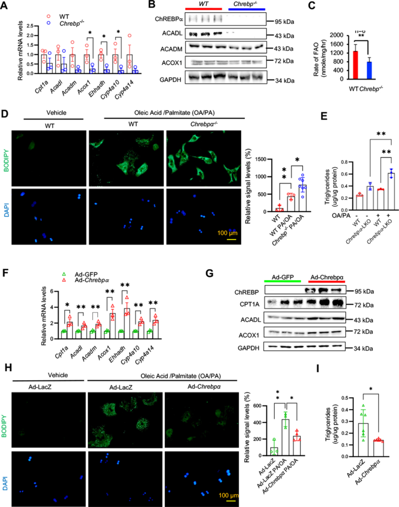
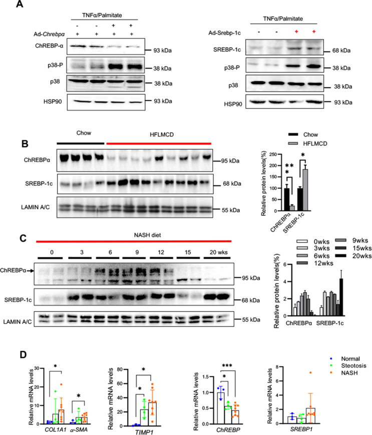
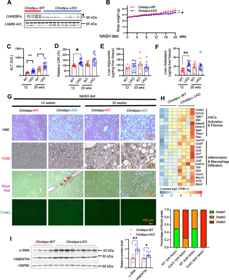
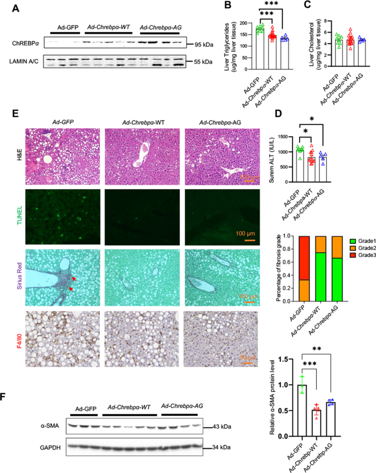
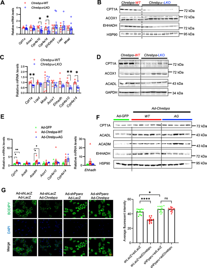
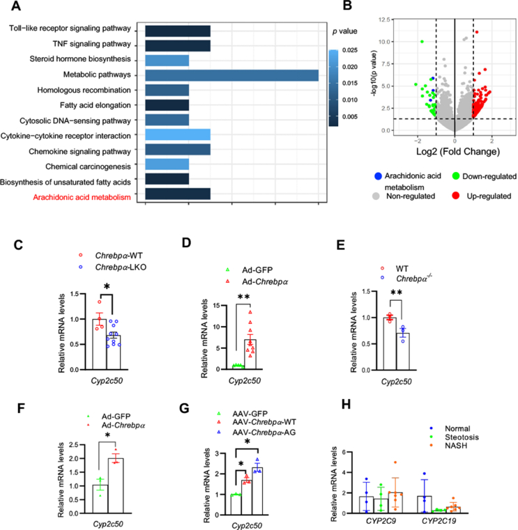
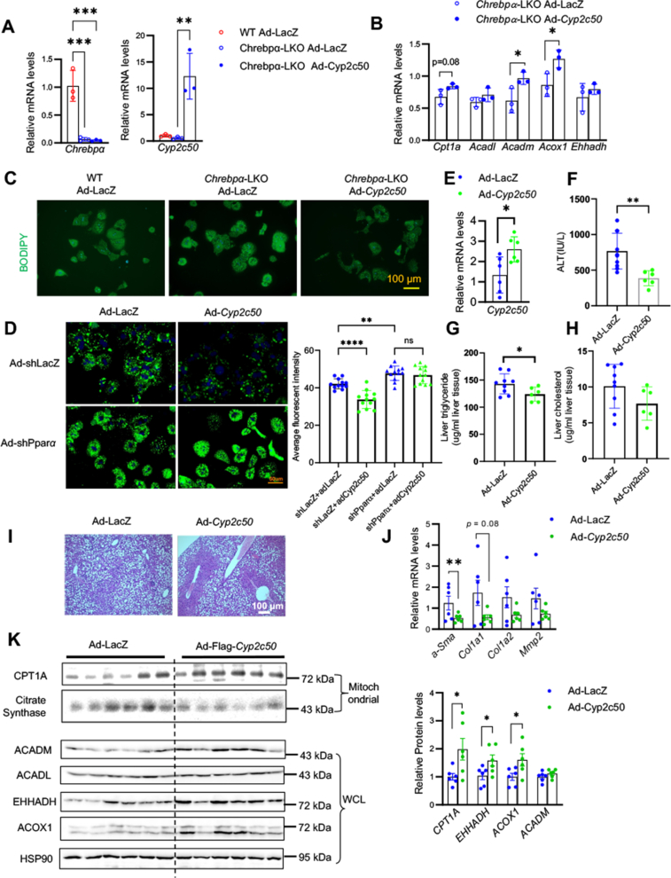
Results: We discovered that hepatocyte ChREBP⍺ is both necessary and sufficient to maintain FAO in a cell-autonomous manner independently of its DNA-binding activity. Supplementation of synthetic PPAR⍺/δ agonist is sufficient to restore FAO in Chrebp-/- primary mouse hepatocytes. Hepatic ChREBP⍺ was decreased in mouse models of diet-induced MAFSLD/MASH and in patients with MASH. Hepatocyte-specific Chrebp⍺ knockout impaired FAO, aggravated liver steatosis and inflammation, leading to early-onset fibrosis in response to diet-induced MASH. Conversely, liver overexpression of ChREBP⍺-WT or its non-lipogenic mutant enhanced FAO, reduced lipid deposition, and alleviated liver injury, inflammation, and fibrosis. RNA-seq analysis identified the CYP450 epoxygenase (CYP2C50) pathway of arachidonic acid metabolism as a novel target of ChREBP⍺. Over-expression of CYP2C50 partially restores hepatic FAO in primary hepatocytes with Chrebp⍺ deficiency and attenuates preexisting MASH in the livers of hepatocyte-specific Chrebp⍺-deleted mice.

Conclusions: Our findings support the protective role of hepatocyte ChREBPa against diet-induced MASLD/MASH in mouse models in part via promoting CYP2C50-driven FAO.

Keywords: Carbohydrate-response element binding protein (ChREBP); Fatty acid oxidation; Lipid metabolism; Metabolic-Associated Steatohepatitis.


Conflict of interest statement

Declaration of competing interest We declare there is no conflict interest for this manuscript.

Figures

Figure 1
Figure 1
ChREBPα is both necessary and sufficient to promote fatty acid oxidation in mouse hepatocytes. Primary mouse hepatocytes (PMHs) were isolated from 8wk-old Chrebp−/− and WT littermates and cultured in serum-free medium for 24 h later prior to RT-qPCR for and immunoblotting to assess the expression of genes of FAO (AB) as well as incubation with H3-palmitate for 4 h before the measurement of the rate of FAO (C). WT and Chrebp−/− PMH were cultured in MEM medium with 300 μM palmitate and 600 μM oleate for 8 h, then switched to serum-free MEM medium for 16 h before lipid droplet detection by BODIPY staining (D) and cellular triglycerides assay (E). WT PMHs were isolated and transduced with either Ad-GFP control vs. Ad-Chrebpα. FAO enzyme expression was assessed by RT-qPCR and immunoblotting (F-G), while lipid content was measured by BODIPY staining and cellular TG assay after palmitate/oleate incubation (H–I). The data were plotted as Mean ± SEM (n = 3). ∗p < 0.05, ∗∗p < 0.01 by the Student's t-test.
Figure 2
Figure 2
Hepatic ChREBPα is suppressed in the liver of mouse models with diet-induced NASH and human patients of NASH. WT PMHs from male mice were firstly transduced with either Ad-Chrebp⍺ (A) or Ad-Srepb-1c (B) prior to treatment with TNF-⍺ (10 ng/ml) plus palmitate (300 μM). The protein levels of ChREBP⍺ or SREBP-1c were determined by immunoblotting. (C) Nuclear fractions of the liver samples of mice fed either regular chow vs. HFLMCD diet for 10 weeks or (D) mice fed NASH diet for 0, 3, 6, 9, 12, 15, or 20 weeks. The nuclear abundance of ChREBP⍺ and SREBP-1c were determined by immunoblotting. (E-F) Livers samples form patients at different stages of NAFLD (Normal, Steatosis, and NASH) were subjected to RT-qPCR to exam the expression levels of fibrosis markers (COL1A1, ⍺-SMA, and TIMP1) and ChREBP and SREBP-1. The data were plotted as mean ± SEM. ∗p < 0.05, ∗∗∗∗p < 0.0001 by the Student's t-test for B, and by one-way ANOVA for D.
Figure 3
Figure 3
Adult-onset deletion of hepatic Chrebp⍺ sensitizes mice to NASH diet-induced liver steatosis. 8-wk old Chrebpαflox/flox mice on regular chow were injected with AAV-TBG-Cre (Chrebpα-LKO) or AAV-TBG-GFP (Chrebpα-WT) via tail vein, and 2 weeks later, the mice were switched to NASH diet. (A) Western blot against ChREBPα was used to confirm Chrebpα deletion. (B) Body weight was monitored weekly. (CD) Serum ALT and LDH at 12 weeks and 20 weeks of NASH diet feeding, (E-F) liver triglyceride and cholesterol. (G) H&E staining, F4/80 immunohistochemistary, Sirius Red staining, and TUNEL staining were used to detect lipid droplets, macrophage, fibrosis and apoptosis, respectively. (H) RNAs-seq analysis of liver tissue from Chrebpα-LKO and Chrebpα-WT mice fed NASH diet for 20 weeks. Heat-map of DGE associated with liver fibrosis and inflammation/macrophage infiltration was generated. (I) Western blot against α-SMA and Vimentin was used to assess fibrosis. The data were plotted as Mean ± SEM. ∗p < 0.05, ∗∗p < 0.01, & ∗∗∗∗p < 0.0001 by linear regression for B, by one-way ANOVA for C, D &F, and by the Student's t-test for I.
Figure 4
Figure 4
Hepatic ChREBPα overexpression ameliorates liver steatosis and hepatocyte injury in mice with pre-existing NASH independently of its lipogenic action. 8-wk old wildtype male mice fed HFLMCD diet for 2 weeks, and then injected with or Ad-GFP (n = 8), Ad-Chrebpα-WT (n = 17), or Ad-Chrebp⍺-AG (n = 5) via tail vein. After another week of HFLMCD feeding, (A) Confirmation of hepatic ChREBPα overexpression by immunoblotting, (B–C) liver triglycerides and cholesterol, (D) serum ALT. (E) H&E staining, F4/80 immunohistochemistry, Sirius Red staining and TUNEL staining were used to detect lipid droplets, macrophage infiltration, fibrosis, and apoptosis, respectively. (F) Protein levels of ⍺-SMA in the liver by immunoblotting. The data were plotted as Mean ± SEM. ∗p < 0.05, ∗∗p < 0.01 & ∗∗∗p < 0.001 by one-way ANOVA.
Figure 5
Figure 5
Manipulations of hepatic ChREBP⍺ impact FAO pathway in NASH. (A-D) Hepatic Chrebpα deficiency impairs FAO in the liver. Liver tissues from Chrebpα-WT and Chrebpα-LKO mice fed NASH diet for 12 weeks (A&B) or 20 weeks (C&D) were subjected to RT-qPCR and western blot to assess the expression of FAO enzymes. (E-F) ChREBPα overexpression promotes FAO in the liver. Liver tissues from mice injected with Ad-GFP, Ad-Chrebpα-WT, or Ad-Chrebpα-AG and fed HFLMCD diet for 3 weeks were subjected to RT-qPCR and immunoblotting to assess FAO enzyme expression. (G) PMHs were transduced with either Ad-shLacZ or Ad-shPpar⍺ with or without Ad-Chrebp⍺ prior to BODIPY staining. The data were plotted as Mean ± SEM. ∗p < 0.05, ∗∗p < 0.01 by the Student's t-test for A&C, by one-way ANOVA for E.
Figure 6
Figure 6
Identification of epoxogenase pathway of arachidonic acid metabolism as a novel pathway of ChREBPα during diet-induced NASH. Liver tissues from Chrebpα-LKO vs. Chrebpα-WT mice on NASH diet for 20 weeks, as well as liver tissues from WT mice fed 2-wk HFLMCD diet and injected Ad-Chrebpα vs. Ad-GFP were subjected to RNA-seq analysis (n = 4). (A-B) Regulation of epoxygenase pathway of the arachidonate metabolism by hepatic Chrebpα via Pathway Analysis. (C) Reduced hepatic Cyp2c50 expression in Chrebpα-LKO mice following NASH diet; (D) Elevated hepatic Cyp2c50 expression in Ad-GFP vs. Ad-Chrebp⍺-injected mice. (E) Decreased expression of hepatic Cyp2c50 in Chrebp−/− mice. (F) Cyp2c50 expression in Ad-GFP vs. Ad-Chrebp⍺ PMHs, (G) Cyp2c50 expression in PMHs from mice injected with AAV-TBG-GFP, AAV-TBG-Chrebpα-WT, or Chrebpα-AG. (H) Expression levels of CYP2C9 and CYP2C19 (the human ortholog of mouse Cyp2c50) in the liver of normal human vs. liver steatosis or NASH patients.
Figure 7
Figure 7
Restoring CYP2c50 expression rescues FAO and alleviates diet-induced liver steatosis in Chrebpα deficient condition. PMHs from Chrebpα-WT and Chrebpα-LKO mice were transduced with adenovirus overexpressing LacZ or Cyp2c50. The cells were harvested 24 h later for RT-qPCR to exam the expression levels of Cyp2c50 and genes involved in FAO (A-B) and BODIPY (C) staining to assess lipid accumulation. (D) PMHs were transduced with either Ad-shLacZ or Ad-shPpar⍺ with or without Ad-Cyp2c50 prior to BODIPY staining. Chrebpα-LKO mice were injected with Ad-Cyp2c50 or Ad-LacZ. 2 days later, the mice were fed HFLMCD diet for 9 days, and then dissected. Liver Cyp2c50 mRNA were measured by RT-qPCR (E). Liver injury was assessed by serum ALT (F), hepatic lipid content by liver triglycerides (G), cholesterol (H), and H&E staining (I), and the gene expression of hepatic fibrosis markers by RT-qPCR (J) and FAO enzymes by immunoblotting with quantification (K).

Similar articles

Cited by

References

    1. Alves-Bezerra M., Cohen D.E. Triglyceride metabolism in the liver. Compr Physiol. 2017;8(1):1–8. - PMC - PubMed
    1. Rui L. Energy metabolism in the liver. Compr Physiol. 2014;4(1):177–197. - PMC - PubMed
    1. Loomba R., Friedman S.L., Shulman G.I. Mechanisms and disease consequences of nonalcoholic fatty liver disease. Cell. 2021;184(10):2537–2564. - PubMed
    1. Savage D.B., Petersen K.F., Shulman G.I. Disordered lipid metabolism and the pathogenesis of insulin resistance. Physiol Rev. 2007;87(2):507–520. - PMC - PubMed
    1. Softic S., Cohen D.E., Kahn C.R. Role of dietary fructose and hepatic de novo lipogenesis in fatty liver disease. Dig Dis Sci. 2016;61(5):1282–1293. - PMC - PubMed

MeSH terms

Substances