. 2024 May 13;16(1):39.
doi: 10.1038/s41368-024-00301-3.

Unveiling the oral-gut connection: chronic apical periodontitis accelerates atherosclerosis via gut microbiota dysbiosis and altered metabolites in apoE-/- Mice on a high-fat diet

Affiliations

Unveiling the oral-gut connection: chronic apical periodontitis accelerates atherosclerosis via gut microbiota dysbiosis and altered metabolites in apoE-/- Mice on a high-fat diet

Guowu Gan et al. Int J Oral Sci. .

Abstract

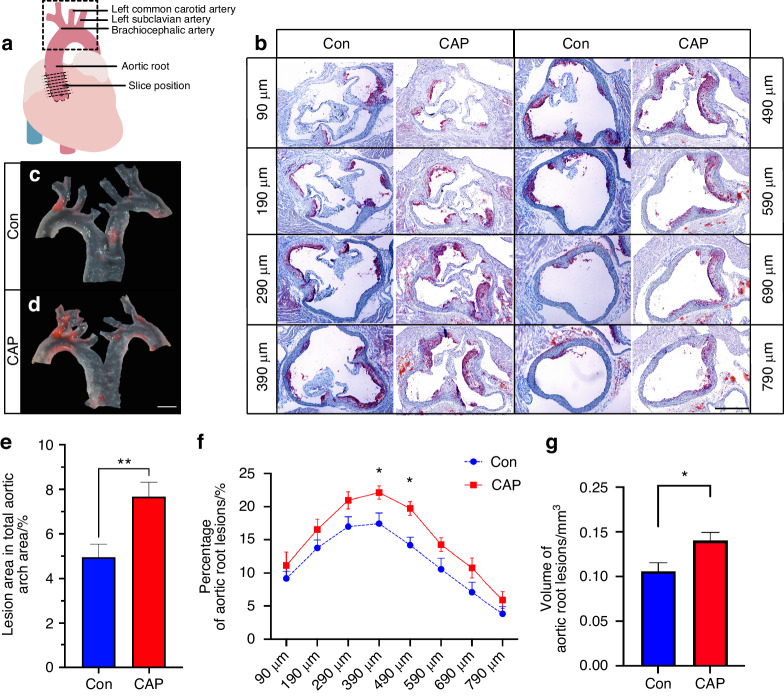
The aim of this study was to explore the impact of chronic apical periodontitis (CAP) on atherosclerosis in apoE-/- mice fed high-fat diet (HFD). This investigation focused on the gut microbiota, metabolites, and intestinal barrier function to uncover potential links between oral health and cardiovascular disease (CVD). In this study, CAP was shown to exacerbate atherosclerosis in HFD-fed apoE-/- mice, as evidenced by the increase in plaque size and volume in the aortic walls observed via Oil Red O staining. 16S rRNA sequencing revealed significant alterations in the gut microbiota, with harmful bacterial species thriving while beneficial species declining. Metabolomic profiling indicated disruptions in lipid metabolism and primary bile acid synthesis, leading to elevated levels of taurochenodeoxycholic acid (TCDCA), taurocholic acid (TCA), and tauroursodeoxycholic acid (TDCA). These metabolic shifts may contribute to atherosclerosis development. Furthermore, impaired intestinal barrier function, characterized by reduced mucin expression and disrupted tight junction proteins, was observed. The increased intestinal permeability observed was positively correlated with the severity of atherosclerotic lesions, highlighting the importance of the intestinal barrier in cardiovascular health. In conclusion, this research underscores the intricate interplay among oral health, gut microbiota composition, metabolite profiles, and CVD incidence. These findings emphasize the importance of maintaining good oral hygiene as a potential preventive measure against cardiovascular issues, as well as the need for further investigations into the intricate mechanisms linking oral health, gut microbiota, and metabolic pathways in CVD development.


Conflict of interest statement

The authors declare no competing interests.

Figures

Fig. 1
Fig. 1
CAP exacerbates atherosclerosis in apoE−/− mice. a Schematic of the dissection of the aortic root and aortic arch of the heart. The dotted straight line indicates the location of slicing at the aortic root, and the dashed rectangle indicates the location of the aortic arch sampling. b Representative images of frozen sections of the aortic root from frozen Oil Red O-stained sections from eight locations. Scale bar, 500 μm. c and d Representative images of Oil Red O-stained aortic arches. Scale bar, 1 mm. e Measurement of the percentage of atherosclerotic plaque at the aortic arch, n = 18–19; independent sample t-test; mean ± SEM; **P < 0.01. f and g Measurement of the percentage and volume of atherosclerotic plaques at the aortic root; n = 12; independent sample t-test; mean ± SEM, *P < 0.05
Fig. 2
Fig. 2
CAP leads to dysbiosis of the gut microbiota in apoE−/− mice. a Comparison of the 20 richest abundances at the bacterial genus level between the two groups. bi CAP leads to differentially expressed genera of gut microbes; n = 10, independent sample t-test; mean ± SEM, * indicates P < 0.05. j Enriched taxa in the CAP group (red) are indicated by negative log discriminant analysis (LDA) scores, and enriched taxa in the Con group have positive scores (blue). Only taxa with an LDA score (log 10) > ± 2 are shown. n = 10, LDA score > ±2 indicates P < 0.05. k Beta diversity of the gut microbiota from the two groups determined by Bray‒Curtis distance, ANOSIM test, P < 0.01, n = 10. Alpha diversity was demonstrated by the Chao1 (l), observed OTUs (m), Shannon (n), and Simpson (o) indices between the two groups
Fig. 3
Fig. 3
LC‒MS detection of altered metabolites in CAP-treated mice. a OPLS-DA score plot of the intestinal metabolites from the CAP and Con groups, n = 10, permutation test P ≤ 0.05, Q2 = 0.378. b Stacked column chart of the percentage of metabolites that play a biological role. c Overview of the metabolite set enrichment analysis based on the intestinal metabolites. d Volcano plot of the univariate analysis showing the differentially abundant metabolites. e Correlation clustering heatmap analysis of metabolites and the abundance of gut microbiota at the genus level. The columns show the different metabolites or the percentage of the lesion area, and each row indicates the different genera of the gut microbiota. The red and blue colors in the plot indicate high and low intensities, respectively; n = 10; *P < 0.05; **P < 0.01; ***P < 0.001
Fig. 4
Fig. 4
Altered bile acid metabolism in CAP-treated mice. a OPLS-DA score plot of the bile acid metabolites from the two groups; n = 10, P ≤ 0.01 in permutation test, Q2 = 0.767. b Volcano plot of univariate analysis showing the differentially abundant metabolites. c Box plot of differentially abundant metabolites, n = 10; *P < 0.05; **P < 0.01. d and e Clustered heatmap of correlations between 36 bile acids; the red and green colors in the plot indicate high and low intensities, respectively. f Correlation clustering heatmap analysis of bile acid levels and the abundance of the gut microbiota at the genus level. Each column refers to the different bile acids or the percentage of the lesion area, and each row indicates the different genera of the gut microbiota. The red and blue colors in the plot indicate high and low intensities, respectively; n = 10; *P < 0.05; **P < 0.01; ***P < 0.001
Fig. 5
Fig. 5
The intestinal mucus barrier was impaired in the apoE−/− mice after CAP treatment. a Representative images of H&E staining, AB-PAS staining and Muc-2 immunofluorescence staining; scale bar, 500 μm; b Measurement of average Muc-2 fluorescence intensity, n = 9; independent sample t-test, mean ± SEM; *P < 0.05; c Measurement of the area of positive AB-PAS staining, n = 12; independent sample t-test, mean ± SEM, *P < 0.05
Fig. 6
Fig. 6
Increased intestinal permeability in apoE−/− mice after CAP treatment. a Representative images of Zo-1, occluding, and claudin immunofluorescence staining. Scale bar, 100 μm; b Differences in protein expression in mouse colon tissue. c Intestinal permeability assay for FD-4 in serum; n = 5; paired samples t-test, P < 0.05
Fig. 7
Fig. 7
Schematic description of chronic apical periodontitis exacerbating atherosclerosis by affecting the gut microbiota and bile acid metabolism to promote intestinal permeability

References

    1. Ricucci D, Siqueira JF., Jr. Biofilms and apical periodontitis: study of prevalence and association with clinical and histopathologic findings. J. Endod. 2010;36:1277–1288. doi: 10.1016/j.joen.2010.04.007. - DOI - PubMed
    1. Persoon IF, Ozok AR. Definitions and epidemiology of endodontic infections. Curr. Oral Health Rep. 2017;4:278–285. doi: 10.1007/s40496-017-0161-z. - DOI - PMC - PubMed
    1. Sasaki H, et al. Interrelationship between periapical lesion and systemic metabolic disorders. Curr. Pharm. Design. 2016;22:2204–2215. doi: 10.2174/1381612822666160216145107. - DOI - PMC - PubMed
    1. Segura-Egea JJ, Martin-Gonzalez J, Castellanos-Cosano L. Endodontic medicine: connections between apical periodontitis and systemic diseases. Int. Endod. J. 2015;48:933–951. doi: 10.1111/iej.12507. - DOI - PubMed
    1. Cotti E, Mercuro G. Apical periodontitis and cardiovascular diseases: previous findings and ongoing research. Int. Endod. J. 2015;48:926–932. doi: 10.1111/iej.12506. - DOI - PubMed

Publication types