. 2024 May 13;15(1):4036.
doi: 10.1038/s41467-024-48232-1.

Targeting bacterial nickel transport with aspergillomarasmine A suppresses virulence-associated Ni-dependent enzymes

Affiliations

Targeting bacterial nickel transport with aspergillomarasmine A suppresses virulence-associated Ni-dependent enzymes

David Sychantha et al. Nat Commun. .

Abstract

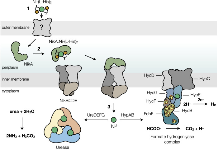
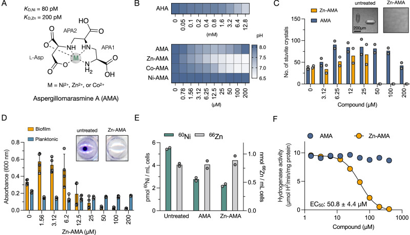
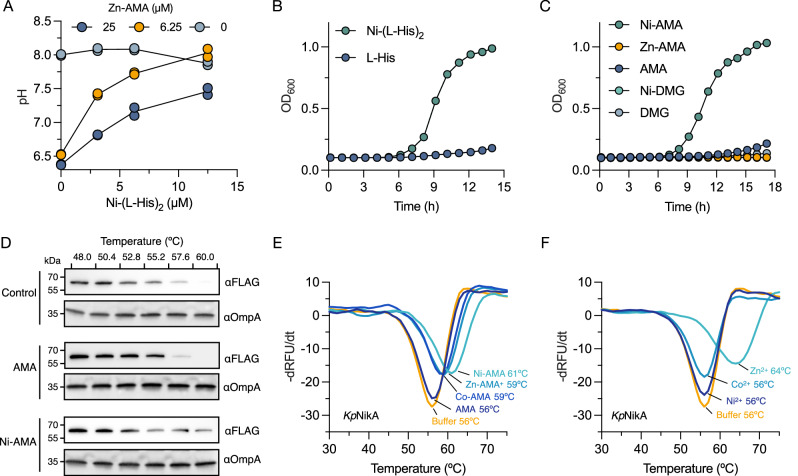
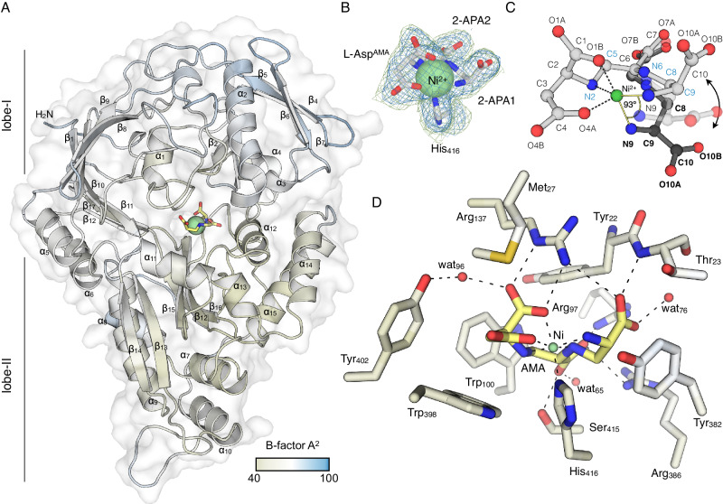
Microbial Ni2+ homeostasis underpins the virulence of several clinical pathogens. Ni2+ is an essential cofactor in urease and [NiFe]-hydrogenases involved in colonization and persistence. Many microbes produce metallophores to sequester metals necessary for their metabolism and starve competing neighboring organisms. The fungal metallophore aspergillomarasmine A (AMA) shows narrow specificity for Zn2+, Ni2+, and Co2+. Here, we show that this specificity allows AMA to block the uptake of Ni2+ and attenuate bacterial Ni-dependent enzymes, offering a potential strategy for reducing virulence. Bacterial exposure to AMA perturbs H2 metabolism, ureolysis, struvite crystallization, and biofilm formation and shows efficacy in a Galleria mellonella animal infection model. The inhibition of Ni-dependent enzymes was aided by Zn2+, which complexes with AMA and competes with the native nickelophore for the uptake of Ni2+. Biochemical analyses demonstrated high-affinity binding of AMA-metal complexes to NikA, the periplasmic substrate-binding protein of the Ni2+ uptake system. Structural examination of NikA in complex with Ni-AMA revealed that the coordination geometry of Ni-AMA mimics the native ligand, Ni-(L-His)2, providing a structural basis for binding AMA-metal complexes. Structure-activity relationship studies of AMA identified regions of the molecule that improve NikA affinity and offer potential routes for further developing this compound as an anti-virulence agent.


Conflict of interest statement

The authors declare no competing interests.

Figures

Fig. 1
Fig. 1. Nickel trafficking in Klebsiella pneumoniae.
1) Ni2+ is sequestered by l-histidine to form a 2:1 complex (Ni-(l-His)2), passing through an unknown porin into the periplasm. 2) Ni-(l-His)2 is bound by NikA once in the periplasm and transported into the cytoplasm via the NikBCDE ABC-type transporter. 3) In K. pneumoniae, cytoplasmic Ni2+ is partitioned between two trafficking systems to mature Ni-dependent enzymes. UreDEFG and HypAB assemble the active site metallocenters of urease and the [NiFe]-hydrogenase (Hyd-3) of the formate hydrogenlyase complex, respectively. The colors represent different protein subunits and are not assigned to different functional classes.
Fig. 2
Fig. 2. Metal complexes of AMA suppress the activity of Ni-dependent enzymes.
A The metal complexed structure, selectivity, and affinity of AMA. The constituents of AMA are labeled l-aspartate (l-Asp) and 2,3-diaminopropionic acid (APA). B Dose-response urease inhibition assays in K. pneumoniae. Urease-dependent pH changes were determined after growth for 24 h at 37 °C in artificial urine. pH is represented by the color scale determined with phenol red. C Dose-response effect of AMA and Zn-AMA on the nucleation of struvite crystals in K. pneumoniae cultures grown in artificial urine. Crystals were examined microscopically in 96-well plates and visually quantified. The inset shows representative images of struvite crystals in the presence and absence of Zn-AMA. The data represent the representative mean of two replicates. D Quantification of planktonic growth and crystal violet-stained biofilms of K. pneumoniae grown in artificial urine and varying concentrations of Zn-AMA. The inset shows representative images of crystal-violet-stained biofilms. Data represent mean ± s.d. of four technical replicates. E ICP-MS quantification of cellular 60Ni and 66Zn content in K. pneumoniae untreated or treated with 200 μM AMA, or Zn-AMA. Data represent the mean of two biological replicates (F) Dose-response curve of [NiFe]-hydrogenase inhibition in K. pneumoniae grown in LB-glucose for 24 h at 37 °C under anaerobic conditions. Whole-cell hydrogenase assays were performed by adding benzyl viologen (BV) at a final concentration of 1 mg/mL and monitoring its reduction at an absorbance of 630 nm. Data represent the mean of two technical replicates.
Fig. 3
Fig. 3. Bacterial nickel uptake is inhibited by competitive binding to the periplasmic Ni-binding protein NikA from K. pneumoinae.
A Zn-AMA complexes compete for uptake with the nickelophore Ni-(l-His)2. K. pneumoniae was cultured in artificial urine for 24 h at 37 °C with varying amounts of Zn-AMA and Ni-(l-His)2 and the pH of the culture was determined with phenol red. The data represent the mean of duplicate experiments. B, C Growth kinetics of K. pneumoniae in Chelex-100 treated nitrogen-limited media containing urea as a sole nitrogen source supplied with Ni-(l-His)2 (25 μM), l-His (25 μM), Ni-AMA (50 μM), Zn-AMA (50 μM), AMA (50 μM), Ni-DMG (50 μM), or DMG. Data represent the mean of duplicate experiments. D Cellular thermal shift assays (CETSA) of K. pneumoniae producing NikA-FLAG. Relative to the untreated control, AMA (250 μM) and Ni-AMA (250 μM) were incubated with cells for 30 min, followed by heating over a gradient of temperatures. Cell lysates were analyzed by immunoblotting with anti-FLAG-HRP (αFLAG) and anti-OmpA (αOmpA) antibodies. Immunoblots are representative of two independent experiments. E, F Negative derivative melt plots using purified recombinant NikA from K. pneumoniae. Metals or complexes (100 μM) were added to NikA (1 μM) with 2× SYPRO orange. The y axis is displayed as negative derivative (−dRFU/dt) plots of the raw output.
Fig. 4
Fig. 4. Metal complexes of AMA bind to NikA with high affinity.
AD Isothermal titration calorimetry analyses of the interactions between various metal complexes and NikA from K. pneumoniae. The metal complexes (250 μM), prepared at a 1:1 molar ratio (AMA) or 2:1 molar ratio (l-His), were titrated into NikA (15 μM) in 25 mM Tris-HCl, 150 mM NaCl, 0.1% (v/v) Triton X-100, pH 7.5 at 37 °C. Top, raw titration; middle, Wiseman isotherm of integrated peaks; bottom, binding signatures of free energy (∆G), enthalpy (∆H), and entropy (−T∆S). Ni-(l-His)2 and Ni-AMA structures represent the NikA-bound form (PDB 4I8C) and free complex, respectively. Zn-AMA and Co-AMA structures are predicted based on the known structure of Ni-AMA.
Fig. 5
Fig. 5. Crystal structure of E. coli NikA in complex with Ni-AMA.
A Cartoon/surface representation of NikA colored by B-factor. AMA and Ni2+ are shown in stick and sphere representation, respectively. The color bar depicts the B-factor scale. B The 2mFo-DFc map and mFo-DFc omit map are shown in blue and green mesh and contoured around the Ni-AMA complex and His416 at 1.0 σ and 3.0 σ, respectively. C Ball and stick structures of NikA-bound Ni-AMA (gray) and free Ni-AMA (black) are shown superposed and depict the possible intermediate between the two states (transparent). Atomic labels corresponding to NikA-bound AMA and free AMA are shown in light and bold font, respectively. D Detailed interactions between the residues of NikA’s ligand binding pocket within 5 Å of Ni-AMA are shown in stick representation. NikA residues are colored by B-factor according to the color bar in (A), with AMA colored in yellow. Non-bonded solvent atoms (red) and Ni2+ (green) are depicted as spheres. All possible hydrogen bonds are shown as black dashed lines. In all panels, nitrogen and oxygen atoms are colored blue and red, respectively.
Fig. 6
Fig. 6. Structural comparison of NikA:Ni-AMA to known ligand-bound structures of NikA.
A Surface representation of NikA apoprotein in its open form (PDB 4UIU) compared to the Ni-AMA bound form of NikA. AMA and Ni2+ are shown as sticks (gray) and spheres (green). B Structural superposition of NikA:Ni-AMA (light blue), NikA:Ni(l-His)2 (PDB ID 4I8C; yellow), NikA:Ni-BTC (PDB ID 3DP8; teal), NikA:Fe-EDTA (PDB 1ZLQ; orange), and NikA apoprotein (PDB 1UIU; white) aligned relative to lobe-I (gray) shown in ribbon representation. C Structural comparison of Ni2+ complexes from B. Complexes are colored according to the scheme from B, except His416, which is shown as white sticks. D Cartoon detail of the lobe-II overlay from B showing NikA:Ni-AMA (blue) and NikA:Ni-(l-His)2 (yellow). Ni-AMA (white) and Ni-(l-His)2 (yellow) are shown in stick representation.
Fig. 7
Fig. 7. Zn-AMA attenuates virulence in vivo.
The probability of G. mellonella larvae survival infected with (A) K. pneumoniae ATCC 33495, or (B) S. aureus USA300, treated with vehicle (PBS; blue) or Zn-AMA (7.5 mg/kg; yellow), compared to the uninfected control injected with PBS (black). The results were obtained from two groups (ten larvae per group) representing the mean. C The probability of G. mellonella larvae survival infected with S. aureus USA300 ureC::tn, treated with vehicle (PBS; blue), or Zn-AMA (7.5 mg/kg; yellow). The results were obtained from two groups (ten larvae per group) representing the mean. The P value indicates treatments where survival was statistically significant to the vehicle (Log-rank test). ns shows non-significant differences.

References

    1. Mulrooney SB, Hausinger RP. Nickel uptake and utilization by microorganisms. FEMS Microbiol. Rev. 2003;27:239–261. doi: 10.1016/S0168-6445(03)00042-1. - DOI - PubMed
    1. Boer JL, Mulrooney SB, Hausinger RP. Nickel-dependent metalloenzymes. Arch. Biochem. Biophys. 2014;544:142–152. doi: 10.1016/j.abb.2013.09.002. - DOI - PMC - PubMed
    1. Funck D, et al. Discovery of a Ni2+-dependent guanidine hydrolase in bacteria. Nature. 2022;603:515–521. doi: 10.1038/s41586-022-04490-x. - DOI - PubMed
    1. Alfano M, Cavazza C. Structure, function, and biosynthesis of nickel‐dependent enzymes. Protein Sci. 2020;29:1071–1089. doi: 10.1002/pro.3836. - DOI - PMC - PubMed
    1. Benoit SL, Maier RJ, Sawers RG, Greening C. Molecular hydrogen metabolism: a widespread trait of pathogenic bacteria and protists. Microbiol. Mol. Biol. Rev. 2020;84:e00092–19. doi: 10.1128/MMBR.00092-19. - DOI - PMC - PubMed

LinkOut - more resources