. 2024 Sep;45(9):1793-1808.
doi: 10.1038/s41401-024-01290-z. Epub 2024 May 13.

E3 ubiquitin ligase UBR5 modulates circadian rhythm by facilitating the ubiquitination and degradation of the key clock transcription factor BMAL1

Affiliations

E3 ubiquitin ligase UBR5 modulates circadian rhythm by facilitating the ubiquitination and degradation of the key clock transcription factor BMAL1

Chun-Yan Duan et al. Acta Pharmacol Sin. 2024 Sep.

Erratum in

Abstract

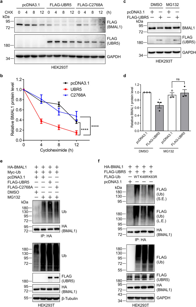
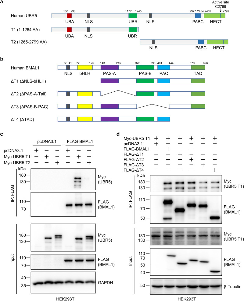
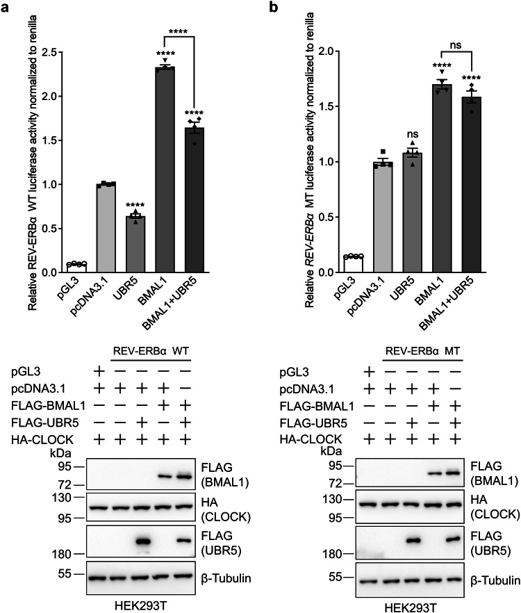
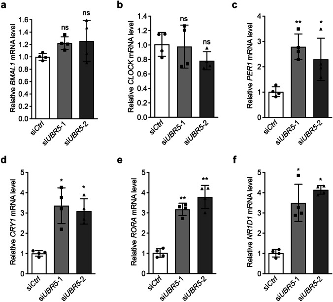
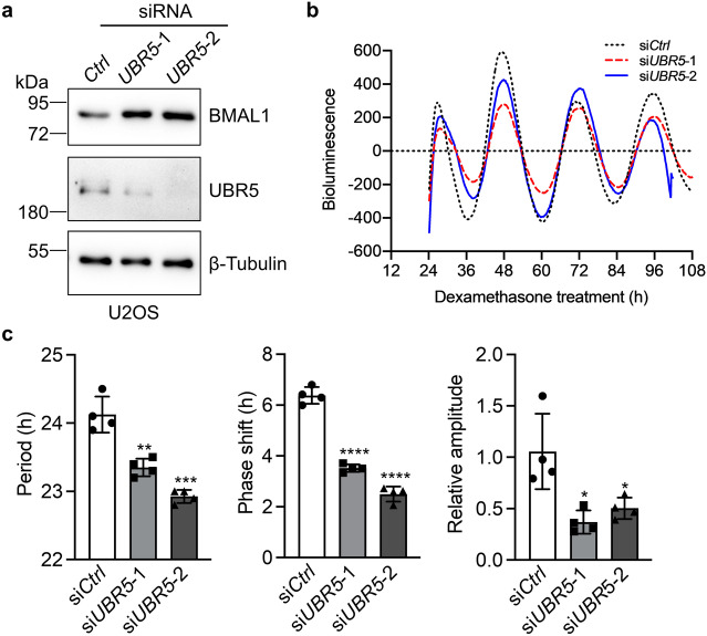
The circadian clock is the inner rhythm of life activities and is controlled by a self-sustained and endogenous molecular clock, which maintains a ~ 24 h internal oscillation. As the core element of the circadian clock, BMAL1 is susceptible to degradation through the ubiquitin-proteasome system (UPS). Nevertheless, scant information is available regarding the UPS enzymes that intricately modulate both the stability and transcriptional activity of BMAL1, affecting the cellular circadian rhythm. In this work, we identify and validate UBR5 as a new E3 ubiquitin ligase that interacts with BMAL1 by using affinity purification, mass spectrometry, and biochemical experiments. UBR5 overexpression induced BMAL1 ubiquitination, leading to diminished stability and reduced protein level of BMAL1, thereby attenuating its transcriptional activity. Consistent with this, UBR5 knockdown increases the BMAL1 protein. Domain mapping discloses that the C-terminus of BMAL1 interacts with the N-terminal domains of UBR5. Similarly, cell-line-based experiments discover that HYD, the UBR5 homolog in Drosophila, could interact with and downregulate CYCLE, the BMAL1 homolog in Drosophila. PER2-luciferase bioluminescence real-time reporting assay in a mammalian cell line and behavioral experiments in Drosophila reveal that UBR5 or hyd knockdown significantly reduces the period of the circadian clock. Therefore, our work discovers a new ubiquitin ligase UBR5 that regulates BMAL1 stability and circadian rhythm and elucidates the underlying molecular mechanism. This work provides an additional layer of complexity to the regulatory network of the circadian clock at the post-translational modification level, offering potential insights into the modulation of the dysregulated circadian rhythm.

Keywords: BMAL1; UBR5; circadian rhythm; proteomics; transcriptional activity; ubiquitination.


Conflict of interest statement

The authors declare no competing interests.

Figures

Fig. 1
Fig. 1. Proteomic identification of UBR5 as a BMAL1-interacting protein.
a Flowchart for the identification of BMAL1-interacting proteins by affinity purification (AP) and mass spectrometry (MS). b Silver staining of BMAL1-interacting proteins purified using FLAG M2 affinity gel from HEK293T cells expressing pcDNA3.1 (control) or FLAG-BMAL1. c Log2(iBAQBMAL1/iBAQCtrl) of BMAL1-interacting proteins from two biological replicates. The right panel is the magnification of the upper right corner of the left panel. BMAL1 and UBR5 are denoted by the red and blue circles, respectively. d MS/MS information for the top 20 peptides identified from UBR5. e A representative MS/MS spectrum of an MS/MS-identified UBR5 tryptic peptide.
Fig. 2
Fig. 2. UBR5 interacts and colocalizes with BMAL1.
a Exogenously expressed BMAL1 co-immunoprecipitated endogenous UBR5. HEK293T cells were transfected with pcDNA3.1 or FLAG-BMAL1 plasmid. FLAG-BMAL1 and its interactors were immunoprecipitated with anti-FLAG M2 affinity gel. The cell lysates and immunoprecipitates were immunoblotted with the indicated antibodies. b Exogenously expressed UBR5 co-immunoprecipitated endogenous BMAL1. HEK293T cells were transfected with pcDNA3.1 or FLAG-UBR5 plasmid. FLAG-UBR5 and its interactors were immunoprecipitated with anti-FLAG M2 affinity gel. The cell lysates and immunoprecipitates were immunoblotted with the indicated antibodies. c BMAL1 interacts with UBR5 endogenously. HeLa cell lysates were immunoprecipitated with control IgG or BMAL1 specific antibody. The cell lysates and immunoprecipitates were immunoblotted for UBR5, BMAL1, and/or GAPDH. d UBR5 colocalizes with BMAL1 in the nucleus. HEK293 cells were co-transfected with HA-BMAL1 plasmid and FLAG, FLAG-UBR5, or FLAG-C2768A plasmid using PEI transfection reagents. At 24 h post-transfection, cells were fixed, permeabilized, and stained with FLAG (UBR5 or C2768A: green) and HA (BMAL1: red) primary antibodies along with the corresponding secondary antibodies and Hoechst (blue). Fluorescence was measured under a microscope. Scale bar: 20 μm.
Fig. 3
Fig. 3. UBR5 downregulates BMAL1.
a, b UBR5 reduces BMAL1 protein levels in a dose-dependent manner. UBR5 plasmid was transfected into HEK293T cells with a gradient concentration for 60 h. The resulting cell lysates were immunoblotted with the indicated antibodies. Quantification (mean ± SDs) was performed for data from triplicates. Student’s t test, **P < 0.01, ***P < 0.001. c UBR5 downregulates endogenous BMAL1 protein. pcDNA3.1 or FLAG-UBR5 plasmid was transfected into HEK293T cells. Cells were harvested at 48, 60, or 72 h after transfection and cell lysates were immunoblotted. d, e The E3 ligase activity of UBR5 is required to destabilize BMAL1. HEK293T cells were transfected with the pcDNA3.1, wild-type (WT), or C2768A mutant (MT) UBR5 plasmid for 48 h and the cell lysates were subjected to immunoblotting analysis. Quantification (mean ± SEMs) was conducted for data from three biological replicates. Student’s t test, **P < 0.01, ns not significant. f, g UBR5 knockdown increases BMAL1 protein level. Control or two pairs of UBR5-specific siRNAs were transfected into HEK293T cells for 48 h. The resulting cell lysates were immunoblotted. Quantification (mean ± SDs) was performed for data from three biological replicates. Student’s t test, *P < 0.05. h UBR5 knockout elevates the BMAL1 protein level. CRISPR/Cas9 technology was used to knock out UBR5 in U2OS cells. Cell lysates were immunoblotted for UBR5, BMAL1, and β-tubulin.
Fig. 4
Fig. 4. UBR5 reduces BMAL1 stability by promoting its ubiquitination and degradation.
a, b UBR5 enhances the degradation of BMAL1. HEK293T cells were co-transfected with FLAG-BMAL1 and control, FLAG-UBR5 WT, or C2768A MT plasmids. At 48 h after transfection, cells were treated with CHX (200 μg/ml) for the indicated time. Cell lysates were immunoblotted for the indicated antibodies. Quantification was performed for data from three biological replicates. Two-way ANOVA, ***P < 0.001. c, d Proteasome inhibitor abolishes the UBR5-mediated reduction of BMAL1. HEK293T cells were transfected with pcDNA3.1 or FLAG-UBR5 plasmid for 48 h and treated with DMSO or MG132 (10 μM) for 12 h. Cell lysates were immunoblotted for the indicated antibodies. The experiment was carried out in triplicates for quantification. Student’s t test, *P < 0.05, ns not significant. e UBR5 increases the polyubiquitination of BMAL1. HEK293T cells were co-transfected with the indicated plasmids for 48 h and then treated with MG132 (10 µM) for 12 h. Anti-HA magnetic beads were used to purify HA-BMAL1. The cell lysates and purified samples were immunoblotted with the indicated antibodies. f UBR5 promotes the formation of K48-linked polyubiquitin chains on BMAL1. HEK293T cells were transfected with the indicated plasmids for 48 h. Cells and samples were prepared and analyzed as described in (e).
Fig. 5
Fig. 5. UBR5 and BMAL1 interact with each other at their N- and C-terminal regions, respectively.
a Schematic illustration of Myc-UBR5 and its truncations. b Sketch of the FLAG-tagged full-length human BMAL1 and its truncating mutants. c BMAL1 interacts with the N-terminal domains of UBR5. HEK293T cells were transfected with the indicated plasmids for 48 h. FLAG-BMAL1 and its interactors were immunoprecipitated with anti-FLAG M2 affinity gel. The samples were immunoblotted for FLAG (BMAL1), Myc (T1, T2), and GAPDH. d BMAL1 interacts with UBR5 mainly through its C-terminal domain. HEK293T cells were transfected with the indicated full-length or truncated FLAG-BMAL1 plasmids along with the Myc-tagged N-terminal domain of UBR5. Co-IP was performed using anti-FLAG M2 affinity gel. Cell lysates and purified samples were immunoblotted.
Fig. 6
Fig. 6. UBR5 reduces the luciferase activity of the WT but not the E-box deleted REV-ERBα plasmid.
a UBR5 reduces the transcription of WT REV-ERBα (pGL3-REV-ERBα::Luciferase plasmid). HEK293T cells were transfected with the indicated plasmids for 36 h. Cells were collected to detect the firefly and renilla luciferase signals. b UBR5 does not affect the transcription of REV-ERBα △E-box MT (pGL3-REV-ERBα △E-box::Luciferase plasmid). The experiments were performed as described in (a) except that the pGL3-REV-ERBα::Luciferase plasmid was replaced with the pGL3-REV-ERBα △E-box::Luciferase plasmid. The relative luciferase activity was normalized to renilla luciferase. Four technical replicates were carried out to obtain mean ± SEMs. Student’s t test, ns not significant, ****P < 0.0001 against the 2nd bar or as indicated. Western blotting was used to confirm the protein expression in cell lysates.
Fig. 7
Fig. 7. UBR5 depletion increases the expression of BMAL1 downstream genes.
UBR5 knockdown does not alter the mRNA level of BMAL1 (a) and CLOCK (b) but elevates that of PER1 (c), CRY1 (d), RORA (e), and NR1D1 (f). β-Actin was used as the internal reference for normalization. U2OS cells were transfected with siCtrl (negative control) or siUBR5 using Lipofectamine 2000. Total RNA was isolated using TRIzol. The first-strand cDNA was synthesized and qPCR was performed. Student’s t test was used to perform statistical analysis for data from four technical replicates and mean ± SDs were plotted. ns not significant; *P < 0.05; **P < 0.01 compared with the sample transfected with control siRNA (siCtrl).
Fig. 8
Fig. 8. UBR5 alters the cellular circadian clock in vitro.
a Validation of UBR5 siRNA silencing in PER2-luciferase U2OS cells. b Baseline subtracted bioluminescence signals obtained from UBR5 knockdown PER2-luciferase U2OS cells after synchronized with dexamethasone. c UBR5 knockdown affects the period, phase, and amplitude of the cellular circadian rhythm. Quantification (mean ± SDs) of the period, phase, and relative amplitude was performed for bioluminescence signals from four biological replicates. Student’s t test, *P < 0.05, **P < 0.01, ***P < 0.001, ****P < 0.0001.
Fig. 9
Fig. 9. HYD regulates CYCLE in cells and the circadian period of fruit flies.
a Illustration of full-length HYD, its C-terminus (HYD-C), and CYCLE. b CYCLE interacts with the C-terminal HYD. Myc-HYD-C was co-transfected with pcDNA3.1 or FLAG-CYCLE into HEK293T cells by PEI transfection reagent. Cells were harvested at 48 h after transfection, and FLAG M2 affinity gel was used to purify FLAG-CYCLE and its interacting proteins. The cell lysates and immunoprecipitates were immunoblotted with the indicated antibodies. c, d UBR5 and HYD-C reduce the CYCLE protein level. HEK293T cells were transfected with FLAG-CYCLE using PEI transfection reagent and equally split into two sets of plates. Then cells were transfected with pcDNA3.1 and FLAG-UBR5 or Myc-HYD-C. Cell lysates were harvested at 48 h after transfection for immunoblotting. Three biological replicates were performed to obtain mean ± SDs. Student’s t test, ****P < 0.0001. e Proteasomal inhibition prevents the HYD-C-induced degradation of CYCLE. HEK293T cells were first transfected with FLAG-CYCLE plasmid and split equally into four plates. Then pcDNA3.1 or Myc-HYD-C plasmid was transfected again at 24 h after the first transfection. At 48 h after the second transfection, cells were treated with the proteasome inhibitor MG132 (20 μM, 10 h). The resulting cell lysates were immunoblotted for FLAG, Myc, and GAPDH. S.E. and L.E. represent short and long exposure. Three biological replicates were used to obtain mean ± SDs. Student’s t test, ***P < 0.001, ns not significant. f HYD-C promotes CYCLE ubiquitination. HEK293T cells were co-transfected with FLAG-CYCLE and HA-Ub, split into three plates, and then transfected again with pcDNA3.1 or Myc-HYD-C. Cells were treated with DMSO or MG132 at 12 h before harvest. FLAG-CYCLE was immunoprecipitated using FLAG M2 affinity gel. Cell lysates and immunoprecipitates were immunoblotted with the indicated antibodies. g The mRNA level of hyd was decreased in elav-Gal4 > cg9484 fruit flies. The total mRNA from the brain tissues of ~50 fruit flies was isolated for the PCR and agarose gel electrophoresis analysis of hyd mRNA level. α-Tubulin was used as the housekeeping gene for normalization. Four biological replicates were used to obtain mean ± SEMs. Student’s t test, ****P < 0.0001. h, i hyd knockdown shortens the circadian period in constant darkness (dark-dark). Representative double-plotted actograms for the locomotor activity of fruit flies with the control or hyd RNAi. Quantification was performed for 53 and 73 fruit flies, respectively. Student’s t test, **P < 0.01.
Fig. 10
Fig. 10. The molecular mechanism by which UBR5 modulates the circadian clock in mammalian cells and fruit flies.
a UBR5 regulates the circadian rhythm in mammalian cells by promoting the ubiquitination and degradation of BMAL1 and diminishing the transcription of the PER and CRY genes. b HYD destabilizes CYCLE by enhancing its ubiquitination and degradation, resulting in a significantly shorter circadian period in fruit flies.

Similar articles

Cited by

References

    1. Brancaccio M, Edwards MD, Patton AP, Smyllie NJ, Chesham JE, Maywood ES, et al. Cell-autonomous clock of astrocytes drives circadian behavior in mammals. Science. 2019;363:187–92. 10.1126/science.aat4104 - DOI - PMC - PubMed
    1. Patke A, Young MW, Axelrod S. Molecular mechanisms and physiological importance of circadian rhythms. Nat Rev Mol Cell Biol. 2020;21:67–84. 10.1038/s41580-019-0179-2 - DOI - PubMed
    1. Zheng Y, Pan L, Wang F, Yan J, Wang T, Xia Y, et al. Neural function of Bmal1: an overview. Cell Biosci. 2023;13:1. 10.1186/s13578-022-00947-8 - DOI - PMC - PubMed
    1. Hirayama J, Sahar S, Grimaldi B, Tamaru T, Takamatsu K, Nakahata Y, et al. CLOCK-mediated acetylation of BMAL1 controls circadian function. Nature. 2007;450:1086–90. 10.1038/nature06394 - DOI - PubMed
    1. Tamaru T, Hattori M, Honda K, Nakahata Y, Sassone-Corsi P, van der Horst GT, et al. CRY drives cyclic CK2-mediated BMAL1 phosphorylation to control the mammalian circadian clock. PLoS Biol. 2015;13:e1002293. 10.1371/journal.pbio.1002293 - DOI - PMC - PubMed

MeSH terms

LinkOut - more resources