. 2024 Jun;9(6):1607-1618.
doi: 10.1038/s41564-024-01697-8. Epub 2024 May 13.

Propionate prevents loss of the PDIM virulence lipid in Mycobacterium tuberculosis

Affiliations

Propionate prevents loss of the PDIM virulence lipid in Mycobacterium tuberculosis

Claire V Mulholland et al. Nat Microbiol. 2024 Jun.

Abstract

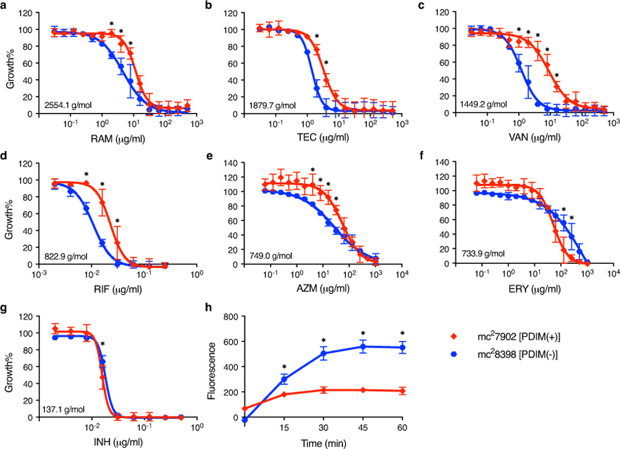
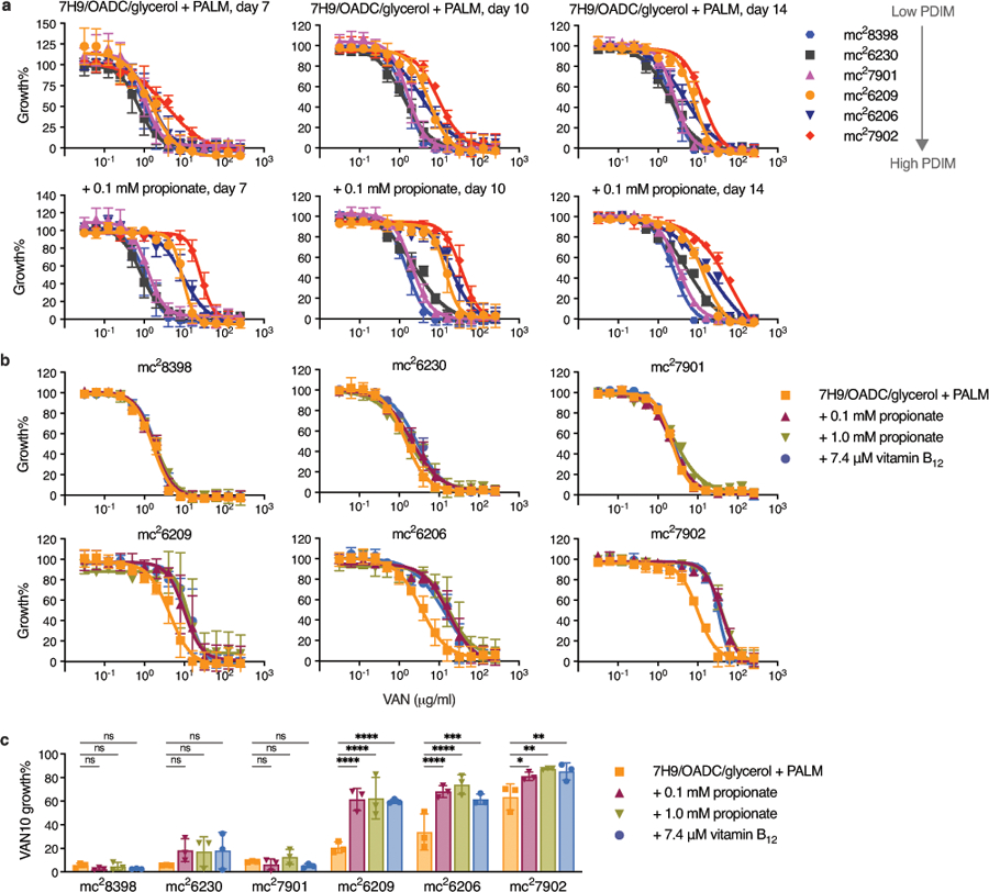
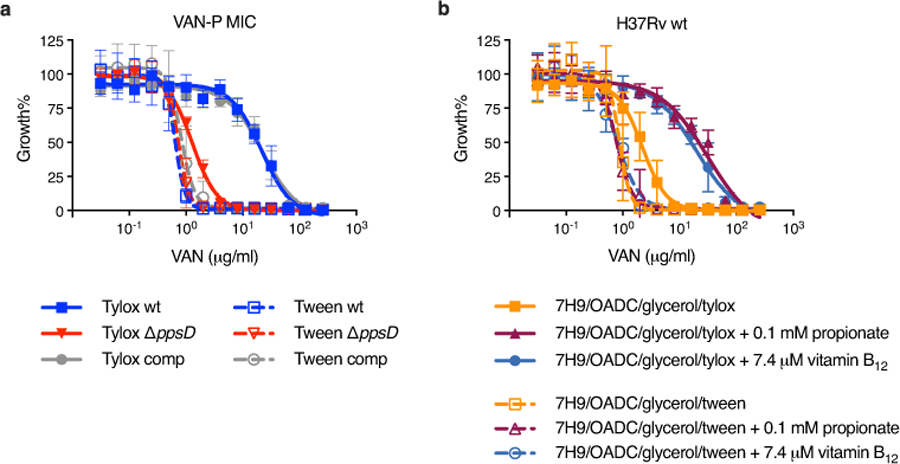
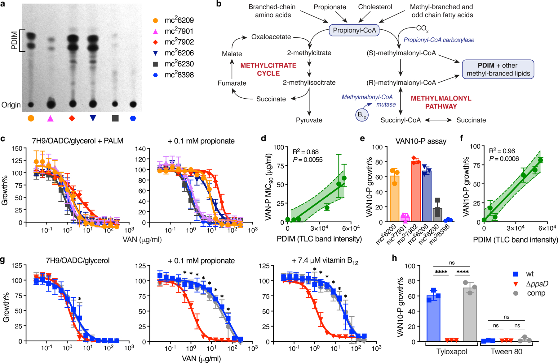
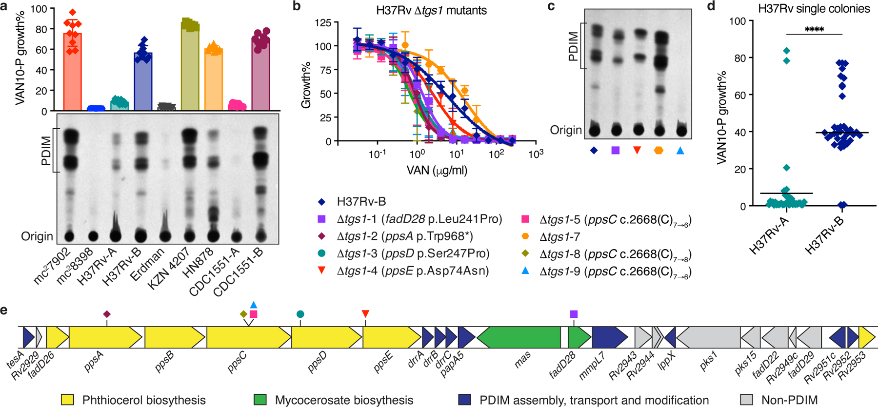
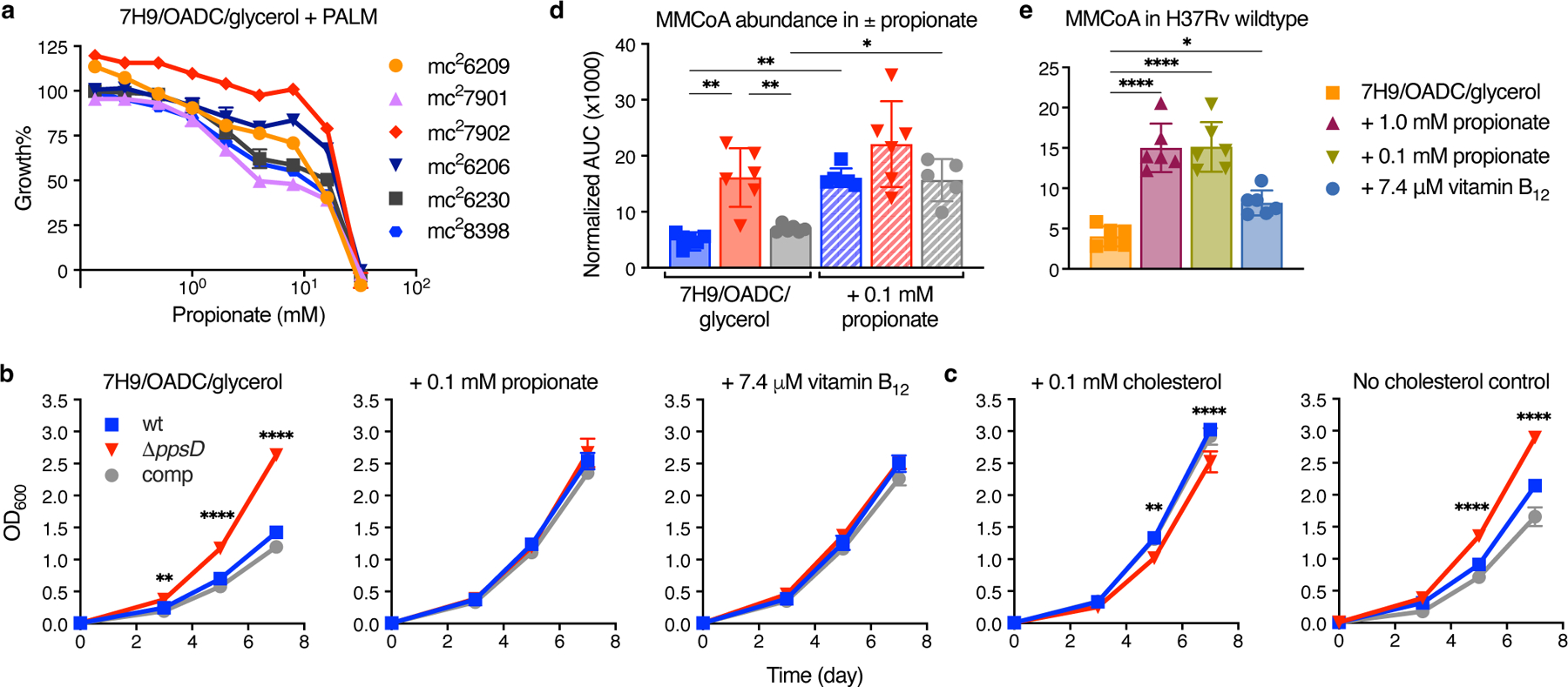
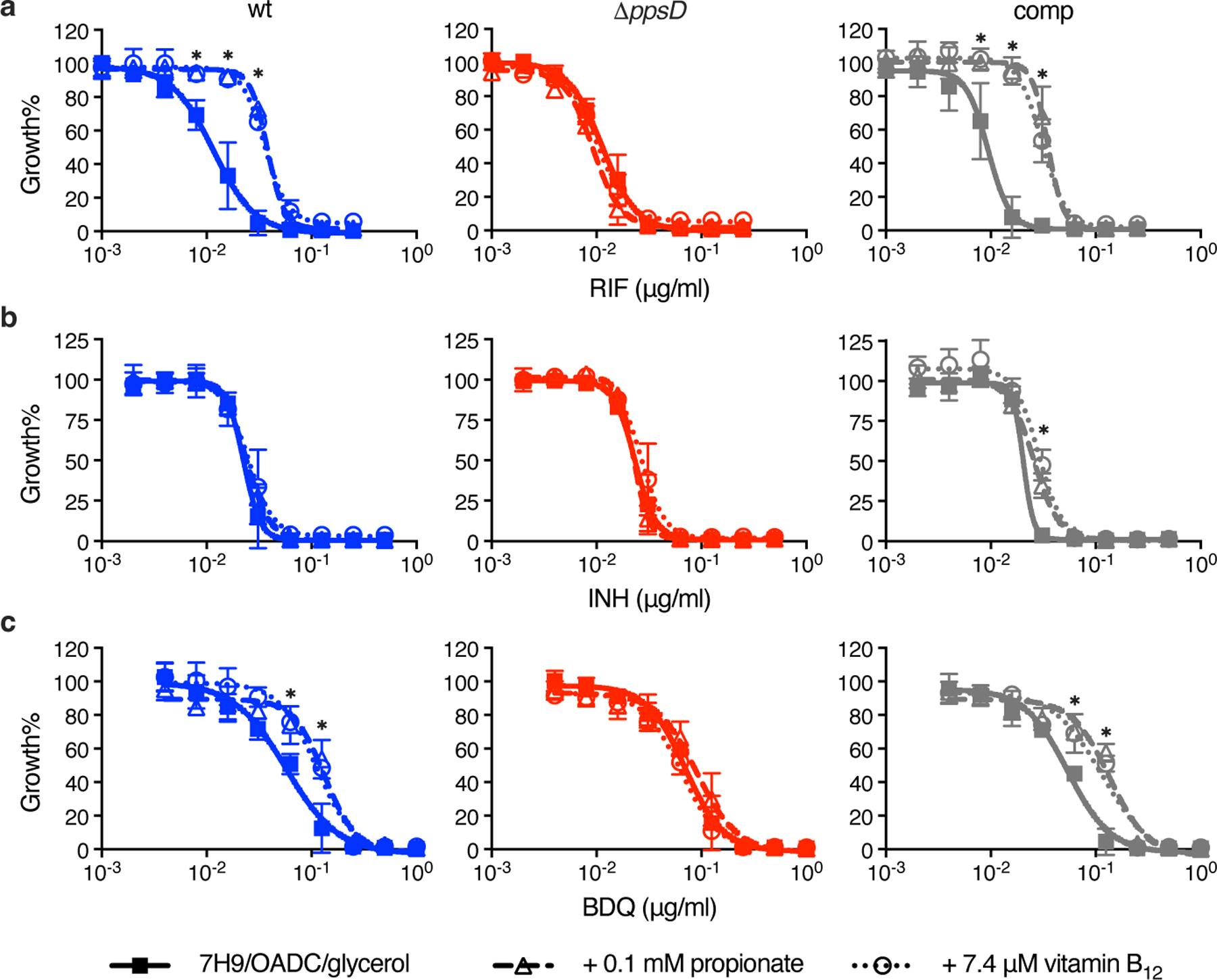
Phthiocerol dimycocerosate (PDIM) is an essential virulence lipid of Mycobacterium tuberculosis. In vitro culturing rapidly selects for spontaneous PDIM-negative mutants that have attenuated virulence and increased cell wall permeability, thus impacting the relevance of experimental findings. PDIM loss can also reduce the efficacy of the BCG Pasteur vaccine. Here we show that vancomycin susceptibility can rapidly screen for M. tuberculosis PDIM production. We find that metabolic deficiency of methylmalonyl-CoA impedes the growth of PDIM-producing bacilli, selecting for PDIM-negative variants. Supplementation with odd-chain fatty acids, cholesterol or vitamin B12 restores PDIM-positive bacterial growth. Specifically, we show that propionate supplementation enhances PDIM-producing bacterial growth and selects against PDIM-negative mutants, analogous to in vivo conditions. Our study provides a simple approach to screen for and maintain PDIM production, and reveals how discrepancies between the host and in vitro nutrient environments can attenuate bacterial pathogenicity.


Conflict of interest statement

Competing interests

C.V.M. and M.B. are inventors on a pending patent related to this work (U.S. Patent Application No. 63/527,831, filed 20 July 2023). The authors declare that they have no other competing interests. The remaining authors declare no competing interests.

Figures

Extended Data Fig. 1
Extended Data Fig. 1. Resistance of PDIM(-) and PDIM(+) Mtb to high molecular weight compounds.
ag, MIC assays of Mtb mc27902 [PDIM(+)] and mc28398 [PDIM(-)] to (a) ramoplanin (RAM), (b) teicoplanin (TEC), (c) vancomycin (VAN), (d) rifampicin (RIF), (e) azithromycin (AZM), (f) erythromycin (ERY), and (g) isoniazid (INH). Compounds are arranged by descending molecular weight, which is shown on the MIC plots. MICs were performed in 7H9/OADC/glycerol/tyloxapol + PALM media and bacterial growth was measured after 10 days of incubation and normalized to drug-free controls. Mean ± SD for n = 4 biological replicates from two independent experiments. h, Ethidium Bromide uptake of mc27902 and mc28398. Uptake in whole cell suspensions was monitored by fluorescence (Ex 355 nm/Em 590 nm). Mean ± SD for n = 4 biological replicates, each measured in five technical replicates. Uptake data are representative of two independent experiments. *P < 0.001; two-way ANOVA with Šidák’s multiple comparison test.
Extended Data Fig. 2
Extended Data Fig. 2. Propionate and vitamin B12 supplementation selectively increase vancomycin resistance of PDIM(+) Mtb improving assay robustness and reducing time to result.
a, Vancomycin MICs for the PDIM reference strain set in standard 7H9/OADC/glycerol/tyloxapol + PALM media and additionally supplemented with 0.1 mM propionate. Growth was measured after 7, 10, and 14 days as indicated. Mean ± SD for n = 4 biological replicates from two independent experiments. b, Vancomycin MICs in standard media and additionally supplemented with 0.1 or 1.0 mM propionate or 7.4 μM vitamin B12. Growth was measured after 10 days. Mean ± SD for n = 4 biological replicates from two independent experiments. c, VAN10 assays in standard and supplemented media. Growth was measured after 10 days. Mean ± SD for n = 3 independent experiments, each performed in triplicate. *P < 0.05, **P < 0.01, ***P < 0.001, ****P < 0.0001; two-way ANOVA with Šidák’s multiple comparison test. The day seven data in (a) are also shown in Fig. 1c and are shown here alongside additional time points. The data in (b) includes one of the same experiments shown in (a), together with data from an independent experiment. The VAN10-P (+ 0.1 mM propionate) data in (c) are also shown in Fig. 1e and are shown here alongside additional conditions.
Extended Data Fig. 3
Extended Data Fig. 3. Tween 80 decreases vancomycin resistance and abolishes PDIM-related differences in MIC.
a, VAN-P MICs for PDIM(+) and PDIM(-) Mtb H37Rv using either tyloxapol or Tween 80 as the culture detergent. b, Vancomycin MICs for PDIM(+) H37Rv wildtype in standard 7H9/OADC/glycerol media and supplemented with propionate or vitamin B12 using either tyloxapol or Tween 80 as the detergent. Mean ± SD for n = 4 biological replicates from two independent experiments.
Extended Data Fig. 4
Extended Data Fig. 4. VAN-P assays predict PDIM levels across different Mtb strains and lineages and enable re-isolation of single PDIM(+) clones.
a, VAN10-P assays for a range of virulent Mtb strains belonging to different lineages. Bacterial growth was measured after 7, 10, and 14 days of incubation as indicated. Data are from the same experiment in Fig. 2a and show additional time points plus an independent experimental repeat measured on day 10 (hatched bars with unfilled symbols). b, VAN10-P assays for strains with an H37Rv background including mc27902 and mc28398. H37Rv-SC is a single PDIM(+) clone isolated from H37Rv-B by VAN10-P colony screening. This clone was used as our PDIM(+) H37Rv wildtype strain throughout this work and was used to construct H37Rv ΔppsD and ΔppsD::comp mutants (Supplementary Table 2). Data in (a,b) show mean ± SD for n = 9 pairwise comparisons between triplicate wells, except for the day 10 repeat in (a) where n = 4 pairwise comparisons between duplicate wells. c, VAN-P MICs of H37Rv stocks and H37Rv-SC. d, VAN-P MICs of non-H37Rv strains from (a). eg, VAN-P MICs of single PDIM(+) clones isolated from Rag-/- mice using VAN10-P colony screening for (e) Erdman, (f) HN878, and (g) CDC1551 (see also Supplementary Table 3). Data are plotted together with MIC data from (d) for comparison. MIC data in (cg) show mean ± SD for n = 4 biological replicates from two independent experiments. hj, Determination that our Mtb mc26230 stock is a mixed population and re-isolation of a single PDIM(+) clone by VAN10-P screening. (h) VAN10-P assay of single colonies isolated from our mc26230 stock (n = 40) and (i) following a single passage in 10 μg/ml vancomycin (n = 20). Vancomycin significantly enriched for PDIM(+) bacilli (P < 0.0001 two-tailed Mann-Whitney test), facilitating re-isolation of low-frequency PDIM(+) clones. Each colony was assayed in triplicate and data points represent mean VAN10-P growth%. Lines indicate the median. j, VAN-P MICs of PDIM(+) (AE1601) and PDIM(−) (AE1611) mc26230 clones identified by VAN10-P colony screening (see also Supplementary Table 3). Mean ± SD for n = 6 biological replicates from two independent experiments.
Extended Data Fig. 5
Extended Data Fig. 5. Assessment of ppsC homopolymeric tract mutations.
a, Schematic showing the location of a homopolymeric tract region in the ppsC gene. Sequence inserts show two adjacent 7-cytosine homopolymeric tracts (c.2668 and c.2685) ± 5 bp on either side. Numbers in black indicate the position in the ppsC gene and numbers in red the genomic position in the H37Rv genome. bd, Analysis of the ppsC homopolymeric tract region in Δtgs1 mutants and identification of frameshift mutations. WGS variant calling failed to identify PDIM mutations in Δtgs1-5, Δtgs1-8 and Δtgs1-9 despite a PDIM(-) result in VAN-P MICs (Fig. 2b) and validation of Δtgs1-9 as PDIM(-) by TLC (Fig. 2c). Close manual inspection of WGS reads showed the ppsC homopolymeric tract region is poorly covered by Illumina MiSeq and identified potentially missed variant calls. PCR and Sanger sequencing confirmed the presence of a 2668(C)76 frameshift mutation in both Δtgs1-5 (b) and Δtgs1-9 (d) and identified a 2668(C)78 mutation in Δtgs1-8 that was not covered at all by WGS (c). (bd) were created with Geneious Prime® 2022.2.2 and Illustrator 26.4.1. Coverage has been cropped to a read depth of 60 ×. See also Supplementary Table 5.
Extended Data Fig. 6
Extended Data Fig. 6. Effect of different media supplements on growth of PDIM(+) and PDIM(−) Mtb.
a, Growth of PDIM(+) and PDIM(-) Mtb H37Rv in standard 7H9/OADC/glycerol/tyloxapol and bj, the same media with additional supplements as indicated. km, Growth using Tween 80 instead of tyloxapol as the culture detergent with additional supplements as indicated. Mean ± SD for n = 3 biological replicates. Data are representative of at least two independent experiments. (a,b,d,f) show independent experimental repeats for the conditions in Fig. 3. *P < 0.001 for both wt and comp versus ΔppsD; two-way ANOVA with Tukey’s multiple comparison test. For some data points the SD is smaller than the data symbols.
Extended Data Fig. 7
Extended Data Fig. 7. Effects of propionate and vitamin B12 supplementation on MMCoA and propionyl-CoA metabolic pathways in Mtb.
a, Abundance of metabolites in propionyl-CoA and MMCoA metabolism in PDIM(+) Mtb H37Rv wildtype grown in standard 7H9/OADC/glycerol/tyloxapol media and supplemented with propionate or vitamin B12, and b, in PDIM(+) and PDIM(-) H37Rv grown in 7H9/OADC/glycerol/tyloxapol ± 0.1 mM propionate. Abundances are shown as normalized area under the curve (AUC) (see Methods). Mean ± SD for n = 6 biological replicates from two independent experiments. *P < 0.05, **P < 0.01, ***P < 0.001, ****P < 0.0001; one-way ANOVA with Tukey’s multiple comparison test. Significant differences compared to unsupplemented media are indicated in (a), and between ± propionate for each strain and between strains for each condition in (b). PROP, propionate; PROP-CoA, propionyl-CoA; MMCoA, methylmalonyl-CoA; SUC-CoA, succinyl-CoA; SUC, succinate; 2MC/2MIC, 2-methyl(iso)citrate; and PYR, pyruvate. Succinyl-CoA and methyl(iso)citrate were not able to be detected in samples by our method. Propionyl-CoA was close to the detection limit and was not detected in all samples (n.d. = not detected). The data for MMCoA are also shown in Fig. 3d,e. See also Supplementary Fig. 8.
Extended Data Fig. 8
Extended Data Fig. 8. Propionate and vitamin B12 supplementation prevent PDIM loss in Mtb.
a, Schematic overview of in vitro evolution experiments. Triplicate inkwells containing standard 7H9/OADC/glycerol/tyloxapol or media supplemented with propionate or vitamin B12 were inoculated with frozen Mtb culture stock (P0) and incubated for 7–10 days (P1). Cultures were then diluted into fresh media every 7 days for serial passage (P2 to PX). Selected passages were input into VAN10-P assays at the time of passage to assess PDIM production over the course of the experiment. For TLC lipid analysis, frozen stocks were first outgrown in media without propionate or vitamin B12 for a single passage to allow the strains to recover before 14C-labelling. Figure created with BioRender.com. b, TLC lipid analysis of H37Rv-B before and after six serial passages in ± 0.1 or 1.0 mM propionate. This figure shows the full TLC plate from Fig. 4a with results for both biological replicates analysed by TLC. c, VAN10-P assays for H37Rv-SC [PDIM(+) H37Rv wildtype] passaged in ± 0.1 mM propionate. d, H37Rv-A and e, H37Rv-B passaged in ± 7.4 μM vitamin B12. Mean ± SD for n = 3 biological replicates, each assayed in triplicate. *P < 0.05, **P < 0.01, ***P < 0.001, ****P < 0.0001; two-way ANOVA with Šidák’s (c) or Tukey’s (d,e) multiple comparison test. Significant differences between conditions are indicated in (c) and between timepoints in (d,e).
Fig. 1 |
Fig. 1 |. Vancomycin resistance is enhanced by propionate or vitamin B12 supplementation and is predictive of PDIM production in Mtb.
a, TLC lipid analysis of the PDIM reference strain set (see Supplementary Table 1). b, Metabolic pathways of methylmalonyl-CoA production and propionyl-CoA catabolism. c, Vancomycin resistance of the Mtb PDIM reference strain set in 7H9/OADC/glycerol/tyloxapol + PALM (pantothenate, arginine, leucine, and methionine) media, and additionally supplemented with 0.1 mM propionate (‘VAN-P’ MIC), measured after 7 days incubation. d, Correlation between VAN-P MIC90 from the curve fit in (c) (+ 0.1 mM propionate) and PDIM band intensity from (a). The solid line indicates the linear regression best-fit, and the error bands the 95% CI. e, ‘VAN10-P’ assay comparing growth in 10 μg/ml vancomycin with 0.1 mM propionate to drug-free controls (VAN10 OD / VAN0 OD × 100 = VAN10-P growth%). f, Correlation between VAN10-P growth% from (e) and PDIM from (a). The solid line indicates the linear regression best-fit, and the error bands the 95% CI. g, Vancomycin resistance of PDIM(+) and PDIM(-) Mtb H37Rv strains in standard 7H9/OADC/glycerol/tyloxapol media and supplemented with 0.1 mM propionate or 7.4 μM vitamin B12 (10 μg/ml). *P < 0.001 for both wt and comp versus ΔppsD; two-way ANOVA with Tukey’s multiple comparison test. h, VAN10-P assay of H37Rv strains with tyloxapol or Tween 80. ****P < 0.0001; one-way ANOVA with Tukey’s multiple comparison test. MIC data show mean ± SD for n = 4 biological replicates from two independent experiments. VAN10-P data show mean ± SD for n = 3 three independent experiments, each performed in triplicate.
Fig. 2 |
Fig. 2 |. VAN-P assays accurately predict PDIM status during genetic manipulations and across different Mtb strains and lineages.
a, TLC lipid analysis and VAN10-P assays of different laboratory stocks of virulent Mtb strains alongside Mtb mc27902 and mc28398. Mean ± SD for n = 9 pairwise comparisons between triplicate wells. b, VAN-P MIC assays of eight Δtgs1 mutants and the parent H37Rv-B. Mean ± SD for n = 3 (Δtgs1-3, -5, -7 and -9) or n = 4 (H37Rv-B, Δtgs1-1, -2, -4, -8) biological replicates from two independent experiments. Mutations in PDIM biosynthetic genes are indicated in brackets in the legend. No mutations were detected in H37Rv-B or Δtgs1-7. c, TLC lipid analysis of four Δtgs1 mutants and H37Rv-B. Lipid extracts in (a) and (c) were run on the same TLC plate. d, VAN10-P screening of single colonies isolated from H37Rv-A (n = 38) and H37Rv-B (n = 37). Each colony was assayed in triplicate and data points represent mean VAN10-P growth%. Lines indicate the median. P < 0.0001; unpaired two-tailed Mann-Whitney test. e. Schematic showing the PDIM gene cluster and location of secondary PDIM mutations in Δtgs1 mutants.
Fig. 3 |
Fig. 3 |. Propionate and vitamin B12 supplementation restore the growth of PDIM(+) Mtb.
a, Relative growth of the PDIM reference strain set in 7H9/OADC/glycerol/tyloxapol + PALM media with increasing concentrations of propionate compared to no propionate controls. Mean ± SD for n = 3 biological replicates. b, Growth curves of PDIM(+) and PDIM(−) Mtb H37Rv in standard 7H9/OADC/glycerol/tyloxapol media and supplemented with 0.1 mM propionate or 7.4 μM vitamin B12 (10 μg/ml). c, Growth in 7H9/OADC/glycerol/tyloxapol media + 0.1 mM cholesterol and no cholesterol controls (see Methods). The growth of ΔppsD was significantly lower in cholesterol compared to the no cholesterol control (P < 0.0001; days 7 and 10). Mean ± SD for n = 3 biological replicates. **P < 0.01, ****P < 0.0001 for both wt and comp versus ΔppsD; two-way ANOVA with Šidák’s multiple comparison test. Data in (ac) are representative of at least two independent experiments. For some data points the SD is smaller than the data symbols. d, Abundance of methylmalonyl-CoA (MMCoA) in PDIM(+) and PDIM(-) H37Rv grown in standard 7H9/OADC/glycerol/tyloxapol media ± 0.1 mM propionate, and e, PDIM(+) H37Rv wildtype in standard media and supplemented with either propionate or vitamin B12. The legend for (d) is the same as that for (bc). Abundances are shown as normalized area under the curve (AUC) (see Methods). Mean ± SD for n = 6 biological replicates from two independent experiments. *P < 0.05, **P < 0.01, ****P < 0.0001; one-way ANOVA with Tukey’s multiple comparison test. Significant differences between ± propionate for each strain and between strains for each condition are indicated in (d), and compared to unsupplemented media in (e).
Fig. 4 |
Fig. 4 |. Propionate and vitamin B12 supplementation prevent PDIM loss in Mtb.
a, VAN10-P and TLC lipid analysis of PDIM levels in Mtb H37Rv-B following serial passage in 7H9/OADC/glycerol/tyloxapol media ± 0.1 or 1.0 mM propionate. ****P < 0.0001; one-way ANOVA with Tukey’s multiple comparison test. A representative result is shown for one of two biological replicates analysed by TLC (see also Extended Data Fig. 8b). b, VAN10-P assays of H37Rv-A passaged in ± 0.1 mM propionate. Significant differences between successive timepoints for each condition are indicated (**P < 0.01, ****P < 0.0001); two-way ANOVA with Tukey’s multiple comparison test. c, VAN10-P screening of single colonies of H37Rv-A before (n = 38; same data as Fig. 2d) and after propionate passage in (b) (n = 30). Each colony was assayed in triplicate and data points represent mean VAN10-P growth%. Lines indicate the median. P = 0.0047; unpaired two-tailed Mann-Whitney test. d, VAN10-P assays of H37Rv-A passaged in media supplemented with ± 0.1 mM propionate and 7.4 μM vitamin B12 (10 μg/ml) alone and in combination. Significant differences compared to + 0.1 mM propionate are indicated (*P < 0.05, **P < 0.01, ***P < 0.001, ****P < 0.0001); two-way ANOVA with Tukey’s multiple comparison test. P > 0.05 for vitamin B12 versus vitamin B12 + propionate and P < 0.0001 for standard media versus each supplemented condition at all timepoints. VAN10-P data in (a,b,d) show mean ± SD for n = 3 biological replicates, each assayed in triplicate. For some data points the SD is smaller than the data symbols.
Fig. 5 |
Fig. 5 |. Propionate and vitamin B12 supplementation increase rifampicin and bedaquiline resistance of Mtb in a PDIM-dependent manner.
Sensitivity of PDIM(+) and PDIM(-) Mtb H37Rv to a, rifampicin (RIF), b, isoniazid (INH), and c, bedaquiline (BDQ), in standard 7H9/OADC/glycerol/tyloxapol media and supplemented with either 0.1 mM propionate or 7.4 μM vitamin B12 (10 μg/ml). *P < 0.001 for both propionate and vitamin B12 versus unsupplemented; two-way ANOVA with Tukey’s multiple comparison test. Mean ± SD for n = 4 biological replicates from two independent experiments.
Fig. 6 |
Fig. 6 |. Tween 80 increases the sensitivity of Mtb to rifampicin and abolishes PDIM-dependent differences in MIC.
Sensitivity of PDIM(+) and PDIM(-) Mtb H37Rv to rifampicin (RIF) in 7H9/OADC/glycerol media ± 0.1 mM propionate using either tyloxapol or Tween 80 as the culture detergent. Mean ± SD for n = 4 biological replicates from two independent experiments.

Update of

References

    1. Daffe M & Laneelle MA Distribution of phthiocerol diester, phenolic mycosides and related compounds in mycobacteria. J Gen Microbiol 134, 2049–2055 (1988). - PubMed
    1. Rens C, Chao JD, Sexton DL, Tocheva EI & Av-Gay Y Roles for phthiocerol dimycocerosate lipids in Mycobacterium tuberculosis pathogenesis. Microbiology (Reading) 167 (2021). - PubMed
    1. Domenech P & Reed MB Rapid and spontaneous loss of phthiocerol dimycocerosate (PDIM) from Mycobacterium tuberculosis grown in vitro: implications for virulence studies. Microbiology (Reading) 155, 3532–3543 (2009). - PMC - PubMed
    1. Manjunatha UH et al. Identification of a nitroimidazo-oxazine-specific protein involved in PA-824 resistance in Mycobacterium tuberculosis. Proc Natl Acad Sci U S A 103, 431–436 (2006). - PMC - PubMed
    1. Kirksey MA et al. Spontaneous phthiocerol dimycocerosate-deficient variants of Mycobacterium tuberculosis are susceptible to gamma interferon-mediated immunity. Infect Immun 79, 2829–2838 (2011). - PMC - PubMed

MeSH terms

LinkOut - more resources