. 2024 May 13;24(1):490.
doi: 10.1186/s12879-024-09375-0.

The role of microRNA-142a in Toxoplasma gondii infection-induced downregulation of Foxp3: implications for adverse pregnancy outcomes

Affiliations

The role of microRNA-142a in Toxoplasma gondii infection-induced downregulation of Foxp3: implications for adverse pregnancy outcomes

Yue Zhong et al. BMC Infect Dis. .

Abstract

Background: Toxoplasma gondii (T. gondii) is capable of infecting nearly all warm-blooded animals and approximately 30% of the global population. Though most infections are subclinical in immunocompetent individuals, congenital contraction can lead to severe consequences such as spontaneous abortion, stillbirth, and a range of cranio-cerebral and/or ocular abnormalities. Previous studies reported that T. gondii-infected pregnancy mice unveiled a deficit in both the amount and suppressive functions of regulatory T (Treg) cells, accompanied with reduced levels of forkhead box p3 (Foxp3). Recently, accumulative studies have demonstrated that microRNAs (miRNAs) are, to some extent, relevant to T. gondii infection. However, the link between alterations in miRNAs and downregulation of Foxp3 triggered by T. gondii has been only sporadically studied.

Methods: Quantitative reverse transcription polymerase chain reaction (RT-qPCR), protein blotting and immunofluorescence were employed to evaluate the impact of T. gondii infection and antigens on miRNA transcription and Foxp3 expression. Dual-luciferase reporter gene assays were performed to examine the fluorescence activity in EL4 cells, which were transfected with recombinant plasmids containing full-length/truncated/mutant microRNA-142a-3p (miR-142a) promoter sequence or wild type/mutant of Foxp3 3' untranslated region (3' UTR).

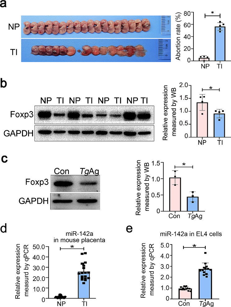
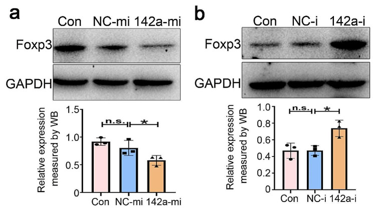
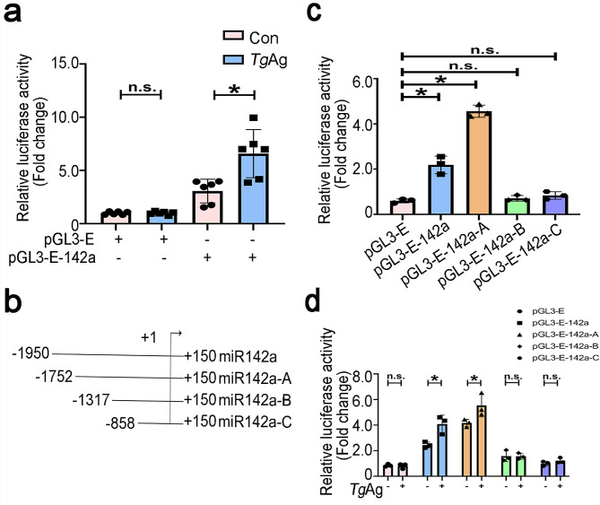
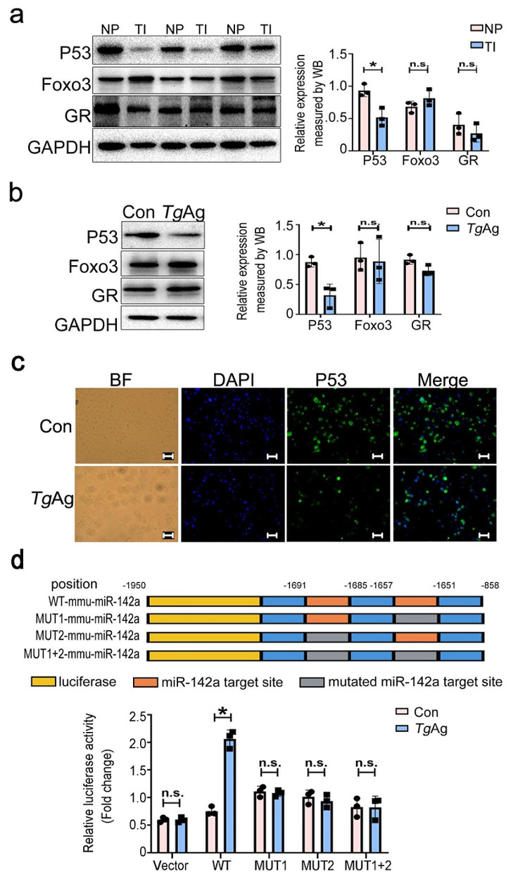
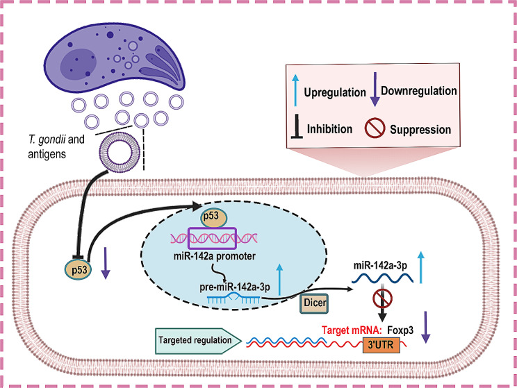
Results: We found a pronounced increase in miR-142a transcription, concurrent with a decrease in Foxp3 expression in T. gondii-infected mouse placental tissue. Similarly, comparable findings have been experimentally confirmed through the treatment of EL4 cells with T. gondii antigens (TgAg) in vitro. Simultaneously, miR-142a mimics attenuated Foxp3 expression, whereas its inhibitors markedly augmented Foxp3 expression. miR-142a promoter activity was elevated upon the stimulation of T. gondii antigens, which mitigated co-transfection of mutant miR-142a promoter lacking P53 target sites. miR-142a mimics deceased the fluorescence activity of Foxp3 3' untranslated region (3' UTR), but it did not affect the fluorescence activity upon the co-transfection of mutant Foxp3 3' UTR lacking miR-142a target site.

Conclusion: In both in vivo and in vitro studies, a negative correlation was discovered between Foxp3 expression and miR-142a transcription. TgAg enhanced miR-142a promoter activity to facilitate miR-142a transcription through a P53-dependent mechanism. Furthermore, miR-142a directly targeted Foxp3 3' UTR, resulting in the downregulation of Foxp3 expression. Therefore, harnessing miR-142a may be a possible therapeutic approach for adverse pregnancy caused by immune imbalances, particularly those induced by T. gondii infection.

Keywords: Toxoplasma Gondii; Forkhead box P3; P53; microRNA-142a-3p.


Conflict of interest statement

The authors declare no competing interests.

Figures

Fig. 1
Fig. 1
Effects of T. gondii infection on miR-142a transcription and Foxp3 expression In vivo and in vitro. a Diagram of mouse fetus at E18.5. In the normal pregnancy group (n = 4 mice), the embryonic mice developed normally, while in the T. gondii infection group (n = 5 mice), mouse fetuses appeared to be stillborn, with the abortion rate up to 60%. b Western blot analysis detected the changes in Foxp3 expression in mouse placental tissues. c Western blot was performed to assay the changes of Foxp3 protein expression in EL4 cells after 24 h of in vitro stimulation with TgAg. d RT-qPCR was used to assay the alterations of miR-142a expression in the placenta. e RT-qPCR was used to analyze the alterations of miR-142a expression in the EL4 cells stimulated with TgAg. NP: normal pregnant mice; TI: Toxoplasma-infected pregnant mice. TgAg: T. gondii antigens. Data were presented as mean ± SD. Statistical analysis was conducted using two-tailed unpaired Student’s t-test (B, C, D and E). *: P < 0.05
Fig. 2
Fig. 2
miR-142a attenuated Foxp3 expression. a miR-142a mimics were transfected into EL4 cells, and Foxp3 expressions were assayed by using immunoblotting. NC-mi: negative controls of miRNA mimics; miR-142a-mi: microRNA-142a mimics. b miR-142a inhibitors were transfected into EL4 cells, and Foxp3 expressions were detected by using immunoblotting. NC-i: negative controls of miRNA inhibitors. miR-142a-i: microRNA-142a inhibitors. Data were representative of the results of three independent experiments (mean ± SD). Statistical analysis was conducted using one-way ANOVA Tukey’s multiple comparisons test. *: P < 0.05; n.s.: P > 0.05
Fig. 3
Fig. 3
Antigens of T. gondii elevated the miR-142a promoter activity. a Luciferase reporter assay was conducted to measure the luciferase activity of EL4 cells that were electroporated with pGL3-E or pGL3-E-142a and then stimulated by TgAg for 24 h, respectively. b Schematic diagram showed the truncation sequences at the miR-142a promoter region. c The luciferase activities of EL4 cells, transfected respectively with recombinant plasmids comprising pGL3-E, pGL3-E-142a, and pGL3-E-142a-A/B/C, were evaluated at 48 h-post transfection. d The luciferase activities of EL4 cells that were transfected respectively with recombinant plasmids including pGL3-E, pGL3-E-142a, and pGL3-E-142a-A/B/C, and subsequently treated with TgAg for 24 h, were measured by luciferase reporter assay at 48 h-post transfection. Data were representative of the results of three independent experiments (mean ± SD). Statistical analysis was conducted using one-way ANOVA Tukey’s multiple comparisons test. *: P < 0.05; n.s.: P > 0.05
Fig. 4
Fig. 4
The inhibitory effects of T. gondii and its antigens on P53. a The expressions of P53, GR, and Foxo3 in mouse placentas infected with T. gondii were assessed by western blot (n = 3 mice). b Western blot analysis was conducted to validate the expressions of P53, GR, and Foxo3 in EL4 cells stimulated by TgAg for 24 h in vitro. c Immunofluorescence was conducted to visualize P53 expression in EL4 cells stimulated with TgAg for 24 h. As for fluorescence microscope image, Dye-green and Hoechst was used to label Foxp3 (green) and nuclei (blue). Scale: 40 μm. d The luciferase activity in EL4 cells that were transfected respectively with recombinant plasmids containing the WT or mutations (Mut 1, Mut 2, and Mut 1 + 2) sequences of miR-142a promoter was detected by luciferase reporter assay at 48 h-post transfection. TgAg: T. gondii antigens. NP: normal pregnant mice; TI: Toxoplasma-infected pregnant mice. Data were representative of the results of three independent experiments (mean ± SD). Statistical analysis was conducted using two-tailed unpaired Student’s t-test. *: P < 0.05; n.s.: P > 0.05
Fig. 5
Fig. 5
miR-142a targeted Foxp3 3’UTR. a Database prediction of miR-142a binding site on Foxp3 3’UTR and schematic representation of the mutation site. b Effect of miR-142a mimics and inhibitors on the fluorescence activity in EL4 cells transfected with recombinant plasmids containing wild type Foxp3 3’UTR. c Effect of miR-142a mimics and inhibitors on fluorescence activity of EL4 cells that were transfected with recombinant plasmids containing mutant Foxp3 3’UTR. Data were representative of the results of three independent experiments (mean ± SD). Statistical analysis was conducted using one-way ANOVA Tukey’s multiple comparisons test. *: P < 0.05; n.s.: P > 0.05
Fig. 6
Fig. 6
A schematic of the proposed model depicting a P53/miR-142a /Foxp3-mediated regulatory pathway in T. gondii-triggered adverse pregnancy outcomes in mice. During intracellular infection, T. gondii antigens lead to the downregulation of P53, which binds to miR-142a promoter region. miR-142a then targets Foxp3 3’UTR, therefore repressing Foxp3 expression

Similar articles

Cited by

References

    1. Ragab MF, Yoshifumi N. Genetic disruption of Toxoplasma Gondii peroxiredoxin (TgPrx) 1 and 3 reveals the essential role of TgPrx3 in protecting mice from fatal consequences of Toxoplasmosis. Int J Mol Sci. 2022;23(6):3076. doi: 10.3390/ijms23063076. - DOI - PMC - PubMed
    1. Xiaofeng W, Miao C, Shuqi Y, Chen X, Qian L, Yating Z, Yongsheng J, Yinan D. CRISPR/Cas12a combined with RPA for detection of T. Gondii in mouse whole blood. Parasit Vectors. 2023;16(1):256. doi: 10.1186/s13071-023-05868-0. - DOI - PMC - PubMed
    1. Aline Cristina Abreu M-S, Thuany Prado R, Sthefani Rodrigues Batista da S, Vanessa Ribeiro F, Luiz Eduardo Baggio S, Felipe S, Christina Maeda T, Angela TSW, Rossiane Claudia V, Robson C-S. Disruption of Purinergic Receptor P2X7 Signaling Increases Susceptibility to Cerebral Toxoplasmosis. Am J Pathol. 2019; 189(4):730–738. - PubMed
    1. Tina M, Andreas S, Ji Youn Y, Samia Valeria Ozorio D, Ming J, Adetola L-J, Karen B, Stephanie P, Kami K, Maureen G. Adverse pregnancy outcomes in Toxoplasma Gondii seropositive hispanic women. J Obstet Gynaecol Res. 2022;49(3):893–903. - PMC - PubMed
    1. Jingfan Q, Yanci X, Chenlu S, Tianye S, Min Q, Rong Z, Xinjian L, Zhipeng X, Yong W. Toxoplasma Gondii microneme protein MIC3 induces macrophage TNF-α production and Ly6C expression via TLR11/MyD88 pathway. PLoS Negl Trop Dis. 2023;17(2):e0011105. doi: 10.1371/journal.pntd.0011105. - DOI - PMC - PubMed