Review
. 2024 May 13;11(1):29.
doi: 10.1186/s40779-024-00532-9.

Biomaterials science and surface engineering strategies for dental peri-implantitis management

Affiliations
Review

Biomaterials science and surface engineering strategies for dental peri-implantitis management

Ya-Meng Yu et al. Mil Med Res. .

Abstract

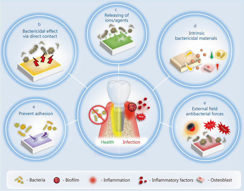
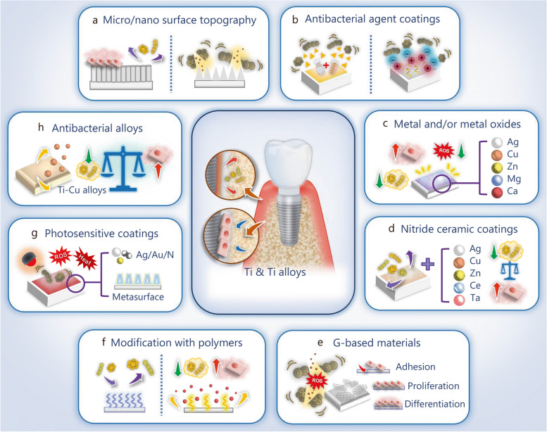
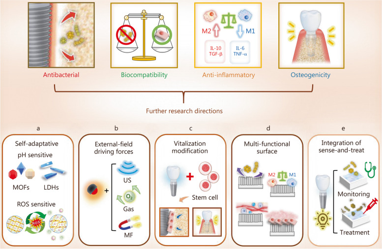
Peri-implantitis is a bacterial infection that causes soft tissue inflammatory lesions and alveolar bone resorption, ultimately resulting in implant failure. Dental implants for clinical use barely have antibacterial properties, and bacterial colonization and biofilm formation on the dental implants are major causes of peri-implantitis. Treatment strategies such as mechanical debridement and antibiotic therapy have been used to remove dental plaque. However, it is particularly important to prevent the occurrence of peri-implantitis rather than treatment. Therefore, the current research spot has focused on improving the antibacterial properties of dental implants, such as the construction of specific micro-nano surface texture, the introduction of diverse functional coatings, or the application of materials with intrinsic antibacterial properties. The aforementioned antibacterial surfaces can be incorporated with bioactive molecules, metallic nanoparticles, or other functional components to further enhance the osteogenic properties and accelerate the healing process. In this review, we summarize the recent developments in biomaterial science and the modification strategies applied to dental implants to inhibit biofilm formation and facilitate bone-implant integration. Furthermore, we summarized the obstacles existing in the process of laboratory research to reach the clinic products, and propose corresponding directions for future developments and research perspectives, so that to provide insights into the rational design and construction of dental implants with the aim to balance antibacterial efficacy, biological safety, and osteogenic property.

Keywords: Anaerobic bacteria; Antibacterial activity; Dental implant; Osteogenic property; Peri-implantitis.


Conflict of interest statement

The authors declare that they have no competing interests.

Figures

Fig. 1
Fig. 1
Schematic illustration of structure composition of dental implant and the classification of dental implant biomaterials with their respective advantages. PEEK poly-ether-ether-ketone
Fig. 2
Fig. 2
Schematic representation illustrating the distinctions among prosthetic infection, peri-implantitis, and periodontitis in terms of infection site and surrounding tissue structure
Fig. 3
Fig. 3
The biological events around dental implants with or without bacterial infection. a Biological events at different stages after implantation. b “Race for the surface” triggered by bacterial infection following implantation
Fig. 4
Fig. 4
Different antibacterial actions involved in the management of bacterial infections. The antibacterial actions can be divided into 5 categories. a Material/surface can inhibit bacterial adhesion. b Engineered surfaces can cause bacterial death via direct contact. c Engineered surface can release antibacterial ions/agents to achieve bactericidal effects. d Material/surface with intrinsic bactericidal effects. e Material/surface can be activated by external driving forces to initiate antibacterial activities
Fig. 5
Fig. 5
Biomaterials and modification strategies for titanium (Ti)-based dental implants to enhance the osteogenic activities and antibacterial properties. a Modification of the surface with micro/nano topography. b Coating the surface with an antibacterial agent. c Modification the surface with metal and/or metal oxides. d Coating the surface with nitride ceramic. e Modification of the surface with graphene (G)-based materials. f Modification of the surface with functional polymers. g Modification of the surface with photosensitive coatings. h Application antibacterial alloys. Ag silver, Cu copper, Zn zinc, Ce cerium, Ta tantalum, Mg magnesium, Ca calcium
Fig. 6
Fig. 6
Schematic diagram of titanium substrate coated with different nanostructured CeO2 (nanorod, nanocube, and nanooctahedron) with the aim of enhancing the antibacterial and anti-inflammatory performance. The antibacterial effects can be attributed to the electrostatic interaction between nanostructured CeO2 and bacterial cell surface. And the anti-inflammatory effects can be attributed to the SOD and CAT mimetic activities [121]. Copyright 2019, Elsevier. Ce cerium, SOD superoxide dismutase, CAT catalase, LPS lipopolysaccharide, ROS reactive oxygen species, IL-1β interleukin-1β, IL-6 interleukin-6, TNF-α tumor necrosis factor-α
Fig. 7
Fig. 7
The preparation and in vivo assessments of osseointegration and anti-infection ability of Ti-PAA-NCl. a Schematic diagram of the synthesis of Ti-PAA-NCl coating on the Ti substrates. b Time period of experiments. c New bone formation via Van Gieson’s staining after 4 weeks of implantation (scale bar is 500 μm in the top image and 250 μm in the bottom one). d Micro-CT images of the bone height surrounding the implants after osseointegration for 4 weeks, peri-implantitis for 8 weeks, and re-osseointegration for 4 weeks (scale bars is 500 μm). e Micro-CT three-dimensional reconstructions of the implants and surrounding bone tissues (scale bars is 500 μm) [105]. Copyright 2021, the author(s). Ti titanium, N-Cl nitrogen-halamine, Ti-OH alkali-heated titanium disks, Ti-PAA-Cl polymeric coating loaded with chloramine
Fig. 8
Fig. 8
The preparation and in vivo anti-bacterial performance of quasi-periodic titanium oxide metasurface. a Schematic diagram of the design principle for the aaBH method to use metasurfaces to endow the implant with potent NIR-responsive antibacterial activity. b The aaBH method to construct quasi-periodic titanium oxide metasurface on Ti alloy implants. c The in vivo animal model with one and two injections of bacteria. Copyright 2021 [111]. TiO2 titanium dioxide, Ti titanium, TN titanium dioxide nanorods, NIR near-infrared, S. aureus Staphylococcus aureus, ROS reactive oxygen species
Fig. 9
Fig. 9
Biomaterials and modification strategies for zirconia dental implant to enhance the antibacterial properties and osteogenic activities. a Modification of the surface with micro-patterns. b Coating the surface with metal or metal oxides. c Modification of the surface with bioactive ceramic coatings. d Coating the surface with GO. Ag silver, ZnO zinc oxide, GO graphene oxide, CaP calcium phosphate
Fig. 10
Fig. 10
ALD of ZnO on microrough zirconia. a Schematic diagram of the preparation and biological properties of different samples. b The optical and SEM images of four samples. c Cross-sectional SEM images of ZnO layers prepared by 500 and 1000 ALD cycles [287]. Copyright 2019, Elsevier. ALD atomic layer deposition, ZnO zinc oxide, SEM scanning electron microscopy, ZrO2 zirconium dioxide, SA and blasted and acid etching, S. aureus Staphylococcus aureus
Fig. 11
Fig. 11
Biomaterials and modification strategies for PEEK dental implant to enhance the osteogenic activities and antibacterial properties. a Physical modification techniques. b Chemical modification techniques. c Composites. PEEK polyether-ether-ketone, TiO2 titanium dioxide, HA hydroxyapatite, ZrP zirconium phosphate, SiN silicon nitride, GO graphene oxide, ZnO zinc oxide, HF hydrofluoric acid
Fig. 12
Fig. 12
The preparation and in vivo assessments of osseointegration of PEEK/nano-FHA implants. a Schematic diagram of the preparation and evaluation of PEEK/nano-FHA composite samples. b Micro-CT three-dimensional reconstruction models showing the regenerated bone of about 0.5 mm width ring around PEEK and PEEK/nano-FHA implants surface at 8 weeks. c Histotomy of bone contact immunostained by toluidine blue-fuchsine at 8 weeks of bare PEEK (i-ii) and PEEK/nano-FHA implants (iii-iv) postoperatively. (ii) and (iv) refer to the higher-magnification images of (i) and (iii), respectively. The dark red area represents the newly formed bone, and the dark black area represents the PEEK-based implant. White scale: 200 μm, black scale: 100 μm; d In histological analysis, new bone formation around bare PEEK (i-ii) and PEEK/nano-FHA implants (iii-iv) were detected by bone labeling (calcein, calcein blue, and tetracycline). (ii) and (iv) refer to the higher-magnification images of (i) and (iii), respectively [314]. Copyright 2014, Elsevier. S sample, NgB newly grown bone deposition and remodeling zone, PeB pre-existing bone tissue zone
Fig. 13
Fig. 13
The further research directions of dental implants. a Developing self-adaptative antibacterial coatings that function according to the microenvironment. b Developing intelligent antibacterial surfaces that can be activated by multiple external-field driving drivers. c Developing lively surface to promote osseointegration by using MSCs-based therapy. d Developing multi-functional surfaces. Combination of the design rules of osteogenesis-angiogenesis regulation and osteoimmunomodulation with existing antibacterial strategies. e Developing versatile surfaces for integrated diagnosis and treatment. M1 M1 type macrophage, M2 M2 type macrophage, IL-10 interleukin-10, TGF-β transforming growth factor-β, IL-6 interleukin-6, TNF-α tumor necrosis factor-α, MOFs metal-organic frameworks, LDHs layered double hydroxides, ROS reactive oxygen species, US ultrasonic, MF magnetic field

Similar articles

Cited by

References

    1. Schwarz F, Derks J, Monje A, Wang HL. Peri-implantitis. J Clin Periodontol. 2018;45(Suppl 20):S246–66. - PubMed
    1. Wilson V. An insight into peri-implantitis: a systematic literature review. Prim Dent J. 2013;2(2):69–73. doi: 10.1308/205016813806144209. - DOI - PubMed
    1. Belibasakis GN, Manoil D. Microbial community-driven etiopathogenesis of peri-implantitis. J Dent Res. 2021;100(1):21–8. doi: 10.1177/0022034520949851. - DOI - PMC - PubMed
    1. Berglundh T, Jepsen S, Stadlinger B, Terheyden H. Peri-implantitis and its prevention. Clin Oral Implants Res. 2019;30(2):150–55. doi: 10.1111/clr.13401. - DOI - PubMed
    1. Nguyen-Hieu T, Borghetti A, Aboudharam G. Peri‐implantitis: from diagnosis to therapeutics. J Investig Clin Dent. 2012;3(2):79–94. doi: 10.1111/j.2041-1626.2012.00116.x. - DOI - PubMed

MeSH terms

LinkOut - more resources