. 2024 Jul;30(3):539-560.
doi: 10.3350/cmh.2024.0060. Epub 2024 May 14.

Novel role of MHC class II transactivator in hepatitis B virus replication and viral counteraction

Affiliations

Novel role of MHC class II transactivator in hepatitis B virus replication and viral counteraction

Mehrangiz Dezhbord et al. Clin Mol Hepatol. 2024 Jul.

Erratum in

Abstract

Background/aims: The major histocompatibility class II (MHC II) transactivator, known as CIITA, is induced by Interferon gamma (IFN-γ) and plays a well-established role in regulating the expression of class II MHC molecules in antigen-presenting cells.

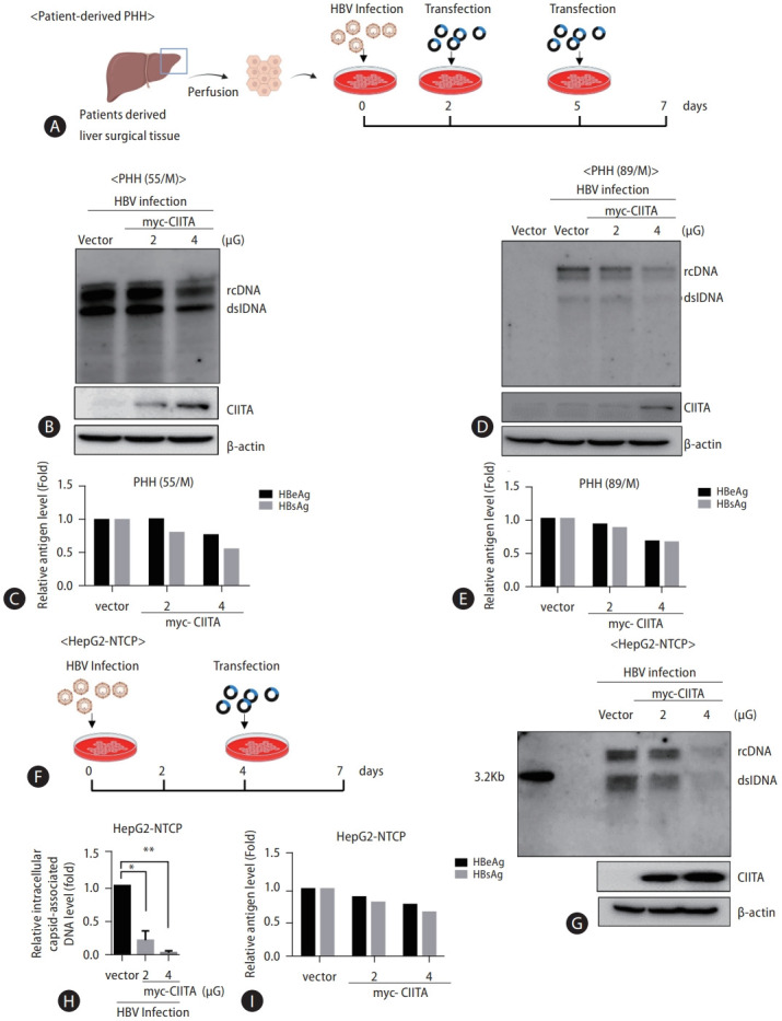
Methods: Primary human hepatocytes (PHH) were isolated via therapeutic hepatectomy from two donors. The hepatocellular carcinoma (HCC) cell lines HepG2 and Huh7 were used for the mechanistic study, and HBV infection was performed in HepG2-NTCP cells. HBV DNA replication intermediates and secreted antigen levels were measured using Southern blotting and ELISA, respectively.

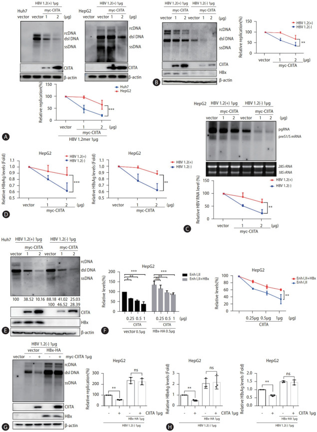
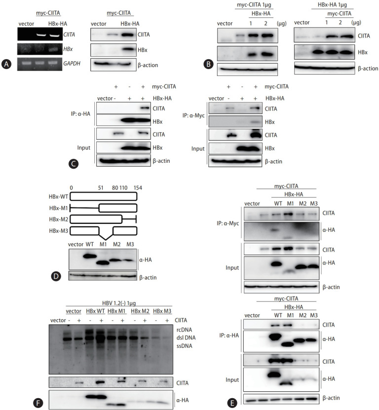
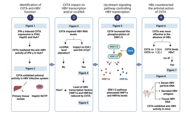
Results: We identified a non-canonical function of CIITA in the inhibition of hepatitis B virus (HBV) replication in both HCC cells and patient-derived PHH. Notably, in vivo experiments demonstrated that HBV DNA and secreted antigen levels were significantly decreased in mice injected with the CIITA construct. Mechanistically, CIITA inhibited HBV transcription and replication by suppressing the activity of HBV-specific enhancers/promoters. Indeed, CIITA exerts antiviral activity in hepatocytes through ERK1/2-mediated down-regulation of the expression of hepatocyte nuclear factor 1α (HNF1α) and HNF4α, which are essential factors for virus replication. In addition, silencing of CIITA significantly abolished the IFN-γ-mediated anti-HBV activity, suggesting that CIITA mediates the anti-HBV activity of IFN-γ to some extent. HBV X protein (HBx) counteracts the antiviral activity of CIITA via direct binding and impairing its function.

Conclusion: Our findings reveal a novel antiviral mechanism of CIITA that involves the modulation of the ERK pathway to restrict HBV transcription. Additionally, our results suggest the possibility of a new immune avoidance mechanism involving HBx.

Keywords: HBV X protein; Hepatitis B virus; Hepatocyte nuclear factor; Interferon-gamma; MHC class II transactivator.


Conflict of interest statement

Conflicts of Interest

The authors have no conflicts to disclose.

Figures

Figure 1.
Figure 1.
CIITA inhibits HBV replication in hepatoma cells and mediates the anti-HBV activity of IFN-γ. (A) Comparison of the gene expression of CIITA in liver biopsies between normal and patients with chronic hepatitis B (CHB) (accession no. GSE14668); data were obtained from a previously published gene expression profiling analysis. (B) Primary human hepatocytes (PHH) were isolated from a 64-year-old female patient donor. PHH, HepG2, and Huh7 cells were seeded in 6-well plates for 24 h, then type I IFNs (IFN-α, IFN-β, and IFN-γ) and TNFα were added at 1,000 units/mL and 50 ng/mL, respectively. After 48 h, cells were harvested and analyzed using semi-quantitative RT-PCR. IL32 gene expression levels were measured as the control. (C) Huh7 cells were transfected with HBV 1.2-mer (HBV 1.2 (+)), myc-CIITA and empty vector plasmids (pcDNA3.1) using lipofectamine 2000. After 72 h, cells and supernatants were harvested. (D) HBV replication was determined using Southern blot analysis (left side) and the expression levels of CIITA were determined using western blot analysis of the same samples. β-actin was used as the loading control. DNA was quantified, and the data was plotted (right side). (E) Relative HBeAg and HBsAg were measured using ELISA of the cell supernatant. (F) Huh7 cells were transfected with siCIITA using lipofectamine RNA iMAX. IFN-γ was added at 1,000 U/mL twice and total RNA was extracted with TRIzol. Cell lysates were subjected to semi-quantitative RT-PCR. GAPDH was used as the control for each lane. (G) Huh7 cells were transfected with HBV 1.2 (+) for 6 h, and then siRNAs were transfected using lipofectamine RNA iMAX. After 12 h, IFN-γ was added twice at 24-h intervals. After 60 h, cells and supernatants were harvested. (H) Southern blot analysis was conducted to confirm the presence of capsid-associated HBV DNA and the signals were quantified. (I) Huh7 supernatants were collected, and relative HBeAg and HBsAg levels were measured using ELISA. Data were obtained from three independent experiments (mean±SD). The statistical significance was assessed using the Student’s t-test: *P<0.05; **P<0.01; ***P<0.001; ****P<0.0001. CIITA, class II transactivator; TNF-α, tumor necrosis factor-alpha; HBV, hepatitis B virus; HBsAg, hepatitis B surface antigen; HBeAg, hepatitis B e antigen; ELISA, enzyme-linked immunosorbent assay.
Figure 2.
Figure 2.
HBV replication is restricted by CIITA in an HBV infection system. (A) PHH cells were isolated from two patient donors and seeded in 6-well plates. HBV infection was induced with 4% PEG and 2% DMSO. The plasmid expressing Myc-CIITA and the mock vector plasmid were transfected at 2 dpi and 5 dpi, respectively. Cells and the supernatant were harvested at 7 dpi. (B, D) HBV replication was determined using Southern blot analysis. CIITA protein levels were measured using Western blot analysis of the same sample. (C, E) HBeAg and HBsAg levels in the supernatant were measured using ELISA. (F) HepG2-NTCP cells were seeded in 6-well plates and were infected using the same protocol. Myc-CIITA was transfected into the cells at 4 dpi and cells were harvested three days later. (G) HBV intracellular-capsid-associated levels were determined using Southern blotting and (H) real-time PCR. (I) HBV-secreted antigen levels in the cell supernatants were quantified. dpi: days post-infection. HBV, hepatitis B virus; CIITA, class II transactivator; PHH, primary human hepatocytes; ELISA, enzyme-linked immunosorbent assay; HBsAg, hepatitis B surface antigen; HBeAg, hepatitis B e antigen. *P<0.05, **P<0.01.
Figure 3.
Figure 3.
CIITA suppresses HBV transcription but does not regulate HBV cccDNA levels. (A and C) Huh7 cells were co-transfected with HBV 1.2 (+), myc-CIITA, and the mock vector control (pcDNA3.1) plasmid using lipofectamine RNAiMAX reagent. (C) Cells were treated with IFN-γ for 8 h before harvesting. HBV RNA was extracted and analyzed using northern blotting. The signals were normalized to the quantity of 18S and 28S ribosomal RNA. (B and D) Relative HBV RNA levels were quantified and plotted in a graph. Data were obtained from three independent experiments (mean±SD). The statistical significance of the differences was assessed using the Student’s t-test: *P<0.05; **P<0.01; ***P<0.001. (E) HepAD38 cells were seeded into a 6-well plate. Tetracycline (Tet) was removed from the media at the indicated time points. (F) HBV DNA was obtained using hirt DNA extraction. The samples shown in the left panel were electrophoresed immediately. (G) Samples were boiled at 88℃ for 5 min and were incubated with EcoRI restriction enzymes at 37℃ for 30 min prior to electrophoresis. (H and I) The upper panel presents the experimental scheme. (Bottom panel) HepAD38 cells were treated with 2 mM PFA between day 2 to day 6 following Tet removal. Cells were transfected with CIITA plasmid on the indicated days. Lysates from HepAD38 cells were harvested for hirt DNA extraction and Southern blotting. (J) One day after seeding, HepG2-NTCP cells were infected with HBV inoculum and were washed the next day. Plasmid transfection was performed 72 h before harvesting. (K) Samples were prepared for Southern blotting analysis of cccDNA levels. mtDNA served as the loading control. mtDNA: mitochondrial DNA. HBV, hepatitis B virus; CIITA, class II transactivator; PFA, phosphonoformic acid; cccDNA, covalently closed circular DNA.
Figure 4.
Figure 4.
CIITA inhibits HBV enhancer and promoter activity. (A) Schematic diagram of the WT and mutant HBV reporter plasmids used in this study. (B and C) HepG2 and Huh7 cells were seeded in a 12-well plate for 24 h, and cells were co-transfected with a set amount of the indicated enhancer reporter, control vector (pcDNA3.1), and β-galactosidase and increasing amounts of myc-CIITA. In the experiment shown in panel C, 0.5 μg of Myc-CIITA plasmid was used. The relative luciferase activity of each enhancer was determined at 48 h post-co-transfection and normalized to the expression levels of β-galactosidase. Bars represent the standard error of triplicate experiments (mean±SD). One-way ANOVA with Tukey’s multiple comparisons; *P<0.05; **P<0.01. ***P<0.001; ****P<0.0001. HBV, hepatitis B virus; CIITA, class II transactivator; WT, wild type.
Figure 5.
Figure 5.
CIITA reduces the expression of major hepatocyte nuclear factors through the ERK1/2 pathway. (A) Representation of the HBV enhancers and their specific binding regions for key transcription factors. (B) Huh7 cells were co-transfected with HBV 1.2 (+), myc-CIITA, and the mock plasmid (pcDNA3.1). The protein expressions of transcription factors were determined using western blot analysis, and related protein levels were quantified by the intensity of each band and plotted. (C and D) The mRNA levels of transcription factors were determined using (C) semi-quantitative RT-PCR and (D) real-time PCR. (E–H) Huh7 cells were co-transfected with HBV 1.2 (+), myc-CIITA, and the mock vector (pcDNA3.1). (E and F) Cell lysis was performed using a lysis buffer with phosphatase inhibitors, and the protein levels of MAPK signaling regulatory factors were determined using Western blot analysis. (G) HBV 1.2 (+) and myc-CIITA were co-transfected into the cells at 1 μg. After 24 h, 10 μM U0126 was added, and, after 2 days, the cells were harvested. HBV replication intermediates were measured using Southern blot analysis and the intensity of each band was quantified, as shown in the graph. (H) HBeAg and HBsAg levels were measured using ELISA. Bars represent the standard error of triplicate values (mean±SD). (A–D) Two-way ANOVA with Tukey’s multiple comparisons; (E–H) One-way ANOVA with Tukey’s multiple comparisons; *P<0.05; **P<0.01; ***P<0.001; ****P<0.0001. CIITA, class II transactivator; ERK1/2, extracellular signal-regulated kinase; HBV, hepatitis B virus; HBsAg, hepatitis B surface antigen; HBeAg, hepatitis B e antigen; ELISA, enzyme-linked immunosorbent assay.
Figure 6.
Figure 6.
The antiviral effect of CIITA is attenuated by HBx. (A) HepG2 and Huh7 cells were co-transfected with HBV 1.2 (+) and myc-CIITA plasmids. Cell lysates were analyzed using Southern blotting. HBV replication levels were detected and compared, as demonstrated in the line graph. (B-D) HepG2 cells were co-transfected with wild type HBV 1.2 (+) or HBx-deletion mutant HBV 1.2 (-), along with the myc-CIITA and mock vectors (pcDNA3.1). (B) HBV replication was measured using Southern blot analysis, and the expression level of CIITA was detected using western blot. The bottom graph shows the comparison of quantified HBV DNA levels. (C) HBV transcription levels were determined using northern blot analysis, and quantified values were graphed. (D) Relative HBeAg and HBsAg levels in the supernatants were measured using ELISA. (E) (top panel) Huh7 cells were transfected with 1 ug of HBV 1.2 (+) or HBV 1.2 (-) plasmids along with increasing doses of CIITA. HBV replication levels were measured using Southern blotting. (Bottom panel) The expression levels of CIITA and HBx protein were determined using Southern blotting. (F) HepG2 cells were seeded in a 12-well plate and transfected with the myc-CIITA, HBx-HA and luciferase reporter plasmids. Relative luciferase activity of each enhancer was determined at 48 h post-co-transfection and normalized to the expression levels of β-galactosidase. (G) HepG2 cells were co-transfected with HBV 1.2 (-) and myc-CIITA with or without HBx-HA plasmids. Southern blot analysis was performed to determine HBV replication levels. The quantified values are indicated in the bar graph (right). (H) Relative HBeAg and HBsAg levels were measured using ELISA. Data were obtained from three independent experiments (mean±SD). The statistical significance of the differences between test groups was assessed by two-way ANOVA with Sidak’s multiple comparisons or (G) One-way ANOVA with Tukey’s multiple comparisons: *P<0.05; **P<0.01; ***P<0.001; ****P<0.0001. CIITA, class II transactivator; HBV, hepatitis B virus; HBsAg, hepatitis B surface antigen; HBeAg, hepatitis B e antigen.
Figure 7.
Figure 7.
HBx interacts with CIITA. (A and B) HepG2 cells were transfected with the Myc-CIITA and HBx-HA plasmids. Cells and supernatants were harvested after 72 h and the mRNA and protein levels of CIITA and HBx were determined using semi-quantitative RT-PCR (left panel) and western blot analysis (right panel). (C) HepG2 cells were transfected with the mock vector or HBx-HA- and myc-CIITA-encoded plasmids. At 48 h post-transfection, cell lysates were immunoprecipitated with anti-HA antibody or anti-myc antibody. CIITA and HBx proteins purified on protein A-agarose beads were measured by immunoblotting. A total of 10% of the lysate was loaded in parallel as the input. (D) (Left panel) The amino acid sequences of the HBx and HBx truncated mutants used in this study. (Right panel) The expression levels of the HBx mutants were examined using western blot. The protein levels of HBx WT and mutant constructs were visualized using an anti-HA tag antibody. β-actin was loaded as the control for each lane. (E) HBx-truncated mutants were transfected into the cells, and cell lysates were subjected to Co-IP. Protein levels were determined by SDS-PAGE following immunoblotting with specific antibodies. (F) The plasmids expressing HBx WT or HBx deletion mutants (HBx M1, M2, and M3) and CIITA were co-transfected with the HBV 1.2 (-) construct as demonstrated. Southern blotting was conducted to test the intracellular capsid-associated HBV DNA levels. The expression levels of CIITA and HBx protein were visualized using anti-CIITA and anti-HA antibodies, respectively. CIITA, class II transactivator; WT, wild type.
Figure 8.
Figure 8.
Hydrodynamic injection of CIITA decreases HBV replication and antigen production in mice. (A) HBV 1.2 (+) and the CIITA plasmid were hydrodynamically injected into the tail vein of mice. After 4 days, the blood and liver tissues of mice were collected. (B and C) The titer of HBsAg and HBeAg in serum was measured using ELISA. (D) HBV DNA was isolated from the mouse liver tissues and serum. Viral gene expression and replication were examined using Southern blot and (E) quantitative real-time PCR, respectively. (D, bottom) A portion of each tissue lysate (10%) was used for protein analysis by Western blot. (F) A hypothetical mechanism of CIITA-mediated suppression of HBV through the downregulation of HNF1α and HNF4α expression. Effects of IFN-γ-induced CIITA expression on HBV replication and transcription. CIITA decreases the expression of HNF4α and HNF1α through activation of the ERK1/2 pathway, thereby inhibiting the transcription of HBV. However, HBx binds to CIITA and counteracts its antiviral activity. This illustration was created with BioRender.com. CIITA, class II transactivator; HBV, hepatitis B virus; ERK1/2, extracellular signal-regulated kinase.
None

Comment in

References

    1. Kim SW, Yoon JS, Lee M, Cho Y. Toward a complete cure for chronic hepatitis B: Novel therapeutic targets for hepatitis B virus. Clin Mol Hepatol. 2022;28:17–30. - PMC - PubMed
    1. Tsukuda S, Watashi K. Hepatitis B virus biology and life cycle. Antiviral Res. 2020;182:104925. - PubMed
    1. Song H, Xu F, Xiao Q, Tan G. Hepatitis B virus X protein and its host partners. Cell Mol Immunol. 2021;18:1345–1346. - PMC - PubMed
    1. Murphy CM, Xu Y, Li F, Nio K, Reszka-Blanco N, Li X, et al. Hepatitis B virus X protein promotes degradation of SMC5/6 to enhance HBV replication. Cell Rep. 2016;16:2846–2854. - PMC - PubMed
    1. Xie S, Yang L, Bi X, Deng W, Jiang T, Lin Y, et al. Cytokine profiles and CD8+ T cells in the occurrence of acute and chronic hepatitis B. Front Immunol. 2022;13:1036612. - PMC - PubMed

MeSH terms

LinkOut - more resources