

Medically responsive amyloidogenic giant sellar-parasellar prolactinoma
- PMID: 38741982
- PMCID: PMC11090579
- DOI: 10.25259/SNI_150_2024
Medically responsive amyloidogenic giant sellar-parasellar prolactinoma
Abstract
Background: Giant prolactinomas are rare; among them, the amyloidogenic variant, prolactinomas with extensive spherical amyloid deposits, are rare, with only 30 cases reported with recommendations of surgical management contrary to the routine prolactinoma's medical management.
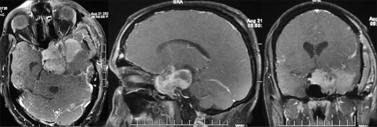
Case description: We report here a case of giant amyloidogenic prolactinoma in a 32-year-old male patient who had a very atypical presentation in terms of clinical, radiological, and pathological features and responded to dopamine agonist therapy like a normal prolactinoma.
Conclusion: Amyloidogenic giant prolactinomas are rare. Contrary to usual belief, even they remain medically responsive; however, more literature is required to decide their ideal management.
Keywords: Amyloidogenic; Macroadenoma; Parasellar; Pituitary; Prolactinoma.
Copyright: © 2024 Surgical Neurology International.
Conflict of interest statement
There are no conflicts of interest.
Figures


Similar articles
-
Giant Prolactinoma Causing Hydrocephalus and Intracranial Hypertension as First Manifestations of Multiple Endocrine Neoplasia Type 1.Front Endocrinol (Lausanne). 2019 Aug 28;10:582. doi: 10.3389/fendo.2019.00582. eCollection 2019. Front Endocrinol (Lausanne). 2019. PMID: 31555208 Free PMC article.
-
Occurrence of extensive spherical amyloid deposits in a prolactin-secreting pituitary macroadenoma: a radiologic-pathologic correlation.Ann Diagn Pathol. 2013 Aug;17(4):361-6. doi: 10.1016/j.anndiagpath.2013.03.001. Epub 2013 Apr 17. Ann Diagn Pathol. 2013. PMID: 23602507
-
The importance of measuring prolactin prior to surgical management of a pituitary lesion: An illustrative case.Radiol Case Rep. 2023 Aug 26;18(11):3889-3893. doi: 10.1016/j.radcr.2023.08.047. eCollection 2023 Nov. Radiol Case Rep. 2023. PMID: 37670916 Free PMC article.
-
A rare case of a giant prolactinoma with atypical histological features: 5 years of follow-up.Hormones (Athens). 2022 Jun;21(2):323-327. doi: 10.1007/s42000-022-00350-5. Epub 2022 Feb 10. Hormones (Athens). 2022. PMID: 35143036 Review.
-
Treatment Strategy for Giant Invasive Macroprolactinoma with Spontaneous Cerebrospinal Fluid Rhinorrhea: A Case Report and Literature Review.World Neurosurg. 2020 Dec;144:19-23. doi: 10.1016/j.wneu.2020.08.129. Epub 2020 Aug 24. World Neurosurg. 2020. PMID: 32853764 Review.
References
-
- Bononi PL, Martinez AJ, Nelson PB, Amico JA. Amyloid deposits in a prolactin-producing pituitary adenoma. J Endocrinol Invest. 1993;16:339–43. - PubMed
-
- Gul S, Bahadir B, Dusak A, Kalayci M, Edebali N, Acikgoz B. Spherical amyloid deposition in a prolactin-producing pituitary adenoma. Neuropathology. 2009;29:81–4. - PubMed
-
- Gatto F, Perez-Rivas LG, Olarescu NC, Khandeva P, Chachlaki K, Trivellin G, et al. Diagnosis and treatment of parasellar lesions. Neuroendocrinology. 2020;110:728–39. - PubMed
-
- Gupta M, Grover N, Sen R. Spherical amyloid deposition in prolactinoma. Indian J Pathol Microbiol. 2020;63:491–2. - PubMed
-
- Iglesias P, Arcano K, Berrocal VR, Bernal C, Villabona C, Díez JJ. Giant prolactinoma in men: Clinical features and therapeutic outcomes. Horm Metab Res. 2018;50:791–6. - PubMed
Publication types
LinkOut - more resources
Full Text Sources