. 2024 Jun 4;63(11):1369-1375.
doi: 10.1021/acs.biochem.4c00090. Epub 2024 May 14.

Anticancer Drugs of Lysine Specific Histone Demethylase-1 (LSD1) Display Variable Inhibition on Nucleosome Substrates

Affiliations

Anticancer Drugs of Lysine Specific Histone Demethylase-1 (LSD1) Display Variable Inhibition on Nucleosome Substrates

Dulmi Senanayaka et al. Biochemistry. .

Abstract

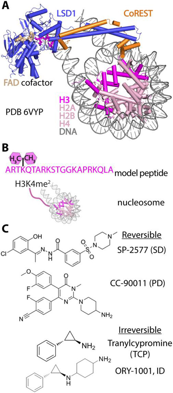
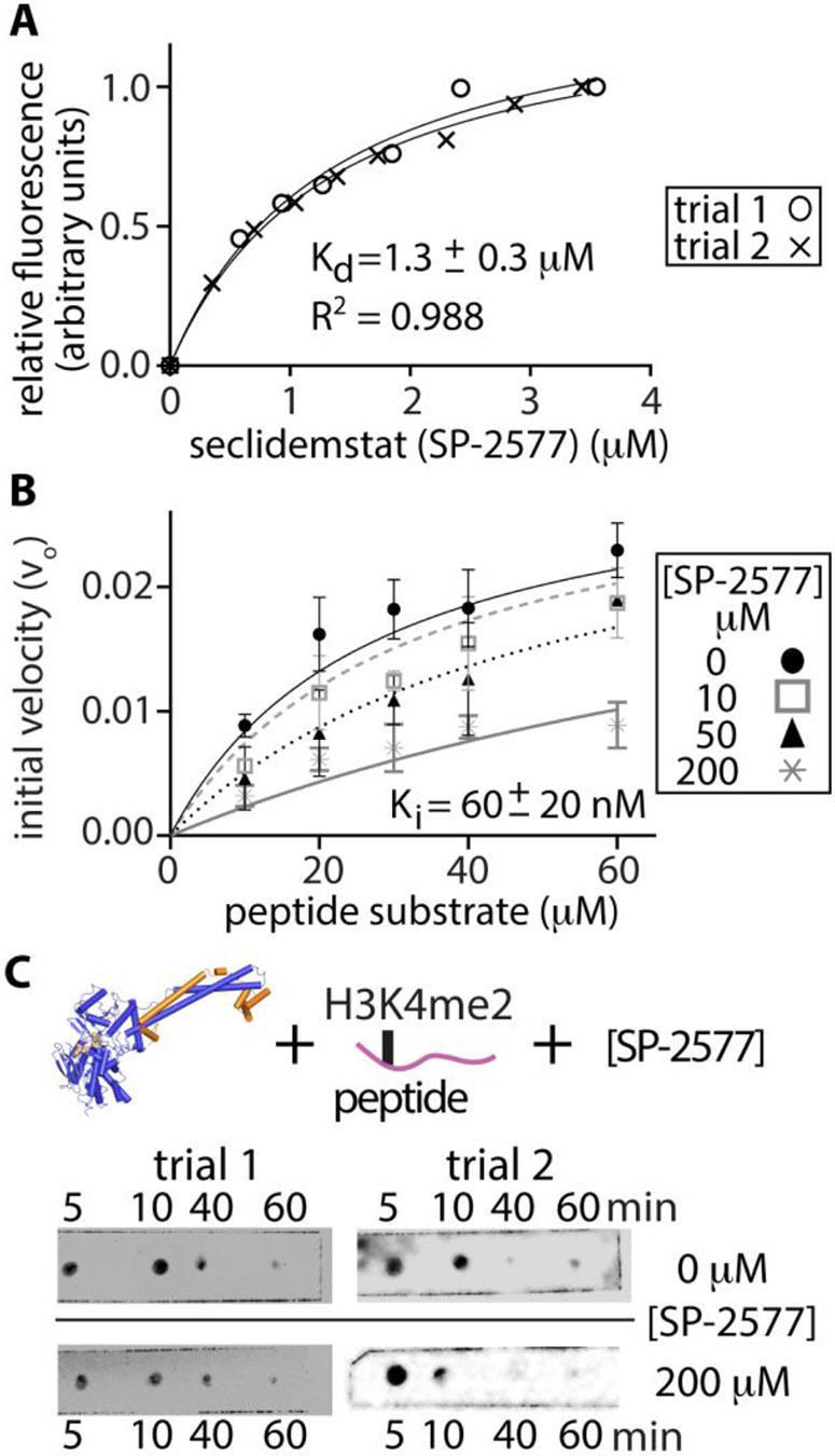
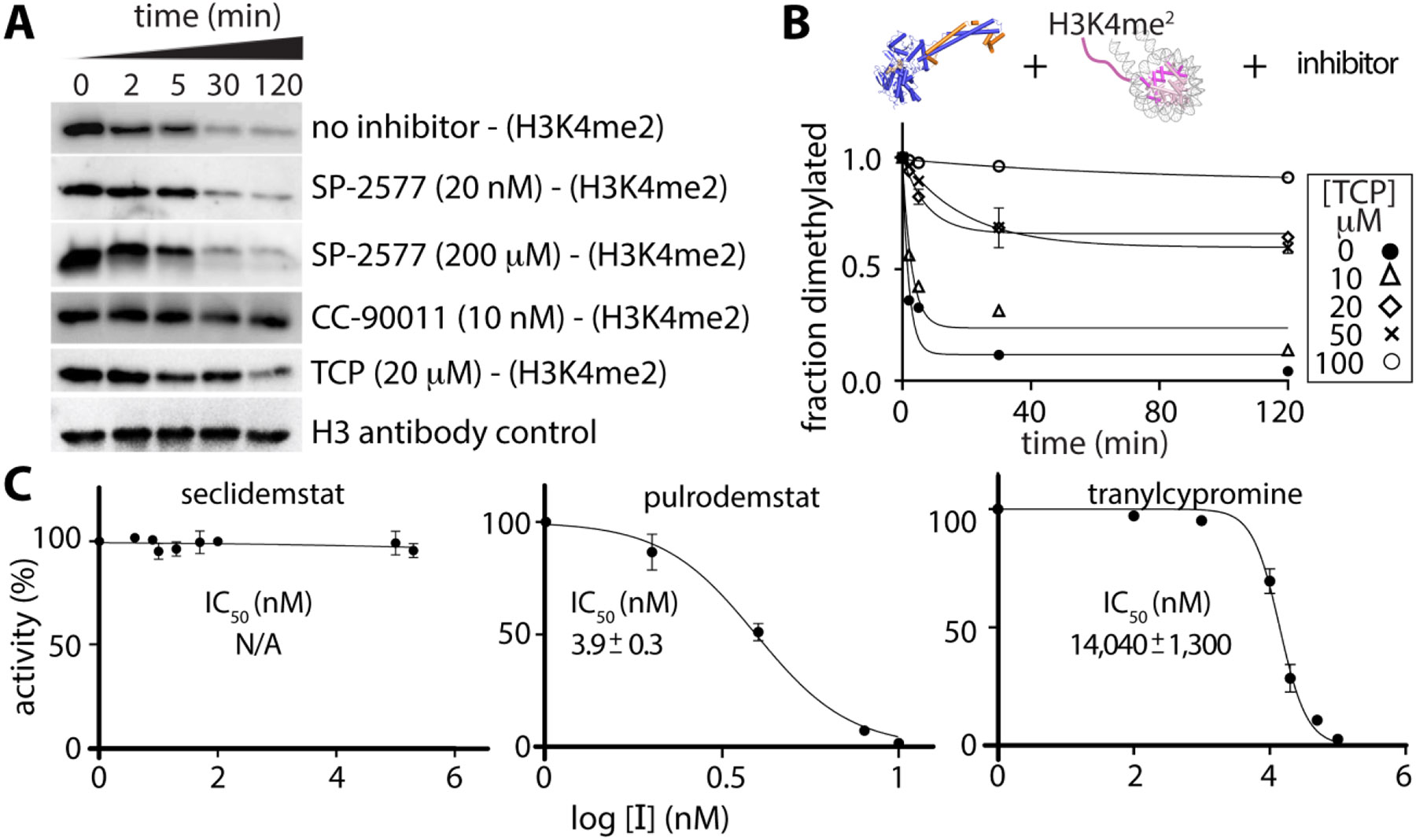
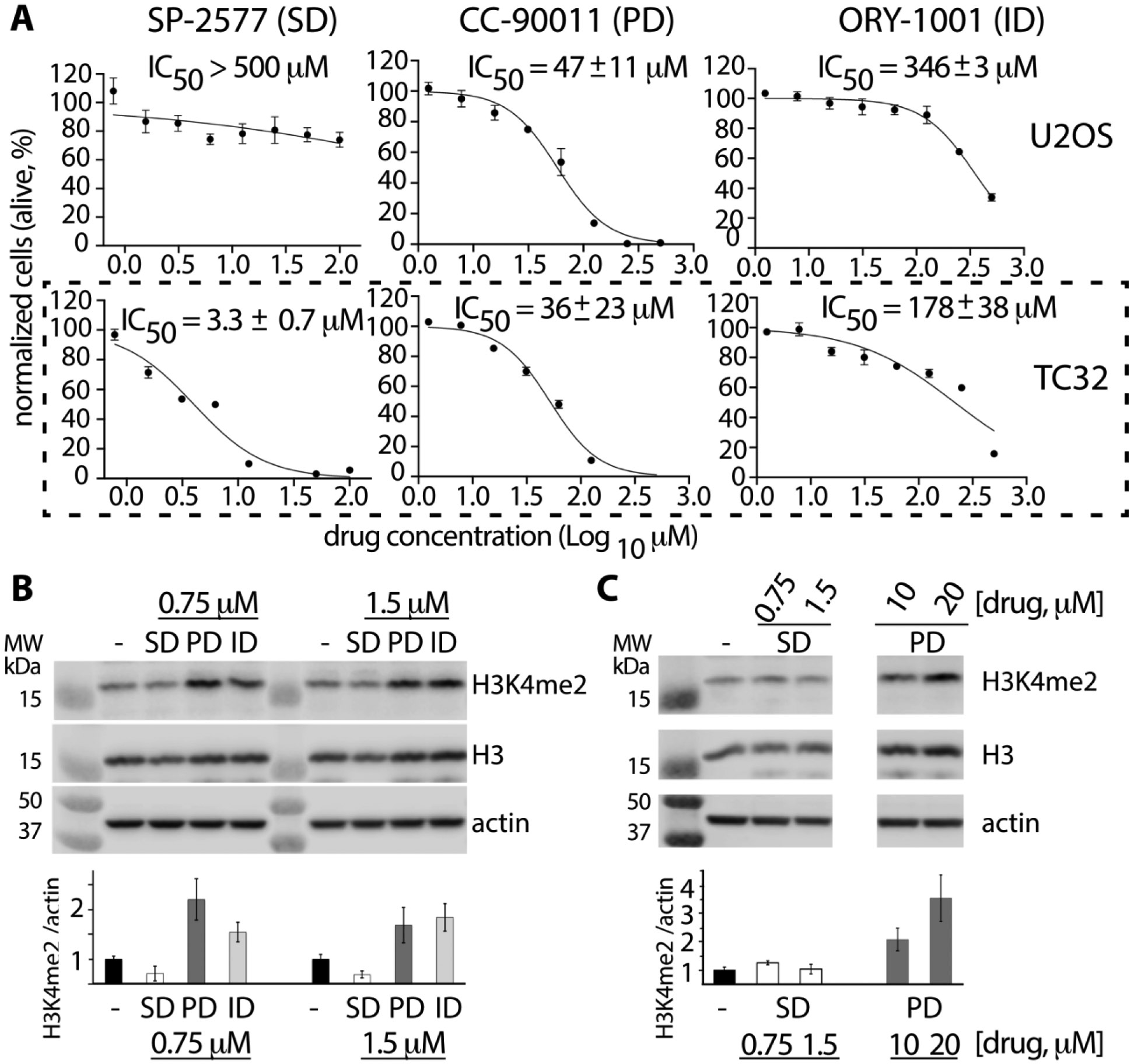
Lysine specific demethylase-1 (LSD1) serves as a regulator of transcription and represents a promising epigenetic target for anticancer treatment. LSD1 inhibitors are in clinical trials for the treatment of Ewing's sarcoma (EWS), acute myeloid leukemia, and small cell lung cancer, and the development of robust inhibitors requires accurate methods for probing demethylation, potency, and selectivity. Here, the inhibition kinetics on the H3K4me2 peptide and nucleosome substrates was examined, comparing the rates of demethylation in the presence of reversible [CC-90011 (PD) and SP-2577 (SD)] and irreversible [ORY-1001 (ID) and tranylcypromine (TCP)] inhibitors. Inhibitors were also subject to viability studies in three human cell lines and Western blot assays to monitor H3K4me2 nucleosome levels in EWS (TC-32) cells, enabling a correlation of drug potency, inhibition in vitro, and cell-based studies. For example, SP-2577, a drug in clinical trials for EWS, inhibits activity on small peptide substrates (Ki = 60 ± 20 nM) using an indirect coupled assay but does not inhibit demethylation on H3K4me2 peptides or nucleosomes using direct Western blot approaches. In addition, the drug has no effect on H3K4me2 levels in TC-32 cells. These data show that SP-2577 is not an LSD1 enzyme inhibitor, although the drug may function independent of demethylation due to its cytotoxic selectivity in TC-32 cells. Taken together, this work highlights the pitfalls of using coupled assays to ascribe a drug's mode of action, emphasizes the use of physiologically relevant substrates in epigenetic drug targeting strategies, and provides insight into the development of substrate-selective inhibitors of LSD1.


Figures

Figure 1.
Figure 1.. Structure of the LSD1–CoREST–nuclesosome complex and the substrates and anticancer drugs used in this study.
(A) Structure of the complex with nucleosomes (Protein Data Bank entry6VYP). LSD1 (blue), CoREST (orange), and cofactor FAD (beige) interact and demethylate H3K4me1/2 nucleosomes, comprised of DNA (gray) and the histone octamer [H3 (magenta), H2A, H2B, and H4 (light pink)]. (B) Schematic of substrates used in this study: H3K4me2 peptide (top) and H3K4me2 nucleosome (bottom). (C) Anticancer drugs that interact with LSD1. SP-2577 [seclidemstat (SD)] and CC-90011 [pulrodemstat (PD)] are reversible binding ligands, whereas tranylcypromine (TCP) and ORY-1001 [iadademstat (ID)] are irreversible inhibitors that react with FAD.
Figure 2.
Figure 2.. SP-2577 binds LSD1 yet displays variable inhibition onH3K4me2 peptide substrates.
(A) Relative fluorescence changes with increasing SP-2577 concentrations enable determination of the apparent dissociation constant (Kd ~ 1.3 ± 0.3 μM). Two independent experiments (× and ○) were both fit to a 1:1 binding isotherm. (B) Demethylation HRP-coupled assays with H3K4me2 peptide substrates and SP-2577. Reactions with increasing concentrations of SP-2577 are shown, allowing for the determination of an apparent inhibition constant (Ki). (C) Dot blot data of LSD1 demethylation on 25 μM H3K4me2 peptide substrate in the absence (0 μM) and presence of 200 μM SP-2577. A schematic of LSD1(blue)–CoREST (orange) with the H3K4me2 peptide substrate(pink) and SP-2577 shows commonalities between contradictory inhibitory data (panels B and C).
Figure 3.
Figure 3.. Anticancer drugs of LSD1 display variable inhibition on H3K4me2 nucleosomes.
(A) Demethylase activity on nucleosomes analyzed using a time-course Western blot assay, with defined inhibitor concentrations (N = 2). The fraction of dimethylated nucleosomes with and without different anticancer drug concentrations was monitored using H3K4me2 antibodies and evaluated using anti-H3 antibodies as a control. A weaker signal intensity corresponds to stronger demethylase activity. (B) Plot of the fraction dimethylated over time with increasing concentrations for tranylcypromine (TCP). Dimethylation profiles reveal individual kobserved values, providing a measure of the potency of a drug to inhibit LSD1’s activity. (C) Half-maximal inhibitory concentrations (IC50) determined on the basis of the analysis of the percent activity (y-axis) and drug concentration (log10, x-axis). See Figures S3-S5 for Western blot images and analysis for SP-2577, CC-90011, and TCP, respectively.
Figure 4.
Figure 4.. Cell viability and ability of anticancer drugs to demethylate H3K4me2 nucleosomes in EWS (TC-32) cancer cells.
(A) Cell viability IC50 determination of the anticancer drugs, SP-2577 (SD), CC-90011 (PD), and ORY-1001 (ID), in U2OS and TC32 cells. Experiments were performed in duplicate (N = 2) with the reported standard error of the mean (SEM). The log-10 scale range of drug concentrations (x-axis) compared to cancer cells (y-axis, normalized alive cells %) was examined for each inhibitor drug. Data from IC50 profiles (black dashed box) were used in the optimization of cell-based H3K4me2 Western blot (WB) assays. Cell viability IC50 profiles in HEK293 cells and for TCP were also determined (panels A and B, respectively, of Figure S6). (B) Western blot (WB) of H3K4me2, H3, and actin levels in TC-32 EWS cancer cells. Drugs were incubated at single-dose concentrations (0.75 and 1.5 μM), and cells were examined after 48 h. Endogenous levels of H3K4me2 at each drug concentration and no drug (−) were determined in the same blot and detected using a H3K4me2 specific antibody. Below the Western blot images, the measured signal of the WB control relative to actin was determined, yielding a H3K4me2/actin signal. Inhibition of LSD1 demethylation on nucleosomes is surmised by an increase in H3K4me2 levels, as previously observed. WB data also allowed for the determination of the H3K4me2/H3 signal (Figure S7). (C) In addition, reversible inhibitor drugs (SP-2577 and CC-90011) were chosen at concentrations according to their comparable cell viability IC50 levels. Here, cells were treated in a manner identical to that described for panel B, except without (−) and with concentrations of the reversible inhibitor drugs SP-2577 (0.75 and 1.5 μM) and CC-90011 (10 and 20 μM). Densitometry data from the images allowed for a qualitative determination of the H3K4me2/actin signal across all conditions.

Similar articles

Cited by

References

    1. Greer EL; Shi Y Histone methylation: a dynamic mark inhealth, disease and inheritance. Nat. Rev. Genet 2012, 13, 343–357. - PMC - PubMed
    1. Marabelli C; Marrocco B; Mattevi A The growing structural and functional complexity of the LSD1/KDM1A histone demethylase. Curr. Opin Struct Biol 2016, 41, 135–144. - PubMed
    1. Burg JM; Link JE; Morgan BS; Heller FJ; Hargrove AE; McCafferty DG KDM1 class flavin-dependent protein lysine demethylases. Biopolymers 2015, 104, 213–246. - PMC - PubMed
    1. Shi Y; Lan F; Matson C; Mulligan P; Whetstine JR; Cole PA; Casero RA; Shi Y Histone demethylation mediated by the nuclear amine oxidase homolog LSD1. Cell 2004, 119, 941–953. - PubMed
    1. Yang M; Gocke CB; Luo X; Borek D; Tomchick DR; Machius M; Otwinowski Z; Yu H Structural basis for CoREST-dependent demethylation of nucleosomes by the human LSD1histone demethylase. Mol. Cell 2006, 23, 377–387. - PubMed

Publication types

LinkOut - more resources