

Regional hippocampal atrophy reflects memory impairment in patients with early relapsing remitting multiple sclerosis
- PMID: 38743090
- PMCID: PMC11319433
- DOI: 10.1007/s00415-024-12290-8
Regional hippocampal atrophy reflects memory impairment in patients with early relapsing remitting multiple sclerosis
Abstract
Background: Research work has shown that hippocampal subfields are atrophic to varying extents in multiple sclerosis (MS) patients. However, studies examining the functional implications of subfield-specific hippocampal damage in early MS are limited. We aim to gain insights into the relationship between hippocampal atrophy and memory function by investigating the correlation between global and regional hippocampal atrophy and memory performance in early MS patients.
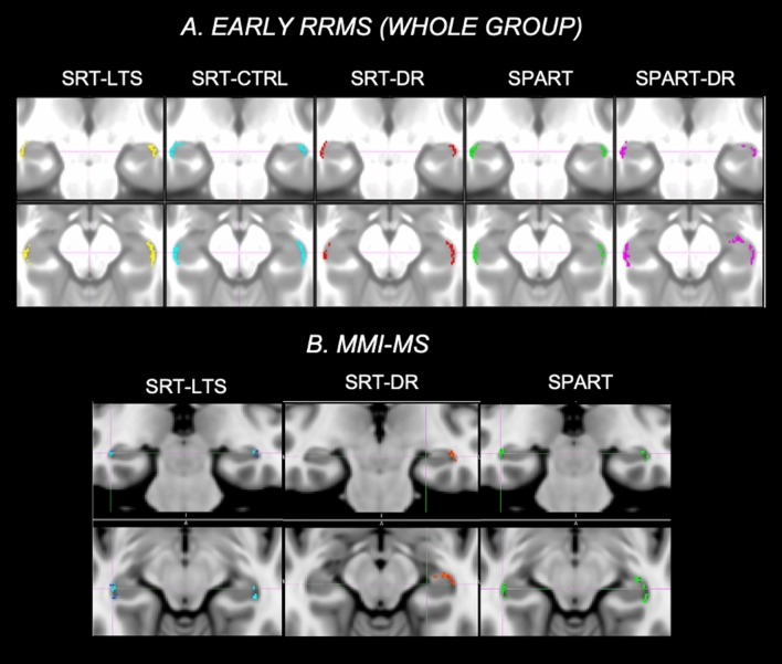
Methods: From the Italian Neuroimaging Network Initiative (INNI) dataset, we selected 3D-T1-weighted brain MRIs of 219 early relapsing remitting (RR)MS and 246 healthy controls (HC) to identify hippocampal atrophic areas. At the time of MRI, patients underwent Selective-Reminding-Test (SRT) and Spatial-Recall-Test (SPART) and were classified as mildly (MMI-MS: n.110) or severely (SMI-MS: n:109) memory impaired, according to recently proposed cognitive phenotypes.
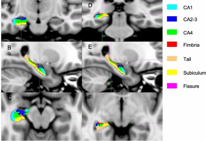
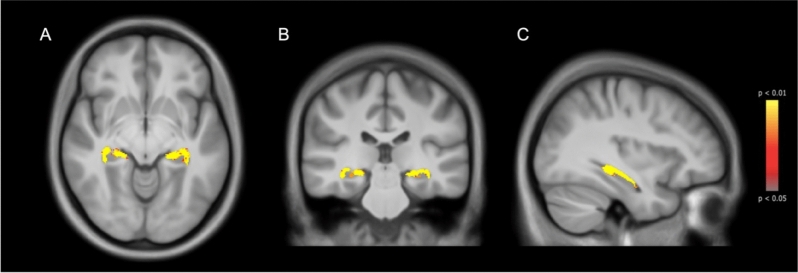
Results: Early RRMS showed lower hippocampal volumes compared to HC (p < 0.001), while these did not differ between MMI-MS and SMI-MS. In MMI-MS, lower hippocampal volumes correlated with worse memory tests (r = 0.23-0.37, p ≤ 0.01). Atrophic voxels were diffuse in the hippocampus but more prevalent in cornu ammonis (CA, 79%) than in tail (21%). In MMI-MS, decreased subfield volumes correlated with decreases in memory, particularly in the right CA1 (SRT-recall: r = 0.38; SPART: r = 0.34, p < 0.01). No correlations were found in the SMI-MS group.
Conclusion: Hippocampal atrophy spreads from CA to tail from early disease stages. Subfield hippocampal atrophy is associated with memory impairment in MMI-MS, while this correlation is lost in SMI-MS. This plays in favor of a limited capacity for an adaptive functional reorganization of the hippocampi in MS patients.
Keywords: Hippocampal atrophy; MRI; Memory impairment; Multiple sclerosis.
© 2024. The Author(s).
Conflict of interest statement
R. Cortese was awarded a MAGNIMS-ECTRIMS fellowship in 2019; she received speaker honoraria from Roche, Merck Serono and Sanofi and travel support for conferences by Novartis. A. Gallo received speaker and consulting fees from Biogen, Genzyme, Merck Serono, Mylan, Novartis, Roche, and Teva, and receives research sup- port from Fondazione Italiana Sclerosi Multipla. E. Pagani received honorarium from Biogen. P. Valsasina received honorarium from Biogen. P. Preziosa received research support from Italian Ministry of Health and Fondazione Italiana Sclerosi Multipla; honoraria from Roche, Biogen, Novartis, Merck Serono, Bristol Myers Squibb, and Genzyme. M.A. Rocca received consulting fees from Biogen, Bristol Myers Squibb, Eli Lilly, Janssen, Roche; and speaker honoraria from AstraZaneca, Biogen, Bristol Myers Squibb, Bromatech, Celgene, Genzyme, Horizon Therapeutics Italy, Merck Serono SpA, Novartis, Roche, Sanofi and Teva. She receives research support from the MS Society of Canada, the Italian Ministry of Health, and Fondazione Italiana Sclerosi Multipla. She is Associate Editor for Multiple Sclerosis and Related Disorders. M. Filippi is Editor-in-Chief of the Journal of Neurology, Associate Editor of Human Brain Mapping, Associate Editor of Radiology, and Associate Editor of Neurological Sciences; received compensation for consulting services from Alexion, Almirall, Biogen, Merck, Novartis, Roche, Sanofi; speaking activities from Bayer, Biogen, Celgene, Chiesi Italia SpA, Eli Lilly, Genzyme, Janssen, Merck-Serono, Neopharmed Gentili, Novartis, Novo Nordisk, Roche, Sanofi, Takeda, and TEVA; participation in Advisory Boards for Alexion, Biogen, Bristol-Myers Squibb, Merck, Novartis, Roche, Sanofi, Sanofi-Aventis, Sanofi-Genzyme, Takeda; scientific direction of educational events for Biogen, Merck, Roche, Celgene, Bristol-Myers Squibb, Lilly, Novartis, Sanofi-Genzyme; he receives research support from Biogen Idec, Merck-Serono, Novartis, Roche, Italian Ministry of Health, Fondazione Italiana Sclerosi Multipla, and ARiSLA (Fondazione Italiana di Ricerca per la SLA). N. De Stefano has received honoraria from Biogen-Idec, Bristol Myers Squibb, Celgene, Genzyme, Immunic, Merck Serono, Novartis, Roche and Teva for consulting services, speaking, and travel support. He serves on advisory boards for Merck, Novartis, Biogen-Idec, Roche, and Genzyme, Immunic and he has received research grant support from the Italian MS Society. M. Battaglini, M.L. Stromillo, L. Luchetti, M. Leoncini, G. Gentile, D. Gasparini, D. Plantone, M. Altieri, A. d’Ambrosio, C. Gianni’, C. Piervincenzi, N. Tedone have nothing to disclose.
Figures
Similar articles
-
Cognitive impairment and memory disorders in relapsing-remitting multiple sclerosis: the role of white matter, gray matter and hippocampus.J Neurol. 2015 Jul;262(7):1691-7. doi: 10.1007/s00415-015-7763-y. Epub 2015 May 10. J Neurol. 2015. PMID: 25957638
-
Thalamic-hippocampal-prefrontal disruption in relapsing-remitting multiple sclerosis.Neuroimage Clin. 2014 Dec 27;8:440-7. doi: 10.1016/j.nicl.2014.12.015. eCollection 2015. Neuroimage Clin. 2014. PMID: 26106524 Free PMC article.
-
Hippocampal atrophy in relapsing-remitting and primary progressive MS: a comparative study.Mult Scler. 2010 Sep;16(9):1083-90. doi: 10.1177/1352458510374893. Epub 2010 Jul 14. Mult Scler. 2010. PMID: 20630904
-
Regional hippocampal atrophy in multiple sclerosis.Brain. 2008 Apr;131(Pt 4):1134-41. doi: 10.1093/brain/awn030. Brain. 2008. PMID: 18375977
-
Pilot investigation of the relationship between hippocampal volume and pattern separation deficits in multiple sclerosis.Mult Scler Relat Disord. 2018 Nov;26:157-163. doi: 10.1016/j.msard.2018.09.016. Epub 2018 Sep 13. Mult Scler Relat Disord. 2018. PMID: 30261457
Cited by
-
Ethnoracial disparities in gray matter atrophy are mediated by structural disconnectivity in multiple sclerosis.Ann Clin Transl Neurol. 2025 Mar;12(3):615-630. doi: 10.1002/acn3.52311. Epub 2025 Feb 17. Ann Clin Transl Neurol. 2025. PMID: 39957675 Free PMC article.
References
-
- Planche V, Gibelin M, Cregut D, Pereira B, Clavelou P (2016) Cognitive impairment in a population-based study of patients with multiple sclerosis: differences between late relapsing-remitting, secondary progressive and primary progressive multiple sclerosis. Eur J Neurol 23(2):282–289. 10.1111/ENE.12715 10.1111/ENE.12715 - DOI - PubMed
MeSH terms
LinkOut - more resources
Full Text Sources
Medical
Miscellaneous
