Multicenter Study
. 2024 May 1;65(5):22.
doi: 10.1167/iovs.65.5.22.

Retinal Dystrophies Associated With Peripherin-2: Genetic Spectrum and Novel Clinical Observations in 241 Patients

Affiliations
Multicenter Study

Retinal Dystrophies Associated With Peripherin-2: Genetic Spectrum and Novel Clinical Observations in 241 Patients

Rachael C Heath Jeffery et al. Invest Ophthalmol Vis Sci. .

Abstract

Purpose: To describe the clinical, electrophysiological and genetic spectrum of inherited retinal diseases associated with variants in the PRPH2 gene.

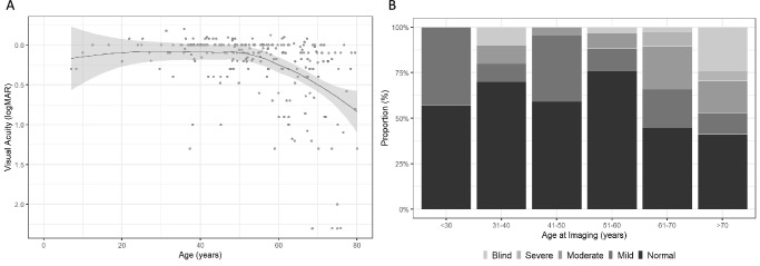
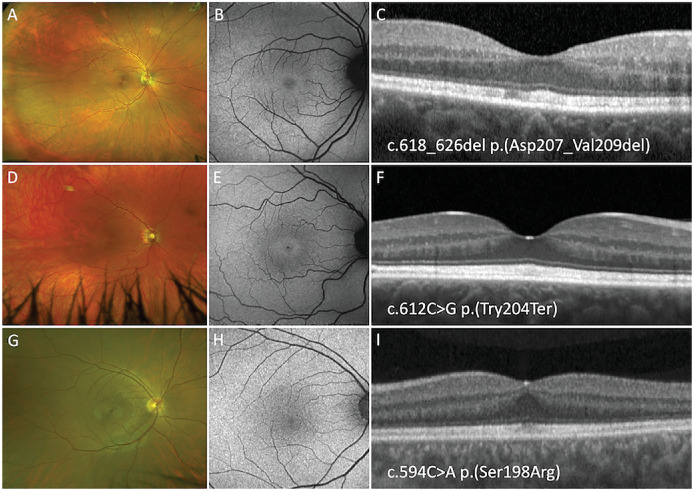
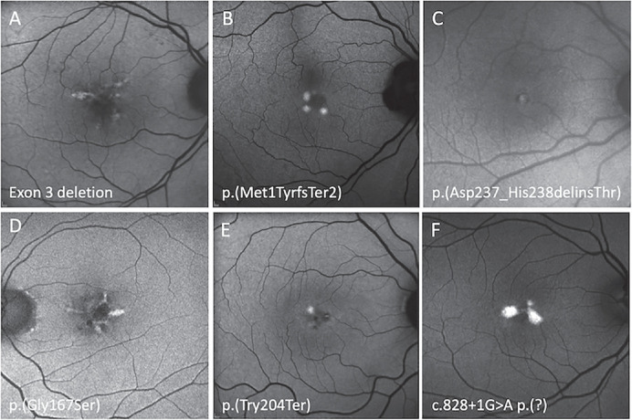
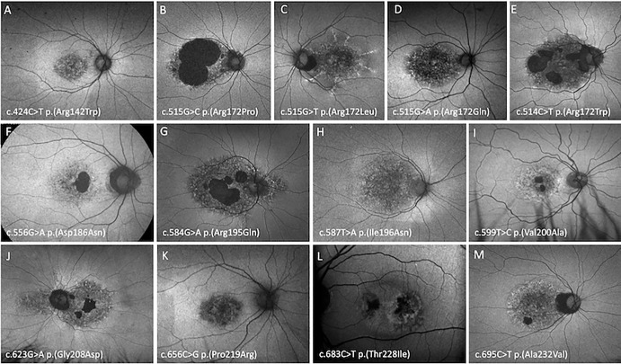
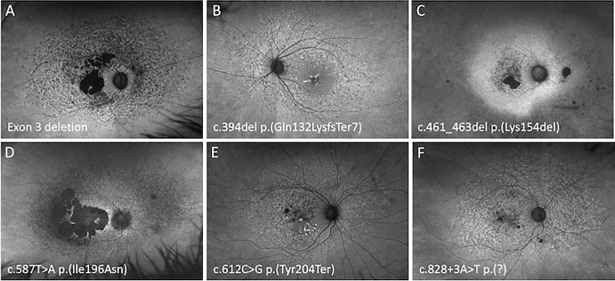
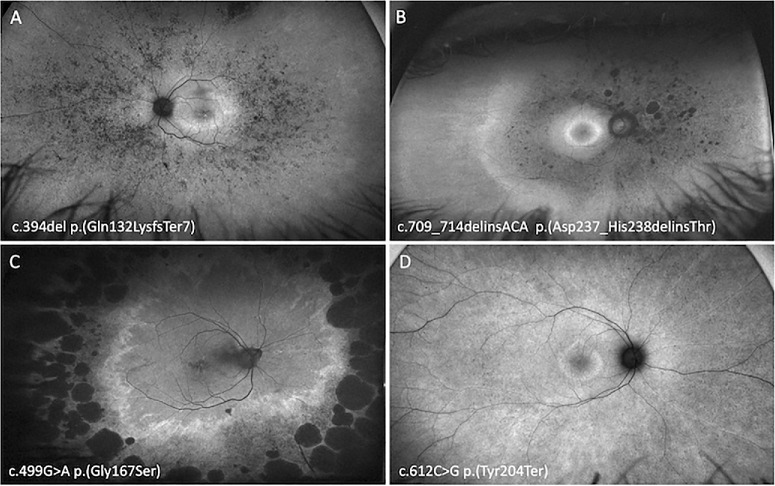
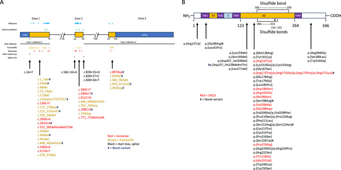
Methods: A total of 241 patients from 168 families across 15 sites in 9 countries with pathogenic or likely pathogenic variants in PRPH2 were included. Records were reviewed for age at symptom onset, visual acuity, full-field ERG, fundus colour photography, fundus autofluorescence (FAF), and SD-OCT. Images were graded into six phenotypes. Statistical analyses were performed to determine genotype-phenotype correlations.

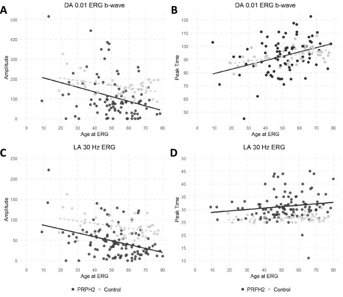
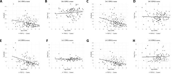
Results: The median age at symptom onset was 40 years (range, 4-78 years). FAF phenotypes included normal (5%), butterfly pattern dystrophy, or vitelliform macular dystrophy (11%), central areolar choroidal dystrophy (28%), pseudo-Stargardt pattern dystrophy (41%), and retinitis pigmentosa (25%). Symptom onset was earlier in retinitis pigmentosa as compared with pseudo-Stargardt pattern dystrophy (34 vs 44 years; P = 0.004). The median visual acuity was 0.18 logMAR (interquartile range, 0-0.54 logMAR) and 0.18 logMAR (interquartile range 0-0.42 logMAR) in the right and left eyes, respectively. ERG showed a significantly reduced amplitude across all components (P < 0.001) and a peak time delay in the light-adapted 30-Hz flicker and single-flash b-wave (P < 0.001). Twenty-two variants were novel. The central areolar choroidal dystrophy phenotype was associated with 13 missense variants. The remaining variants showed marked phenotypic variability.

Conclusions: We described six distinct FAF phenotypes associated with variants in the PRPH2 gene. One FAF phenotype may have multiple ERG phenotypes, demonstrating a discordance between structure and function. Given the vast spectrum of PRPH2 disease our findings are useful for future clinical trials.


Conflict of interest statement

Disclosure: R.C. Heath Jeffery, None; J.A. Thompson, Retina Australia; J. Lo, None; E.S. Chelva, None; S. Armstrong, None; J.S. Pulido, None; R. Procopio, None; A.L. Vincent, None; L. Bianco, None; M. Battaglia Parodi, None; L. Ziccardi, None; G. Antonelli, None; L. Barbano, None; J.P. Marques, None; S. Geada, None; A. L. Carvalho, None; W. Chao Tang, None; C. Mun Chan, None; C.J.F. Boon, None; J. Hensman, None; T.-C. Chen, None; C.-Y. Lin, None; P.-L. Chen, None; A. Vincent, None; A. Tumber, None; E. Heon, None; J.R. Grigg, NHMRC APP1116360, APP1099165, APP1109056; R.V. Jamieson, NHMRC APP1116360, APP1099165, APP1109056; E.E. Cornish, None; B.M. Nash, None; S. Borooah, Foundation Fighting Blindness Grant CD-GT-0918-0746-SEI and Nixon Visions Foundation; L.N. Ayton, Novartis, Apellis, National Health & Medical Research Council (NHMRC) GNT1195713; A.C. Britten-Jones, None; T.L. Edwards, None; J.B. Ruddle, None; A. Sharma, None; R.G. Porter, None; T.M. Lamey, Retina Australia; T.L. McLaren, Retina Australia; S. McLenachan, None; D. Roshandel, None; F.K. Chen, Future Health Research and Innovation Fund, the McCusker Charitable Foundation, Channel 7 Telethon Trust, Retina Australia, NHMRC GNT1116360, GNT1188694, GNT1054712 and MRF1142962

Figures

Figure 1.
Figure 1.
Locally estimated scatterplot smoothing (LOESS) curve using VA from the better-seeing eye illustrating the average evolution of VA with age across the entire cohort (A). Proportion (%) of PRPH2 patients with normal, mild (VA, <20/40 and ≥20/60), moderate (VA, <20/60 and ≥20/200), severe visual impairment (VA, <20/200 and ≥20/400) and blindness (VA, <20/400) by age at imaging (B).
Figure 2.
Figure 2.
Optos pseudocolor, FAF, and SD-OCT imaging of a 40-year-old woman (A, B, C), a 39-year-old woman (D, E, F), and a 40 year-old-man (G, H, I) harboring the p.(Asp207_Val209del), p.(Try204Ter), and p.(Ser198Arg) PRPH2 variants respectively. All three patients show normal pseudocolor and FAF imaging with thickening of band 2 on SD-OCT.
Figure 3.
Figure 3.
Optos FAF imaging of six PRPH2 patients exhibiting focal central macula hyperautofluorescence defined as a BPD or VMD. An 88-year-old woman with a VA of 20/25 harboring exon 3 deletion (A), a 49-year-old woman with a VA of 20/20 harboring p.(Met1TyrfsTer2) (B), a 47-year-old woman with a VA of 20/16 harboring p.(Asp237_His238delinsThr) (C), a 66-year-old man with a VA of 20/25 harboring p.(Gly167Ser) (D), a 57-year-old woman with a VA of 20/20 harboring p.(Tyr204Ter) (E), and a 66-year-old woman with a VA of 20/25 harboring c.828+1G>A (F).
Figure 4.
Figure 4.
Optos FAF imaging of PRPH2 patients demonstrating the CACD phenotype. A 53-year-old woman with VA of 20/25 harboring p.(Arg142Trp) (A), a 45-year-old man with a VA of 20/60 harboring p.(Arg172Pro) (B), an 89-year-old woman with a VA of 20/250 harboring p.(Arg172Leu) (C), a 46-year-old woman with a VA of 20/20 harboring p.(Arg172Gln) (D), a 67-year-old woman with a VA of 20/60 harboring p.(Arg172Trp) (E), a 33-year-old man with a VA of 20/20 harboring p.(Asp186Asn) (F), a 53-year-old man with a VA of 20/60 harboring p.(Arg195Gln) (G), a 27-year-old man with a VA of 20/40 harboring p.(Ile196Asn) (H), a 48-year-old man with a VA of counting fingers harboring p.(Val200Ala) (I), a 68-year-old man with a VA of 20/120 harboring p.(Gly208Asp) (J), a 61-year-old woman with a VA of 20/30 harboring p.(Pro219Arg) (K), a 61-year-old man with a VA of 20/15 harboring p.(Thr228Ile), (L) and a 53-year-old woman with a VA of 20/30 harboring p.(Ala232Val) (M).
Figure 5.
Figure 5.
Optos FAF imaging of PRPH2 patients demonstrating the PSPD phenotype. A 66-year-old woman with a VA of 20/200 harboring an exon 3 deletion (A), a 55-year-old woman with a VA of 20/80 harboring p.(Gln132LysfsTer7) (B), a 59-year-old woman with a VA of 20/20 harboring p.(Lys154del) (C), a 61-year-old woman with a VA of 20/40 harboring p.(Ile196Asn) (D), a 51-year-old woman with a VA of 20/20 harboring p.(Tyr204Ter) (E), and a 67-year-old woman with a VA of 20/90 harboring c.828+3A>T (F).
Figure 6.
Figure 6.
Optos FAF imaging of PRPH2 patients demonstrating the RP phenotype. A 48-year-old woman with VA of 20/40 harboring p.(Gln132LysfsTer7) (A), a 55-year-old woman with a VA of 20/25 harboring p.(Asp237_His238delinsThr) (B), a 71-year-old woman with a VA of 20/30 harboring p.(Gly167Ser) (C), and a 36-year-old woman with a VA of 20/25 harboring p.(Tyr204Ter) (D).
Figure 7.
Figure 7.
Scatter plots for electrophysiologic parameters and age. The full-field ERG component amplitudes and peak times in PRPH2 patients (black circles) and unaffected controls (grey circles) are plotted against age. Data are shown for the DA 0.01 ERG (A and B), LA 30-Hz ERG (C and D). Regression lines are shown for the PRPH2-associated retinopathy (solid line) and control (grey broken line) data.
Figure 8.
Figure 8.
Scatter plots for electrophysiologic parameters and age. The major full-field ERG component amplitudes and peak times in PRPH2 patients (black circles) and unaffected controls (grey circles) are plotted against age. Data are shown for the DA3.0 ERG a-wave (A, B) and b-wave (C, D), LA 3.0 ERG a-wave (E, F) and b-wave (G, H). Regression lines are shown for the PRPH2-associated retinopathy (solid line) and control (grey broken line) data.
Figure 9.
Figure 9.
Position and type of PRPH2 truncating variants (nonsense, frameshift, start-loss, splice site) by exon location (A). Missense and inframe deletion variants and their protein positions within our PRPH2 cohort (B).

References

    1. Heath Jeffery RC, Mukhtar SA, McAllister IL, et al. .. Inherited retinal diseases are the most common cause of blindness in the working-age population in Australia. Ophthalmic Genet. 2021; 42(4): 431–439. - PMC - PubMed
    1. Pontikos N, Arno G, Jurkute N, et al. .. Genetic basis of inherited retinal disease in a molecularly characterized cohort of more than 3000 families from the United Kingdom. Ophthalmology. 2020; 127(10): 1384–1394. - PMC - PubMed
    1. Stenson PD, Mort M, Ball EV, Shaw K, Phillips A, Cooper DN.. The human gene mutation database: building a comprehensive mutation repository for clinical and molecular genetics, diagnostic testing and personalized genomic medicine. Hum Genet. 2014; 133(1): 1–9. - PMC - PubMed
    1. Boon CJ, den Hollander AI, Hoyng CB, Cremers FP, Klevering BJ, Keunen JE.. The spectrum of retinal dystrophies caused by mutations in the peripherin/RDS gene. Prog Retina Eye Res. 2008; 27(2): 213–235. - PubMed
    1. Michaelides M, Holder GE, Bradshaw K, Hunt DM, Moore AT.. Cone-rod dystrophy, intrafamilial variability, and incomplete penetrance associated with the R172W mutation in the peripherin/RDS gene. Ophthalmology. 2005; 112(9), 1592–1598. - PubMed

Publication types

MeSH terms