A fungal protein organizes both glycogen and cell wall glucans
- PMID: 38743622
- PMCID: PMC11126952
- DOI: 10.1073/pnas.2319707121
A fungal protein organizes both glycogen and cell wall glucans
Abstract
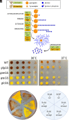
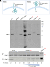
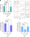
Glycogen is a glucose storage molecule composed of branched α-1,4-glucan chains, best known as an energy reserve that can be broken down to fuel central metabolism. Because fungal cells have a specialized need for glucose in building cell wall glucans, we investigated whether glycogen is used for this process. For these studies, we focused on the pathogenic yeast Cryptococcus neoformans, which causes ~150,000 deaths per year worldwide. We identified two proteins that influence formation of both glycogen and the cell wall: glycogenin (Glg1), which initiates glycogen synthesis, and a protein that we call Glucan organizing enzyme 1 (Goe1). We found that cells missing Glg1 lack α-1,4-glucan in their walls, indicating that this material is derived from glycogen. Without Goe1, glycogen rosettes are mislocalized and β-1,3-glucan in the cell wall is reduced. Altogether, our results provide mechanisms for a close association between glycogen and cell wall.
Keywords: Cryptococcus neoformans; cell wall; glycogen; glycogenin; glycosyltransferase.
Conflict of interest statement
Competing interests statement:The authors declare no competing interest.
Figures


Similar articles
-
Puf4 Mediates Post-transcriptional Regulation of Cell Wall Biosynthesis and Caspofungin Resistance in Cryptococcus neoformans.mBio. 2021 Jan 12;12(1):e03225-20. doi: 10.1128/mBio.03225-20. mBio. 2021. PMID: 33436441 Free PMC article.
-
KRE genes are required for beta-1,6-glucan synthesis, maintenance of capsule architecture and cell wall protein anchoring in Cryptococcus neoformans.Mol Microbiol. 2010 Apr;76(2):517-34. doi: 10.1111/j.1365-2958.2010.07119.x. Epub 2010 Apr 6. Mol Microbiol. 2010. PMID: 20384682 Free PMC article.
-
Glucan Unmasking Identifies Regulators of Temperature-Induced Translatome Reprogramming in C. neoformans.mSphere. 2021 Feb 10;6(1):e01281-20. doi: 10.1128/mSphere.01281-20. mSphere. 2021. PMID: 33568457 Free PMC article.
-
α- and β-1,3-Glucan Synthesis and Remodeling.Curr Top Microbiol Immunol. 2020;425:53-82. doi: 10.1007/82_2020_200. Curr Top Microbiol Immunol. 2020. PMID: 32193600 Review.
-
Fungal beta(1,3)-D-glucan synthesis.Med Mycol. 2001;39 Suppl 1:55-66. doi: 10.1080/mmy.39.1.55.66. Med Mycol. 2001. PMID: 11800269 Review.
Cited by
-
Phenotypic landscape of a fungal meningitis pathogen reveals its unique biology.bioRxiv [Preprint]. 2024 Oct 29:2024.10.22.619677. doi: 10.1101/2024.10.22.619677. bioRxiv. 2024. Update in: Cell. 2025 Jul 24;188(15):4003-4024.e24. doi: 10.1016/j.cell.2025.05.017. PMID: 39484549 Free PMC article. Updated. Preprint.
-
HSP104 and HSP20-L Are Required by Aspergillus nidulans in Response to Attack by Fungivorous Springtail Sinella curviseta.Environ Microbiol Rep. 2025 Aug;17(4):e70147. doi: 10.1111/1758-2229.70147. Environ Microbiol Rep. 2025. PMID: 40619344 Free PMC article.
References
-
- Shearer J., Graham T. E., Novel aspects of skeletal muscle glycogen and its regulation during rest and exercise. Exercise Sport Sci. Rev. 32, 120–126 (2004). - PubMed
-
- Mundkur B., Electron microscopical studies of frozen-dried yeast: I. Localization of polysaccharides. Exp. Cell Res. 20, 28–42 (1960). - PubMed
-
- Mu J., Cheng C., Roach P. J., Initiation of glycogen synthesis in yeast: Requirement of multiple tyrosine residues for function of the self-glucosylating Glg proteins in vivo. J. Biol. Chem. 271, 26554–26560 (1996). - PubMed
MeSH terms
Grants and funding
- F31 AI150194/AI/NIAID NIH HHS/United States
- F31 150194/HHS | NIH | National Institute of Allergy and Infectious Diseases (NIAID)
- R01 AI100272/AI/NIAID NIH HHS/United States
- R01 AI135012/AI/NIAID NIH HHS/United States
- R21 175875/HHS | NIH | National Institute of Allergy and Infectious Diseases (NIAID)
LinkOut - more resources
Full Text Sources
Miscellaneous

