. 2024 Aug 1;30(15):3287-3297.
doi: 10.1158/1078-0432.CCR-24-0426.

Bystander Effects, Pharmacokinetics, and Linker-Payload Stability of EGFR-Targeting Antibody-Drug Conjugates Losatuxizumab Vedotin and Depatux-M in Glioblastoma Models

Affiliations

Bystander Effects, Pharmacokinetics, and Linker-Payload Stability of EGFR-Targeting Antibody-Drug Conjugates Losatuxizumab Vedotin and Depatux-M in Glioblastoma Models

Sonia Jain et al. Clin Cancer Res. .

Abstract

Purpose: Antibody-drug conjugates (ADC) are targeted therapies with robust efficacy in solid cancers, and there is intense interest in using EGFR-specific ADCs to target EGFR-amplified glioblastoma (GBM). Given GBM's molecular heterogeneity, the bystander activity of ADCs may be important for determining treatment efficacy. In this study, the activity and toxicity of two EGFR-targeted ADCs with similar auristatin toxins, Losatuxizumab vedotin (ABBV-221) and Depatuxizumab mafodotin (Depatux-M), were compared in GBM patient-derived xenografts (PDX) and normal murine brain following direct infusion by convection-enhanced delivery (CED).

Experimental design: EGFRviii-amplified and non-amplified GBM PDXs were used to determine in vitro cytotoxicity, in vivo efficacy, and bystander activities of ABBV-221 and Depatux-M. Nontumor-bearing mice were used to evaluate the pharmacokinetics (PK) and toxicity of ADCs using LC-MS/MS and immunohistochemistry.

Results: CED improved intracranial efficacy of Depatux-M and ABBV-221 in three EGFRviii-amplified GBM PDX models (Median survival: 125 to >300 days vs. 20-49 days with isotype control AB095). Both ADCs had comparable in vitro and in vivo efficacy. However, neuronal toxicity and CD68+ microglia/macrophage infiltration were significantly higher in brains infused with ABBV-221 with the cell-permeable monomethyl auristatin E (MMAE), compared with Depatux-M with the cell-impermeant monomethyl auristatin F. CED infusion of ABBV-221 into the brain or incubation of ABBV-221 with normal brain homogenate resulted in a significant release of MMAE, consistent with linker instability in the brain microenvironment.

Conclusions: EGFR-targeting ADCs are promising therapeutic options for GBM when delivered intratumorally by CED. However, the linker and payload for the ADC must be carefully considered to maximize the therapeutic window.


Conflict of interest statement

S. Rathi was a student at the University of Minnesota when the work was conducted. S.K. Gupta reports grants from NCI/NIH during the study, and AbbVie provided the ADCs used in the study. J.E. Eckel-Passow reports grants from NIH during the study. E.B. Reilly reports employment with AbbVie. J.N. Sarkaria reports grants from AbbVie Inc during the conduct of the study, as well as grants from Bayer, Wayshine, Black Diamond, Karyopharm, Boston Scientific, Wugen, Rain Therapeutics, Sumitomo Dainippon Pharma Oncology, SKBP, Boehringer Ingelheim, AstraZeneca, ABL Bio, ModifiBio, Inhibrx, Otomagnetics, and Reglagene outside the submitted work. No disclosures were reported by the other authors.

Figures

Figure 1.
Figure 1.
In vitro sensitivity of GBM PDX lines to MMAE, ABBV-221, and Depatux-M. A, Cytotoxicity of unconjugated MMAE in GBM6, GBM39, GBM108, and GBM10 was determined in a CellTiter-Glo assay. Results are shown as mean ± SEM from at least three independent experiments in each line. B and C, Cytotoxicity of ABBV-221 (B) and Depatux-M (C) to GBM6, GBM39, GBM108, and GBM10 were evaluated using CellTiter-Glo compared with the relevant control ADCs, AB095-MMAE and AB095-MMAF, respectively. A two-sample t-test compared groups at 1 µg/mL. D and E, Using live-cell imaging, the time-dependent internalization of ABBV-221 and Depatux-M was evaluated in GBM6 (D) and GBM10 (E) cells. Results are representative of three independent experiments. P-values were calculated using analysis of covariance at 48 hours. F, The expression of apoptosis markers, cleaved-caspase 3, and cleaved-PARP by Western blot was determined in lysates prepared after treatment of GBM6 and GBM10 with indicated treatments. β-actin was used as a loading control.
Figure 2.
Figure 2.
Bystander effects of ABBV-221 and Depatux-M in EGFR non-amplified lines. A, Concentration of released MMAE was quantified using LC-MS/MS spectrometry in conditioned media collected from GBM6 and GBM108 (EGFRviii-amplified) and GBM10 (EGFR non-amplified) after 4 days (GBM6 and GBM10) and 7 days (GBM108) of treatment with 1 µg/mL AB095-MMAE and ABBV-221. BLQ denotes below the quantification limit (<0.2 ng/mL). B and C, Cytotoxicity of conditioned media on EGFR non-amplified GBM PDX lines, GBM43 (B) and GBM14 (C), harvested from GBM6 (EGFRviii-amplified) and GBM10 (EGFR non-amplified) after 4 days of treatment with the indicated treatments. D, Cytotoxicity on normal human astrocytic cells, SVG-A (EGFR non-amplified), after treatment with GBM6 and GBM10 conditioned media. Symbols represent independent experimental replicates. P-values in B–D were calculated using a two-sample t-test across groups.
Figure 3.
Figure 3.
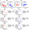
In vivo efficacy of ABBV-221 and Depatux-M. A and B, BLI to monitor the growth of eGFP/fLuc2 transduced GBM39 (A) and GBM6 (B) intracranial tumors over time after treatment with ABBV-221 and Depatux-M administered either intraperitoneally or via CED. Each line represents an individual mouse. P-values were calculated using an analysis of covariance. C and D, Kaplan–Meier curves showing survival of mice after treatment with CED infusions and IP injections of ABBV-221 (C) and Depatux-M (D). AB095 (120 µg/mouse) is an isotype antibody control, AB095-MMAE (GBM6 and GBM108: 48 µg/mouse; GBM39: 88 µg/mouse) and AB095-MMAF (51.2 µg/mouse) are nonspecific ADC controls for ABBV-221 (CED: 66 µg/mouse; IP: 6 mg/kg) and Depatux-M (CED: 60 µg/mouse; IP: 5 mg/kg), respectively. The same data for the AB095 treatment group are shown for C and D for respective GBM PDX lines. Significance between different groups is calculated using the log-rank test. Black arrows along the x-axis indicate when CED infusions were done. IP injections were given weekly from the first infusion to the last CED infusion.
Figure 4.
Figure 4.
Toxicity of AB095-MMAE and ABBV-221 in nontumor-bearing mice brains. A, H&E-stained brain sections representing pathological changes in the ipsilateral and contralateral sides of the brain after a single CED infusion of AB095 (228 µg/mouse), Depatux-M (740 µg/mouse), AB095-MMAE (88 µg/mouse) and ABBV-221 (82 µg/mouse). Images represent changes observed in at least n = 4 mice/group. Scale bars, 50 µm. B, Immunohistochemistry shows CD68 staining in the mouse brain sections after a single CED infusion of indicated treatments. C, NeuN stained brain sections post single CED infusion of indicated drugs. The image inset is marked as a black square and projected on the left for each group. The arrow on the brain represents the side infused with the drug. Images are representative of at least n = 4 mice per group. D, Quantification of neuronal numbers in the ipsilateral side of the brain after CED infusion of each drug. Each symbol represents one mouse. Results are depicted as average neurons per HPF per animal, and four HPFs are analyzed per animal. P-values were calculated using a two-sample t-test.
Figure 5.
Figure 5.
Pharmacokinetics of unconjugated MMAE and ABBV-221 in FVB mice. A, Concentration–time profiles of total MMAE in the plasma and brain of wild-type and triple knockout mice post intravenous injection of unconjugated MMAE (0.5 mg/kg). B, Concentration–time profiles of total MMAE in the plasma, right and left brain of mice post CED infusion of unconjugated MMAE (570 ng/mouse). C, Concentration–time profile of total and released MMAE in plasma and brain of mice after intraperitoneal injection of ABBV-221 (5 mg/kg). D, Concentration–time profiles of total and released MMAE in the plasma, right and left brain of mice post CED infusion of ABBV-221 (60 µg/mouse). Each symbol represents mean of 5 mice. LOQ brain and LOQ plasma denote the limit of quantification in brain and plasma, respectively.
Figure 6.
Figure 6.
Cleavage of MMAE from ABBV-221 in FVB brain homogenate. A, Concentration of released MMAE quantified using LC-MS/MS spectrometry in ABBV-221 samples incubated in buffer control and FVB brain homogenate (50 mmol/L tris-PBS buffer at pH 7.4, 37°C). A two-sample t-test was used to calculate the significance between groups. B, Sensitivity of U87 to unconjugated MMAE. Results are presented as mean ± SEM from three independent experiments. C, Cytotoxicity of ABBV-221 incubated with FVB brain homogenate using CellTiter-Glo assay. P-values were calculated using a two-sample t-test across groups.

References

    1. Carlisle JW, Harvey RD. Tyrosine kinase inhibitors, antibody-drug conjugates, and proteolysis-targeting chimeras: the pharmacology of cutting-edge lung cancer therapies. Am Soc Clin Oncol Educ Book 2021;41:e286–93. - PubMed
    1. Fu Z, Li S, Han S, Shi C, Zhang Y. Antibody drug conjugate: the “biological missile” for targeted cancer therapy. Signal Transduct Target Ther 2022;7:93. - PMC - PubMed
    1. Dumontet C, Reichert JM, Senter PD, Lambert JM, Beck A. Antibody-drug conjugates come of age in oncology. Nat Rev Drug Discov 2023;22:641–61. - PubMed
    1. An Z, Aksoy O, Zheng T, Fan Q-W, Weiss WA. Epidermal growth factor receptor and EGFRvIII in glioblastoma: signaling pathways and targeted therapies. Oncogene 2018;37:1561–75. - PMC - PubMed
    1. Oprita A, Baloi S-C, Staicu G-A, Alexandru O, Tache DE, Danoiu S, et al. . Updated insights on EGFR signaling pathways in glioma. Int J Mol Sci 2021;22:587. - PMC - PubMed

MeSH terms