. 2024 May 14;15(5):338.
doi: 10.1038/s41419-024-06718-2.

METTL8 links mt-tRNA m3C modification to the HIF1α/RTK/Akt axis to sustain GBM stemness and tumorigenicity

Affiliations

METTL8 links mt-tRNA m3C modification to the HIF1α/RTK/Akt axis to sustain GBM stemness and tumorigenicity

Bernice Woon Li Lee et al. Cell Death Dis. .

Erratum in

Abstract

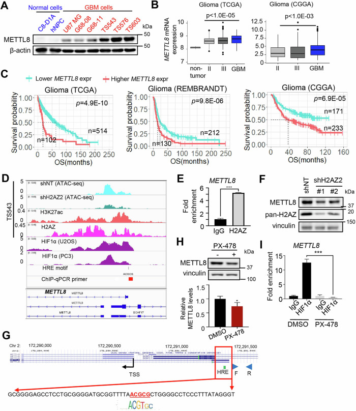
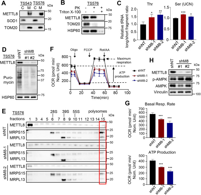
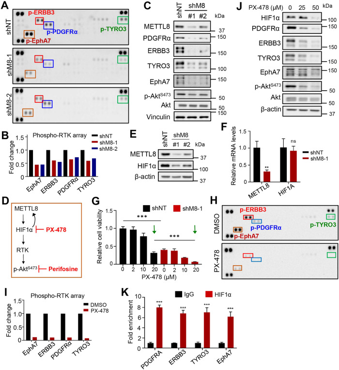
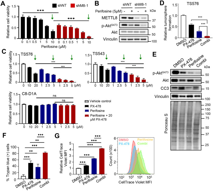
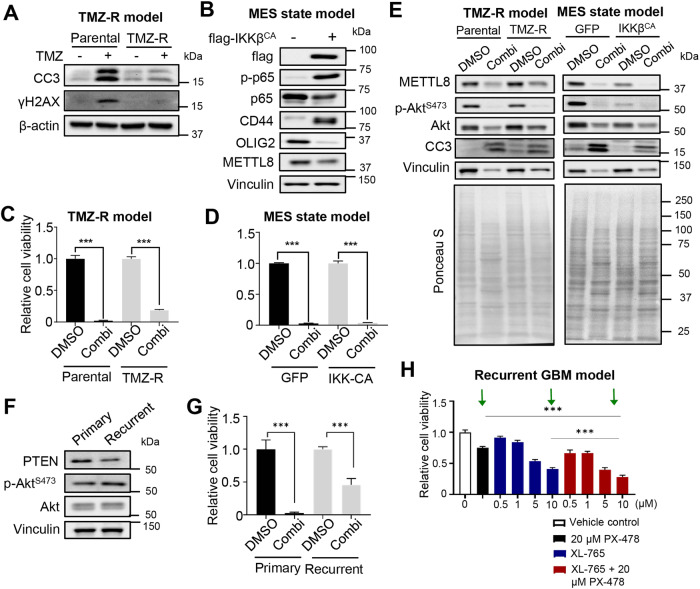
Epitranscriptomic RNA modifications are crucial for the maintenance of glioma stem cells (GSCs), the most malignant cells in glioblastoma (GBM). 3-methylcytosine (m3C) is a new epitranscriptomic mark on RNAs and METTL8 represents an m3C writer that is dysregulated in cancer. Although METTL8 has an established function in mitochondrial tRNA (mt-tRNA) m3C modification, alternative splicing of METTL8 can also generate isoforms that localize to the nucleolus where they may regulate R-loop formation. The molecular basis for METTL8 dysregulation in GBM, and which METTL8 isoform(s) may influence GBM cell fate and malignancy remain elusive. Here, we investigated the role of METTL8 in regulating GBM stemness and tumorigenicity. In GSC, METTL8 is exclusively localized to the mitochondrial matrix where it installs m3C on mt-tRNAThr/Ser(UCN) for mitochondrial translation and respiration. High expression of METTL8 in GBM is attributed to histone variant H2AZ-mediated chromatin accessibility of HIF1α and portends inferior glioma patient outcome. METTL8 depletion impairs the ability of GSC to self-renew and differentiate, thus retarding tumor growth in an intracranial GBM xenograft model. Interestingly, METTL8 depletion decreases protein levels of HIF1α, which serves as a transcription factor for several receptor tyrosine kinase (RTK) genes, in GSC. Accordingly, METTL8 loss inactivates the RTK/Akt axis leading to heightened sensitivity to Akt inhibitor treatment. These mechanistic findings, along with the intimate link between METTL8 levels and the HIF1α/RTK/Akt axis in glioma patients, guided us to propose a HIF1α/Akt inhibitor combination which potently compromises GSC proliferation/self-renewal in vitro. Thus, METTL8 represents a new GBM dependency that is therapeutically targetable.


Conflict of interest statement

The authors declare no competing interests.

Figures

Fig. 1
Fig. 1. GSCs overexpress METTL8 via H2AZ-mediated chromatin accessibility of HIF1α.
A Western blot analysis of METTL8 levels in mouse astrocyte C8-D1A, human neural progenitor cells (NPC), U87 MG and patient-derived GSCs. B Comparison of METTL8 mRNA levels in non-tumor tissues and gliomas of different clinical grades in TCGA and CGGA cohorts. Wilcoxon–Mann Whitney test. C Correlative analysis of METTL8 levels with glioma patient survival in multiple glioma patient cohorts. OS overall survival. Wald test. D Integrative analysis of ATAC-Seq data (shH2AZ2 vs shNT), as well as H3K27ac, H2AZ and HIF1α ChIP-Seq data of METTL8 proximal promoter, along with the location of ChIP-qPCR primers and predicted HRE motif. E ChIP-qPCR analysis of H2AZ occupancy on the METTL8 promoter in GSC (n = 3) (mean ± SD), ***P < 0.001. F Western blot analysis of H2AZ and METTL8 protein levels in H2AZ2 KD GSC. G Zoom-in view of the METTL8 promoter to highlight the predicted HRE motif, along with the location of ChIP-qPCR primers. H Western blot analysis of METTL8 levels in GSC TS576 treated with or without 25 µM PX-478 for 1 day. The relative METTL8 levels was normalized to DMSO control (n = 3) (mean ± SD) *P < 0.05. I ChIP-qPCR analysis of HIF1α occupancy on the METTL8 promoter upon PX-478 (25 µM, 1 day) treatment of GSC (n = 3) (mean ± SD), ***P < 0.001.
Fig. 2
Fig. 2. METTL8 depletion impairs GSC stemness and tumorigenicity.
A Tumorsphere formation of GSCs following METTL8 KD (n = 6) (mean ± SD). ***P < 0.001. B In vitro limiting dilution assays of GSCs transduced with NT/control or METTL8 shRNA calculated with ELDA analysis. C Quantification of EdU+ cells upon METTL8 KD (n = 3) (mean ± SD). Approximately 200 nuclei were counted per replicate. **P < 0.01, ***P < 0.001. D Representative images of EdU immunofluorescence. Scale bar: 50 µm. E Western blot analysis of GFAP and OLIG2 levels in METTL8 KD GSC upon serum-induced differentiation. F, G In vivo bioluminescence-based imaging 35 days post-orthotropic injection of GSC TS576 (7.5 × 10⁵ cells) transduced with NT/control or METTL8 shRNAs. Quantification of tumor volume based on bioluminescence (F) and representative images of the tumor-bearing mice (G) (n = 5) (mean ± SD). ***P < 0.001. Two-tailed unpaired Student’s t-test. H Survival curves of mice implanted with GSC transduced with NT or METTL8 shRNAs. ***P < 0.001. Log-rank test.
Fig. 3
Fig. 3. METTL8 mediates mt-tRNA m3C modification for mitochondrial translation and respiration in GSC.
A Western blot analysis of METTL8 levels in the cytosolic (C) and mitochondrial (M) fractions of GSCs. B Western blot analysis of METTL8 levels upon proteinase K treatment of GSC mitochondrial extracts, with or without detergent. C qRT-PCR analysis of m3C modification on mt-tRNA(Thr/Ser(UCN)) upon METTL8 KD (n = 3) (mean ± SD). *P < 0.05, **P < 0.01, ***P < 0.001. D Western blot analysis of puromycin levels in the mitochondrial extracts of METTL8 KD GSC. E Western blot analysis of METTL8, MRPS15, and MRPL13 levels in different fractions of mitochondrial extracts of METTL8 KD GSC after sucrose gradient ultracentrifugation. F, G Seahorse analysis of GSC upon METTL8 KD (n = 3). ***P < 0.001. H Western blot analysis of p-AMPK and AMPK levels in METTL8 KD GSC.
Fig. 4
Fig. 4. METTL8 loss inactivates RTK signaling via HIF1α downregulation in GSC.
Phospho-RTK array analysis of METTL8 KD (A, B) or PX-478 treated (1 day) (H, I) GSC TS576. Representative blots (n = 2 replicates) (A, H) and quantification of EphA7, ERBB3, PDGFRα and TYRO2 dot intensities when normalized to the intensity of control dots (B, I) are shown. C, J Western blot analysis of PDGFRα, ERBB3, TYRO3, EphaA7, p-AktS473, and Akt levels in METTL8 KD (C) or PX-478 treated (J) GSC TS576. D Schematic diagram of the proposed mechanism. E Western blot analysis of HIF1α levels in METTL8 KD GSC TS576. F qRT-PCR analysis of HIF1A mRNA levels in METTL8 KD GSC. HSP70 serves as the housekeeping genes (n = 3) (mean ± SD). G Cell viability of METTL8 depleted GSC TS576, with or without PX-478 treatment (2 days). The values were normalized to the DMSO control (n = 6) (mean ± SD) ***P < 0.001. K ChIP-qPCR analysis of HIF1α enrichment at the promoters of PDGFRA, ERBB3, TYRO3 and EPHA7 in GSC TS576 (n = 3) (mean ± SD). ***P < 0.001 (n = 3) (mean ± SD).
Fig. 5
Fig. 5. METTL8 depleted GSCs are sensitive to Akt inhibitor, leading to the rational combination of HIF1α/Akt inhibitors to target GSC.
A Cell viability analysis of shNT or shM8 transduced GSC, treated with or without Perifosine at the indicated concentrations for 2 days (n = 6) (mean ± SD). ***P < 0.001. B Western blot of p-AktS473 and Akt levels in shNT or shM8 transduced GSC, with or without Perifosine treatment (5 µM, 2 days). C Cell viability assay of GSCs and mouse astrocytes with 3-day treatment of the indicated drugs (n = 6) (mean ± SD). ***P < 0.001. D Tumorsphere formation of GSC treated with the indicated drugs (5 days) (n = 6) (mean ± SD). ***P < 0.001. E Western blot analysis of p-AktS473, Akt, cleaved-caspase 3 (CC3) levels with the respective drug treatment of GSC. F Trypan blue exclusion assay of GSC treated with the indicated drugs for 3 days. **P < 0.01, ***P < 0.001. G CellTraceTM Violet staining of GSC treated with the indicated drugs (1 day) (n = 3) (mean ± SD). ***P < 0.001.
Fig. 6
Fig. 6. The HIF1α/Akt inhibitor combination also suppresses proliferation of various hard-to-treat GBM cellular models in vitro.
A Western blot analysis of CC3 and γH2AX levels in parental vs TMZ-R GSCs, with or without TMZ treatment (200 µM, 5 days). B Western blot analysis of flag, p-p65, p65, CD44, OLIG2, and METTL8 levels in GSC overexpressing GFP or flag-IKKβCA. C, D Cell viability assay of parental vs TMZ-R GSCs, or GFP vs flag-IKKβCA overexpressing GSCs, with or without PX-478/Perifosine combination treatment for 3 days (n = 6) (mean ± SD). ***P < 0.001. E Western blot analysis of METTL8, p-AktS473, Akt, and CC3 in TMZ-R and mesenchymal GBM cells, with or without PX-478/Perifosine combination treatment for 3 days. F Western blot analysis of PTEN, p-AktS473 and Akt levels in primary (G68-11) vs recurrent (G68-28) patient-derived GSC lines. G Cell viability assay of primary vs recurrent GSC lines, with or without PX-478/Perifosine combination treatment for 3 days (n = 6) (mean ± SD). ***P < 0.001. H Cell viability assay of recurrent GSC line (G68-28) when treated with the indicated drugs for 3 days (n = 6) (mean ± SD). ***P < 0.001.

References

    1. Bao SD, Wu QL, McLendon RE, Hao YL, Shi Q, Hjelmeland AB, et al. Glioma stem cells promote radioresistance by preferential activation of the DNA damage response. Nature. 2006;444:756–60. 10.1038/nature05236 - DOI - PubMed
    1. Hu BL, Wang QH, Wang YA, Hua SJ, Sauve CEG, Ong D, et al. Epigenetic activation of WNT5A drives glioblastoma stem cell differentiation and invasive growth. Cell. 2016;167:1281–95.e18. 10.1016/j.cell.2016.10.039 - DOI - PMC - PubMed
    1. Chen J, Li YJ, Yu TS, McKay RM, Burns DK, Kernie SG, et al. A restricted cell population propagates glioblastoma growth after chemotherapy. Nature. 2012;488:522–6. 10.1038/nature11287 - DOI - PMC - PubMed
    1. Singh SK, Hawkins C, Clarke ID, Squire JA, Bayani J, Hide T, et al. Identification of human brain tumour initiating cells. Nature. 2004;432:396–401. 10.1038/nature03128 - DOI - PubMed
    1. Gimple RC, Bhargava S, Dixit D, Rich JN. Glioblastoma stem cells: lessons from the tumor hierarchy in a lethal cancer. Genes Dev. 2019;33:591–609. 10.1101/gad.324301.119 - DOI - PMC - PubMed

Publication types

MeSH terms

Substances