. 2024 May 15;9(1):133.
doi: 10.1038/s41392-024-01816-1.

2-APQC, a small-molecule activator of Sirtuin-3 (SIRT3), alleviates myocardial hypertrophy and fibrosis by regulating mitochondrial homeostasis

Affiliations

2-APQC, a small-molecule activator of Sirtuin-3 (SIRT3), alleviates myocardial hypertrophy and fibrosis by regulating mitochondrial homeostasis

Fu Peng et al. Signal Transduct Target Ther. .

Abstract

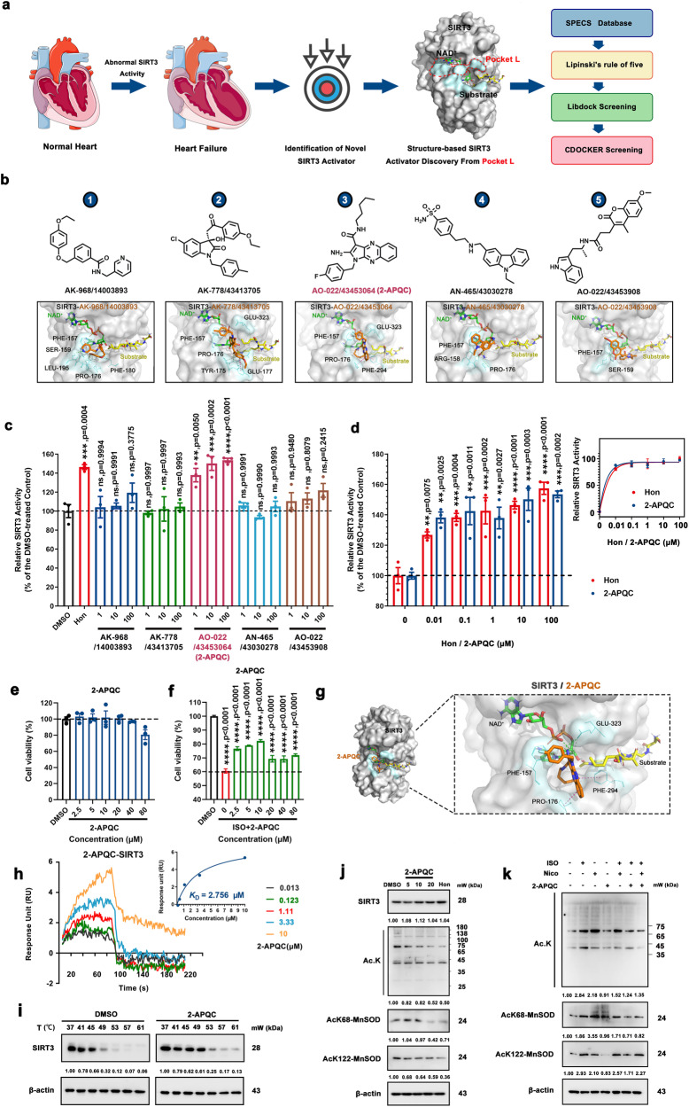
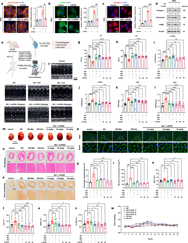
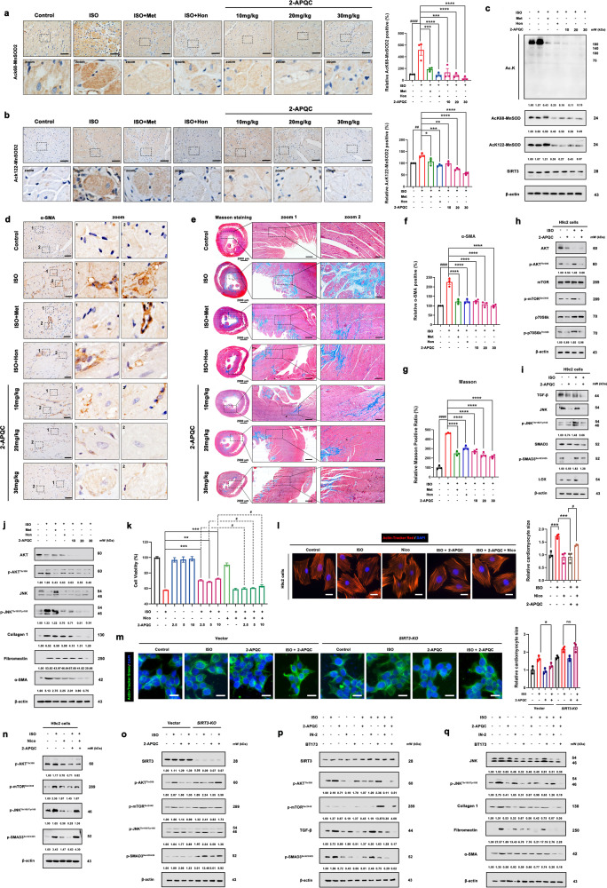
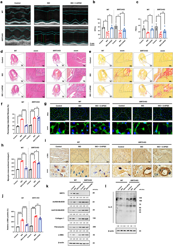
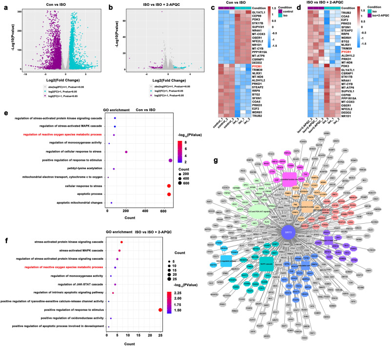
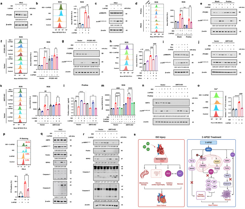
Sirtuin 3 (SIRT3) is well known as a conserved nicotinamide adenine dinucleotide+ (NAD+)-dependent deacetylase located in the mitochondria that may regulate oxidative stress, catabolism and ATP production. Accumulating evidence has recently revealed that SIRT3 plays its critical roles in cardiac fibrosis, myocardial fibrosis and even heart failure (HF), through its deacetylation modifications. Accordingly, discovery of SIRT3 activators and elucidating their underlying mechanisms of HF should be urgently needed. Herein, we identified a new small-molecule activator of SIRT3 (named 2-APQC) by the structure-based drug designing strategy. 2-APQC was shown to alleviate isoproterenol (ISO)-induced cardiac hypertrophy and myocardial fibrosis in vitro and in vivo rat models. Importantly, in SIRT3 knockout mice, 2-APQC could not relieve HF, suggesting that 2-APQC is dependent on SIRT3 for its protective role. Mechanically, 2-APQC was found to inhibit the mammalian target of rapamycin (mTOR)-p70 ribosomal protein S6 kinase (p70S6K), c-jun N-terminal kinase (JNK) and transforming growth factor-β (TGF-β)/ small mother against decapentaplegic 3 (Smad3) pathways to improve ISO-induced cardiac hypertrophy and myocardial fibrosis. Based upon RNA-seq analyses, we demonstrated that SIRT3-pyrroline-5-carboxylate reductase 1 (PYCR1) axis was closely assoiated with HF. By activating PYCR1, 2-APQC was shown to enhance mitochondrial proline metabolism, inhibited reactive oxygen species (ROS)-p38 mitogen activated protein kinase (p38MAPK) pathway and thereby protecting against ISO-induced mitochondrialoxidative damage. Moreover, activation of SIRT3 by 2-APQC could facilitate AMP-activated protein kinase (AMPK)-Parkin axis to inhibit ISO-induced necrosis. Together, our results demonstrate that 2-APQC is a targeted SIRT3 activator that alleviates myocardial hypertrophy and fibrosis by regulating mitochondrial homeostasis, which may provide a new clue on exploiting a promising drug candidate for the future HF therapeutics.


Conflict of interest statement

The authors declare no competing interests. Liang Ouyang is the editorial board member of Signal Transduction and Targeted Therapy, but he has not been involved in the process of the paper handling.

Figures

Fig. 1
Fig. 1
Structure-based screening of small-molecule activators of SIRT3 and identifies 2-APQC activates SIRT3 deacetylase activity in H9c2 cells. a The structure of SIRT3 and virtual screening scheme model for the discovery of SIRT3 activator. Created with biorender.com and https://smart.servier.com. b The 5 candidate compounds based on virtual screening and molecular docking. c The relative SIRT3 deacetylation activity after the candidate compounds (1, 10, 100 μM) and Honokiol (Hon) (10 μM) treatment. Data are present as mean ± s.e.m, n = 3. d The relative SIRT3 deacetylation activity after Hon or 2-APQC (0.01, 0.1 1, 10, 100 μM) treatment. Data are present as mean ± s.e.m, n = 3. e H9c2 cells were treated with indicated concentration of 2-APQC for 24 h, and cell viability was measured by the MTT assay. Data are present as mean ± s.e.m, n = 3. f H9c2 cells were treated with indicated concentration of 2-APQC for 24 h, and then treated with isoproterenol (ISO) for 48 h, the cell viability was measured by the MTT assay. Data are present as mean ± s.e.m, n = 3. g The molecular docking sketch map to indicate the binding mode between SIRT3 and 2-APQC. h Surface plasmon resonance assay detected the Kd value of SIRT3 in H9c2 cells treated with 2-APQC. i Cellular thermal shift assay detected the thermal stability of SIRT3 in H9c2 cells treated with 2-APQC. j Western blot analysis of SIRT3, acetylated-lysine, acetylated manganese superoxide dismutase 2 (MnSOD2) at K68 and K122 protein expression in H9c2 cells treated with 2-APQC for 24 h. k Western blot analysis of acetylated-lysine, acetylated MnSOD2 at K68 and K122 protein expression in H9c2 cells treated with 2-APQC for 24 h, together with or without ISO/nicotinamide (Nico) as the research design. β-actin, loading control. ns no significance; *p < 0.05, **p < 0.01, ***p < 0.001, ****p < 0.0001. Hon Honokiol, Nico nicotinamide, ISO isoproterenol
Fig. 2
Fig. 2
2-APQC improves heart failure (HF) by relieving myocardial hypertrophy and inhibiting myocardial fibrosis in ISO-induced HF model in vitro and in vivo. a H9c2 cells were treated with the indicated concentration 2-APQC for 24 h, and then ISO treated for 48 h. Determine the size of cardiomyocytes by β-actin tracker staining (red). The position of the nucleus is marked by 4’,6-diamidino-2-phenylindole (DAPI) staining. Scale bar = 10 μm. Data are present as mean ± s.e.m, n = 3. b, c Immunostaining of cells with α-SMA and Collagen. The position of the nucleus is marked by DAPI staining. The representative image is shown, and the Scale bar = 10 μm. Data are present as mean ± s.e.m, n = 3. d Detection and quantification of the expression of fibronectin and collagen I by Western blot. e, f Representative image of echocardiogram of indicated groups. gl ejection fraction (EF), fractional shortening (FS), left ventricular posterior wall dimension (LVPWD) (mean ± s.e.m, con group, ISO group,ISO + Met group, ISO + 10 μM 2-APQC group, ISO + 20 μM 2-APQC group, ISO + 30 μM 2-APQC group, n = 6; ISO + Hon group, n = 10), LVESd, HWI (mean ± s.e.m, con group, n = 6; ISO group, ISO + Met group, ISO+Hon group, ISO + 10 μM 2-APQC group, ISO + 20 μM 2-APQC group, ISO + 30 μM 2-APQC group, n = 10), BNP (mean±s.e.m, con group, ISO group, ISO + Met group, ISO + Hon group, ISO + 10 μM 2-APQC group, ISO + 30 μM 2-APQC group, n = 8; ISO + 20 μM 2-APQ group, n = 10) were assessed by quantitative echocardiography, HWI was calculated by heart weight/body weight and Enzyme-linked immunosorbent assay was used to detect BNP levels. m Representative heart tissue in each group. n Represents the result of HE staining in heart tissues, scale bar = 4 mm. o Sirius Red staining in heart tissues, scale bar = 4 mm. n = 3. p Wheat germ agglutinin (WGA) staining in heart tissues, scale bar = 10 μm. q The percentage of fibrosis in heart tissue. The analysis of myocyte cross-sectional area in heart tissue. Data are present as mean ± s.e.m, n = 3. rv Analysis of serum biochemical indicators. Use enzymatic reagent kit to measure serum AST (mean ± s.e.m, con group, ISO group, ISO + Met group, ISO + Hon group, ISO + 10 μM 2-APQC group, ISO + 20 μM 2-APQC group, n = 7; ISO + 30 μM 2-APQ group, n = 6), LDH (mean ± s.e.m, con group, ISO + Hon group, n = 7; ISO group, ISO + 10 μM 2-APQC group, ISO + 20 μM 2-APQC group, n = 9; ISO + Met group, ISO + 30 μM 2-APQ group, n = 6;), α-HBDH (mean ± s.e.m, con group, ISO + Hon group, n = 8; ISO group, n = 9; ISO + Met group, n = 7; ISO + 10 μM 2-APQC group, ISO + 20 μM 2-APQC group, n = 6; ISO + 30 μM 2-APQ group, n = 7;) and creatine kinase-MB (CK-MB) (mean ± s.e.m, con group, ISO + 30 μM 2-APQ group, n = 8; ISO group, n = 10; ISO + Met group, n = 6; ISO + Hon group, ISO + 10 μM 2-APQC group, ISO + 20 μM 2-APQC group, n = 7;). w The body weight changes during the experiment of the indicated groups. β-actin, loading control. ns, no significance, *p < 0.05, **p < 0.01, ***p < 0.001, ****p < 0.0001, compared with ISO group; #p < 0.05, ##p < 0.01, ###p < 0.001, ####p < 0.0001, compared with control group. Nico nicotinamide, Hon Honokiol, ISO isoproterenol
Fig. 3
Fig. 3
2-APQC alleviates myocardial fibrosis to improve HF by SIRT3 activation in vivo. a, b Immunohistochemical detection of ac-MnSOD2 (K68) and ac-MnSOD2 (K122) in rat heart tissues in different groups. Data are present as mean ± s.e.m, n = 3. c The expression levels of Ac-MnSOD2 (K68), ac-MnSOD2 (K122), SIRT3 and acetylated lysine were detected by Western blot. d, f Immunohistochemical detection of α-SMA cells with α-SMA in rat heart tissues in different groups. The position of the nucleus is marked by DAPI staining. Scale bar = 25 μm. Data are present as mean±s.e.m, n = 3. e, g Immunohistochemical detection of masson staining in rat heart tissues in different groups. Data are present as mean±s.e.m, n = 3. h Detection of Akt/ mTOR/p70S6K pathway by Western blot. i Detection and quantification of the expression of TGF-β/Smad3 pathway, TGF-β, JNK, p-JNK, Smad3, p-Smad3 and lysyl oxidase (Lox) by Western blot. j The expression levels of Akt, p-Akt, JNK, p-JNK, α-SMA, fibronectin and collagen I in rat heart tissues in different groups were detected by Western blot. k H9c2 cells were treated with indicated concentration of 2-APQC in the presence of absence of nicotinamide for 24 h, and then treated with ISO for 48 h, the cell viability was measured by the MTT assay. l H9c2 cells were treated with indicated concentration of 2-APQC in the presence of absence of nicotinamide for 24 h, and then treated with ISO for 48 h. Determine the size of cardiomyocytes by β-actin tracker staining (red). The position of the nucleus is marked by DAPI staining. Scale bar = 10 μm. Data are present as mean ± s.e.m, n = 3. m SIRT3 knockdown plasmid transfection was conducted 48 h prior to cell treatment. H9c2 cells were treated with indicated concentration of 2-APQC for 24 h, and then treated with ISO for 48 h. Determine the size of cardiomyocytes by β-actin tracker staining (green). The position of the nucleus is marked by DAPI staining. Scale bar = 10 μm. Data are present as mean ± s.e.m, n = 3. n Detect the expression of p-AKT, p-mTOR, p-JNK and p-SMAD3 by Western blot. o Detect the expression of p-AKT, p-mTOR, p-JNK and p-SMAD3 by Western blot in SIRT3 KO mice. p Detect the expression of p-AKT, p-mTOR, TGF-β and p-SMAD3 by Western blot. q Detect the expression of JNK, p-JNK, α-SMA, fibronectin and collagen I by Western blot. ns, no significance, *p < 0.05, **p < 0.01, ***p < 0.001, ****p < 0.0001, compared with ISO group; #p < 0.05, ##p < 0.01, ###p < 0.001, ####p < 0.0001, compared with control group. Nico nicotinamide, ISO isoproterenol
Fig. 4
Fig. 4
2-APQC could not alleviate ISO-induced HF in SIRT3 knockout mouse model. ac Representative image of echocardiogram of indicated groups. n = 3. d Represents the result of HE staining in heart tissues, scale bar = 1 mm; scale bar = 100 μm. e, f Sirius Red staining in heart tissues. Data are present as mean ± s.e.m, n = 3. g, h wheat germ agglutinin (WGA) staining in heart tissues, scale bar = 20 μm. Data are present as mean ± s.e.m, n = 3. i, j Represents the result of α-SMA staining in heart tissues, scale bar = 25 μm. Data are present as mean ± s.e.m, n = 3. k, l Detection and quantification of the expression of SIRT3, ac-MnSOD2 (K68), ac-MnSOD2 (K122) fibronectin, collagen 1 and α-SMA by Western blot. β-actin, loading control. ns, no significance, *p < 0.05, **p < 0.01, ***p < 0.001, ****p < 0.0001; #p < 0.05, ##p < 0.01, ###p < 0.001, ####p < 0.0001. Nico nicotinamide, ISO isoproterenol, Hon Honokiol, Met metoprolol
Fig. 5
Fig. 5
Transcriptomics-based identification of possible molecular mechanisms of HF improvement effect of 2-APQC in H9c2 cells. a, b The volcano plots showing the differential expression of genes in the heart tissues of CON VS ISO and ISO VS ISO + 2-APQC as shown. c, d Heat map analysis of genes differentially expressed in rat hearts of Con and ISO and ISO and ISO ISO + 2-APQC, grouping all related genes into hierarchical clusters. e, f GO analysis of genes differentially expressed in rat hearts of Con and ISO and ISO and ISO ISO + 2-APQC. g SIRT3-regulated PPI after 2-APQC treatment
Fig. 6
Fig. 6
The SIRT3 activator 2-APQC plays a protective role in ISO-induced heart injury by SIRT3-pyrroline-5-carboxylate reductase 1 (PYCR1) regulated mitochondrial oxidative stress and SIRT3-AMPK-receptor interacting protein kinase 3 (RIPK3)-modulated necrosis. a H9c2 cells were treated with the specified concentration of 2-APQC for 24 h, and then treated with ISO for 48 h. The expression of PYCR1 was analyzed by western blot. β-actin was measured as a load control. The quantification of western blotting analysis is shown. b The content of reactive oxygen species (ROS) was measured by flow cytometry. The quantification of analysis is shown. Data are present as mean ± s.e.m, n = 3. c The expression of p38MAPK and p-p38MAPK was analyzed by western blotting. β-actin was measured as a load control. d H9c2 cells were treated with indicated concentration of 2-APQC in the presence of absence of proline for 24 h, and then treated with ISO for 48 h. The content of ROS was measured by flow cytometry. The quantification of analysis is shown. Data are present as mean ± s.e.m, n = 3. e H9c2 cells were treated with indicated concentration of 2-APQC in the presence of absence of proline for 24 h, and then treated with ISO for 48 h. The expression of PYCR1 and p-p38MAPK was analyzed by western blotting. β-actin was measured as a load control. f PYCR1 knockdown plasmid transfection was conducted 48 h prior to cell treatment. H9c2 cells were treated with the specified concentration of 2-APQC for 24 h, and then treated with ISO for 48 h. The content of ROS was measured by flow cytometry. The quantification of analysis is shown. Data are present as mean ± s.e.m, n = 3. g PYCR1 knockdown plasmid transfection was conducted 48 h prior to cell treatment. H9c2 cells were treated with the specified concentration of 2-APQC for 24 h, and then treated with ISO for 48 h. The expression of PYCR1 and p-p38MAPK was analyzed by western blotting. β-actin was measured as a load control. h The content of ROS was measured by flow cytometry. The quantification of analysis is shown. Data are present as mean ± s.e.m, n = 3. i H9c2 cells were treated with the specified concentration of 2-APQC and nicotinamide for 24 h, and then treated with ISO for 48 h, western blot analysis of p38MAPK and p-p38MAPK expressions. β-actin was measured as a load control. j SIRT3 knockdown plasmid transfection was conducted 48 h prior to cell treatment. H9c2 cells were treated with the specified concentration of 2-APQC for 24 h, and then treated with ISO for 48 h. The expression of PYCR1 and p-p38MAPK was analyzed by western blotting. β-actin was measured as a load control. k SIRT3 knockdown plasmid transfection was conducted 48 h prior to cell treatment. H9c2 cells were treated with the specified concentration of 2-APQC for 24 h, and then treated with ISO for 48 h. The content of ROS was measured by flow cytometry. The quantification of analysis is shown. Data are present as mean ± s.e.m, n = 3. l SIRT3 knockdown plasmid transfection was conducted 48 h prior to cell treatment. H9c2 cells were treated with indicated concentration of 2-APQC in the presence of absence of proline for 24 h, and then treated with ISO for 48 h. Detect the proline content levels of each group, the quantification of analysis is shown. Data are present as mean ± s.e.m, n = 3. m SIRT3 knockdown plasmid transfection was conducted 48 h prior to cell treatment. H9c2 cells were treated with the specified concentration of 2-APQC for 24 h, and then treated with ISO for 48 h. The content of ROS was measured by flow cytometry. The quantification of analysis is shown. Data are present as mean ± s.e.m, n = 3. n SIRT3 knockdown plasmid transfection was conducted 48 h prior to cell treatment. H9c2 cells were treated with indicated concentration of 2-APQC in the presence of absence of proline for 24 h, and then treated with ISO for 48 h. The expression of SIRT3 and p-p38MAPK was analyzed by western blotting. β-actin was measured as a load control. o The content of Ca2+ was measured by flow cytometry. The quantification of analysis is shown. Data are present as mean ± s.e.m, n = 3. p H9c2 cells were treated with the specified concentration of 2-APQC for 24 h, and then treated with ISO for 48 h. Flow cytometry was used to detect the level of cell necrosis after PI staining. The quantification of analysis is shown. Data are present as mean ± s.e.m, n = 3. q The expression of AMPK, p-AMPK, Parkin, RIPK3, Caspase-1, Caspase-8 was analyzed by western blotting. r SIRT3 knockdown plasmid transfection was conducted 48 h prior to cell treatment. H9c2 cells were treated with the specified concentration of 2-APQC for 24 h, and then treated with ISO for 48 h. The expression of p-AMPK, Parkin, RIPK3, Caspase 1, Caspase 8 was analyzed by western blotting. β-actin was measured as a load control. s 2-APQC, a small-molecule activator of SIRT3, alleviates heart failure by regulating SIRT3-mediated mitochondrial proline and ROS metabolic homeostasis. Created with biorender.com and https://smart.servier.com/. Data are expressed as mean ± SEM. All data represent at least three independent experiments. ns, no significance; *p < 0.05, **p < 0.01, ***p < 0.001, ****p < 0.0001, compared with control group; #p < 0.05, ##p < 0.01, ###p < 0.001, ####p < 0.0001, compared with ISO group. ISO isoproterenol

References

    1. Chen J, et al. SIRT3 as a potential therapeutic target for heart failure. Pharmacol. Res. 2021;165:105432. doi: 10.1016/j.phrs.2021.105432. - DOI - PubMed
    1. Wang XY, et al. Puerarin attenuates isoproterenol-induced myocardial hypertrophy via inhibition of the Wnt/beta-catenin signaling pathway. Mol. Med. Rep. 2022;26:306. doi: 10.3892/mmr.2022.12822. - DOI - PMC - PubMed
    1. Liu BY, et al. Baicalein attenuates cardiac hypertrophy in mice via suppressing oxidative stress and activating autophagy in cardiomyocytes. Acta Pharmacol. Sin. 2021;42:701–714. doi: 10.1038/s41401-020-0496-1. - DOI - PMC - PubMed
    1. Cluntun AA, et al. The pyruvate-lactate axis modulates cardiac hypertrophy and heart failure. Cell Metab. 2021;33:629–648.e610. doi: 10.1016/j.cmet.2020.12.003. - DOI - PMC - PubMed
    1. Sun W, et al. SIRT3: a new regulator of cardiovascular diseases. Oxid. Med. Cell. Longev. 2018;2018:7293861. doi: 10.1155/2018/7293861. - DOI - PMC - PubMed

Publication types

MeSH terms