. 2024 May 14;15(1):3939.
doi: 10.1038/s41467-024-48102-w.

Enabling 3D CT-scanning of cultural heritage objects using only in-house 2D X-ray equipment in museums

Affiliations

Enabling 3D CT-scanning of cultural heritage objects using only in-house 2D X-ray equipment in museums

Francien G Bossema et al. Nat Commun. .

Abstract

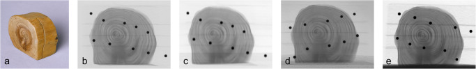
Visualizing the internal structure of museum objects is a crucial step in acquiring knowledge about the origin, state, and composition of cultural heritage artifacts. Among the most powerful techniques for exposing the interior of museum objects is computed tomography (CT), a technique that computationally forms a 3D image using hundreds of radiographs acquired in a full circular range. However, the lack of affordable and versatile CT equipment in museums, combined with the challenge of transporting precious collection objects, currently keeps this technique out of reach for most cultural heritage applications. We propose an approach for creating accurate CT reconstructions using only standard 2D radiography equipment already available in most larger museums. Specifically, we demonstrate that a combination of basic X-ray imaging equipment, a tailored marker-based image acquisition protocol, and sophisticated data-processing algorithms, can achieve 3D imaging of collection objects without the need for a costly CT imaging system. We implemented this approach in the British Museum (London), the J. Paul Getty Museum (Los Angeles), and the Rijksmuseum (Amsterdam). Our work paves the way for broad facilitation and adoption of CT technology across museums worldwide.


Conflict of interest statement

The authors declare no competing interests.

Figures

Fig. 1
Fig. 1
Steps in the workflow for post-scan marker-based parameter derivation method for 3D reconstruction.
Fig. 2
Fig. 2. X-ray imaging facilities.
a The British Museum (London), b the J. Paul Getty Museum (Los Angeles), c the Rijksmuseum (Amsterdam), and d the FleX-ray laboratory (Amsterdam).
Fig. 3
Fig. 3. The wooden test object.
a Wooden object (h 5 cm x w 6 cm x d 3 cm). Zoomed radiographs of the wooden test object at b the British Museum, c the J. Paul Getty Museum, d the Rijksmuseum, and e the FleX-ray laboratory.
Fig. 4
Fig. 4. Scan results of the wooden test object.
Top row. Single horizontal CT slice from reconstructions using the British Museum standard reconstruction workflow based on system feedback (BM system), all three museum systems with marker-based parameter retrieval (BM markers, GM markers, and RM markers), and the FleX-ray setup with system feedback. After reconstruction, the resulting 3D volumes have been scaled and registered in order to show a similar slice through the object using the FleXbox toolbox. The intensities were normalized. The red line measures 1.5 cm. The tree rings and the saw cut are visible in all reconstructions. Middle row. Zoomed-in CT slices. Bottom row. A line profile of the normalized intensities corresponding to the red line in the reconstruction.
Fig. 5
Fig. 5. Results of scanning the case study.
a The sculpture Python Killing a Gnu (1840s–1860s), Antoine-Louis Barye (French, 1796 –1875), the J. Paul Getty Museum collection number 85.SE.48, h 27.9 cm, w 39.1 cm, d 20.5 cm. b Single radiograph of the Barye model Python Killing a Gnu, including the markers used to determine the system parameters for CT reconstruction. c Three orthogonal slices of the CT reconstruction. d Horizontal slice, red arrows indicating the lines that show where the original square base is contained in the sculpture. e Vertical slice, the blue box indicates where gaps in the reconfigured neck were filled with wax instead of plaster. f Enlargement of the red box in e, the arrows indicate the three different layers of plaster used to create the sculpture.
Fig. 6
Fig. 6. Schematic representation of the X-ray setup.
X-ray setup with system components and parameters indicated: source (s), detector (d), source-detector-distance (SOD), object-detector-distance (ODD), coordinate system (x, y, z), rotation angle α, detector pixel δ and detector tilts (η, θ, ϕ).

References

    1. Kalender WA. CT: the unexpected evolution of an imaging modality. Eur. Radiol. Suppl. 2005;15:d21–d24. doi: 10.1007/s10406-005-0128-3. - DOI - PubMed
    1. Van Kaick G, Delorme S. Computed tomography in various fields outside medicine. Eur. Radiol. Suppl. 2005;15:d74–d81. doi: 10.1007/s10406-005-0138-1. - DOI - PubMed
    1. Morigi MP, Casali F, Bettuzzi M, Brancaccio R, D’Errico V. Application of X-ray computed tomography to cultural heritage diagnostics. Appl. Phys. A. 2010;100:653–661. doi: 10.1007/s00339-010-5648-6. - DOI
    1. Vandenbeusch M, O’Flynn D, Moreno B. Layer by layer: the manufacture of Graeco-Roman funerary masks. J. Egypt. Archaeol. 2021;107:281–298. doi: 10.1177/03075133211050657. - DOI
    1. Dorscheid J, et al. Looking under the skin: multi-scale CT scanning of a peculiarly constructed cornett in the Rijksmuseum. Herit. Sci. 2022;10:161. doi: 10.1186/s40494-022-00800-8. - DOI