. 2024 May 14;15(1):4047.
doi: 10.1038/s41467-024-48452-5.

Three-dimensional liquid metal-based neuro-interfaces for human hippocampal organoids

Affiliations

Three-dimensional liquid metal-based neuro-interfaces for human hippocampal organoids

Yan Wu et al. Nat Commun. .

Abstract

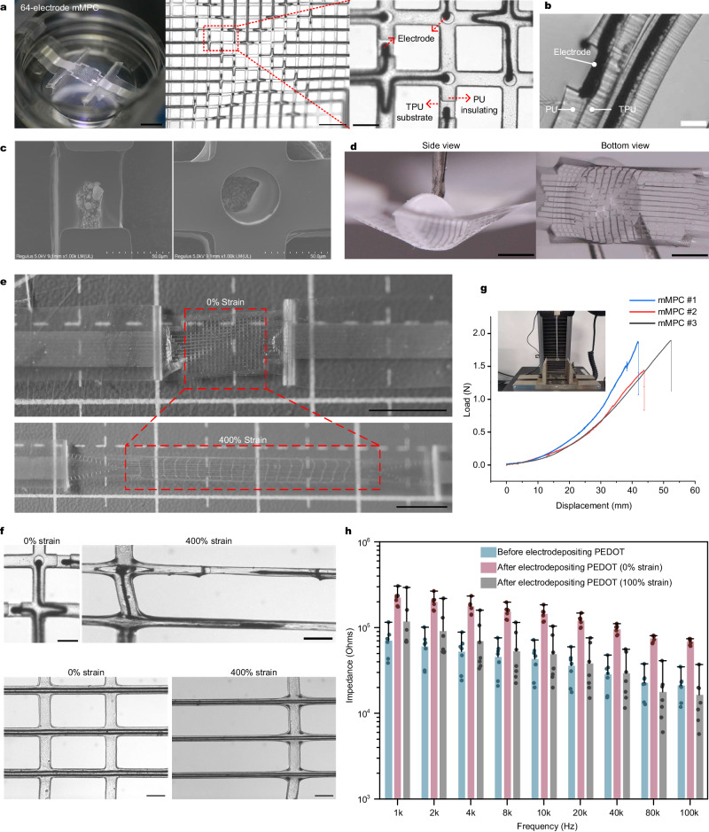
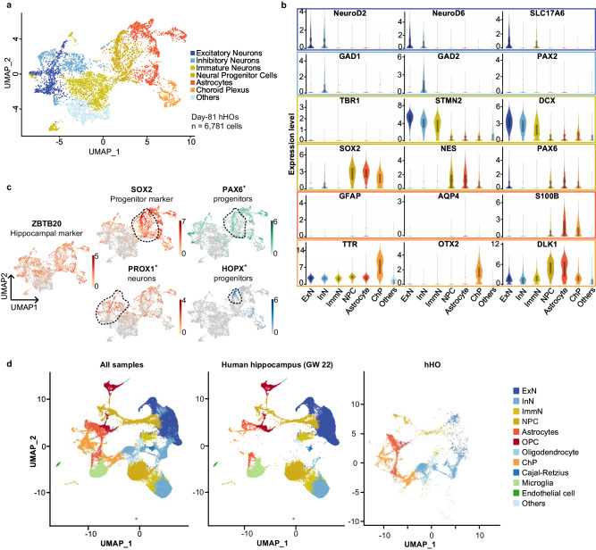
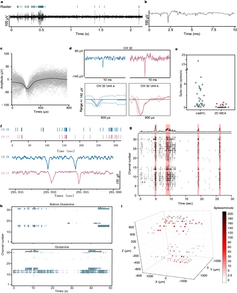
Human hippocampal organoids (hHOs) derived from human induced pluripotent stem cells (hiPSCs) have emerged as promising models for investigating neurodegenerative disorders, such as schizophrenia and Alzheimer's disease. However, obtaining the electrical information of these free-floating organoids in a noninvasive manner remains a challenge using commercial multi-electrode arrays (MEAs). The three-dimensional (3D) MEAs developed recently acquired only a few neural signals due to limited channel numbers. Here, we report a hippocampal cyborg organoid (cyb-organoid) platform coupling a liquid metal-polymer conductor (MPC)-based mesh neuro-interface with hHOs. The mesh MPC (mMPC) integrates 128-channel multielectrode arrays distributed on a small surface area (~2*2 mm). Stretchability (up to 500%) and flexibility of the mMPC enable its attachment to hHOs. Furthermore, we show that under Wnt3a and SHH activator induction, hHOs produce HOPX+ and PAX6+ progenitors and ZBTB20+PROX1+ dentate gyrus (DG) granule neurons. The transcriptomic signatures of hHOs reveal high similarity to the developing human hippocampus. We successfully detect neural activities from hHOs via the mMPC from this cyb-organoid. Compared with traditional planar devices, our non-invasive coupling offers an adaptor for recording neural signals from 3D models.


Conflict of interest statement

The authors declare no competing interests.

Figures

Fig. 1
Fig. 1. Overview of coupling the mMPC with the hHO and design of the mMPC.
a Schematic diagram of coupling the mMPC with the hHO. b Fabrication process of the mMPC. Scale bar: (i) 500 µm; (ii) and (iii) 200 µm; and (vii) 200 µm. hHO human hippocampal organoid, iPSC induced pluripotent stem cell, MPC liquid metal-polymer conductor, mMPC mesh MPC, TPU thermoplastic polyurethane, PU polyurethane.
Fig. 2
Fig. 2. Characterization for the mMPC.
a (Left) the single 64-electrode mMPC. Scale bar: 5 mm. (Middle) the distribution of 64 electrodes in the mesh structure. Scale bar: 500 µm. (Right) the structure of electrodes and encapsulation layers. Scale bar: 100 µm. b Optical image of the sandwich structure. Scale bar: 20 µm. c SEM images of electrodes. d Conformal attachment of the stretchable mMPC to the surface of a TPU ball. Scale bar: 1.5 mm. e The mMPC under 400% strain. Scale bar: 5 mm. f Two electrodes, circuits of the mMPC before and after 400% strain. Scale bar: 100 µm. g The bending test of 3 mMPCs. h Impedance of the mMPC across frequency before and after depositing PEDOT on electrodes and under 100% strain (n = 7, 7 independent electrodes randomly selected from 2 mMPCs; data are presented as median with maxima). mMPC mesh liquid metal-polymer conductor, TPU thermoplastic polyurethane, PU polyurethane. Images in c are representative of n = 4 independent measurement.
Fig. 3
Fig. 3. Generation of hHOs.
a The overall strategy to generate hHOs. b The typical morphology of hHOs at different ages. Scale bar: 100 µm. c The significant markers in the developmental process of the hHO. d Staining images of day-30 hHOs. Upper: Lager-scale image of the whole hHO. Scale bar: 100 µm. Lower: Zoom-in greyscale view of the white box in the upper figures. Scale bar: 40 µm. e The hippocampal PAX6+ and HOPX+ progenitors in day-60 hHOs. Scale bar: (left) 100 µm and (right, zoom-in greyscale view of the white box in the left figures) 40 µm. f The hippocampal PROX1+ cells in day-60 hHO and day-90 hHO. Scale bar: 150 µm. g qPCR for genes expressed in day-117 hHOs, day-71 hHOs versus day-46 hHOs (n = 3, three independent measurements using three independent samples, one-tailed t-test). h, qPCR for genes expressed in day-45 hHOs versus day-45 DMT organoids (n = 3, 3 independent measurements using three samples from different organoids, one-tailed t-test). Images in df are representative of n = 3 independent experiments. Exact sample size and values for h and g are provided in Source Data. Source data are provided as Source data files. hHO human hippocampal organoid, DMT dorsomedial telencephalon.
Fig. 4
Fig. 4. Transcriptomic signature of hHOs.
a UMAP visualization of 7 scRNA-seq clusters from day-81 hHOs. The cluster of oligodendrocytes and oligodendrocyte progenitor cells was presented in day-70 hHOs dataset in Supplementary Fig. 8a–c. b Violin plots of expression levels of markers in six clusters. c Feature plots of hippocampal ZBTB20+ cells, SOX2+HOPX+ progenitors, SOX2+PAX6+ progenitors and PROX1+ neurons. d UMAP visualization of the integrated dataset of the human hippocampus in GW22 and day-81 hHOs. (Left) All samples dataset. (Middle) The human hippocampus. (Right) Day-81 hHOs. The distribution of day-70 hHOs was present in Supplementary Fig. 8c. hHO human hippocampal organoid, GW gestational weeks.
Fig. 5
Fig. 5. The hippocampal cyb-organoid platform.
a Integration of the 128-channel mMPC and the hHO. (Upper) The hHO was stayed in a low-attachment dish. (Lower) The hHO was enveloped in two-layer mMPC. Scale bar: 1 mm. Notably, there was no significant deformation when the hHO was inserted between two layers of mMPC. b The bottom view captured by a microscope. Scale bar: (Left) 500 µm, (right) 100 µm. c Confocal images of cells from suckling mice hippocampus culturing in the mMPC for 30 days. Scale bar: 100 µm. d Confocal images of MAP2+ neurons, GFAP+ astrocytes and EGFP+ cells in the cyb-organoid platform. Scale bar: 100 µm. e The cross-sectional view of the attachment between the mMPC and the hHO. The arrowhead points to the mMPC. Scale bar: 100 µm. Images in ce are representative immunofluorescence staining cross-independent experiments: c (n = 4), d (n = 3), and e (n = 3).
Fig. 6
Fig. 6. Electrical activity recording.
a (Upper) Raster plots of neural spikes, and (lower) the continuous signal in a channel of the mMPC. b Continuous recording for a spike. c The uniform spike waveforms and the average waveform in a channel of the mMPC. d Two different spikes appeared in the same channel (upper) and the sorting results (lower). e The comparison of spike rates detected by the mMPC and the 2D MEA (these channels covered by the hHO). f The synchronous signals between two channels. (Upper) Raster plots of neural spikes in these two channels and (lower) continuous recording for two spikes. The time interval was <1 ms. g The oscillatory network activity of the hHO. (Upper) the population activity histogram and (lower) the raster plot (black) identified bursts of spiking detected on 32 channels and coordinated bursts (red) of activity across channels. h Raster plots before and after Glutamine supplement. i 3D visualization plot showing average spike numbers throughout 128 channels. mMPC mesh liquid metal-polymer conductor, MEA multi-electrode array.

References

    1. Bird CM, Burgess N. The hippocampus and memory: insights from spatial processing. Nat. Rev. Neurosci. 2008;9:182–194. doi: 10.1038/nrn2335. - DOI - PubMed
    1. Andersen, P., Morris, R., Amaral, D., O’Keefe, J. & Bliss, T. The Hippocampus Book (Oxford University Press, USA, 2007).
    1. Small SA, Schobel SA, Buxton RB, Witter MP, Barnes CA. A pathophysiological framework of hippocampal dysfunction in ageing and disease. Nat. Rev. Neurosci. 2011;12:585–601. doi: 10.1038/nrn3085. - DOI - PMC - PubMed
    1. Deng W, Aimone JB, Gage FH. New neurons and new memories: how does adult hippocampal neurogenesis affect learning and memory? Nat. Rev. Neurosci. 2010;11:339–350. doi: 10.1038/nrn2822. - DOI - PMC - PubMed
    1. Yu DX, et al. Modeling hippocampal neurogenesis using human pluripotent stem cells. Stem Cell Rep. 2014;2:295–310. doi: 10.1016/j.stemcr.2014.01.009. - DOI - PMC - PubMed