. 2024 May 14;8(1):28.
doi: 10.1038/s41538-024-00270-1.

First principles modelling of the ion binding capacity of finger millet

Affiliations

First principles modelling of the ion binding capacity of finger millet

Wei Cong Matthew Yong et al. NPJ Sci Food. .

Abstract

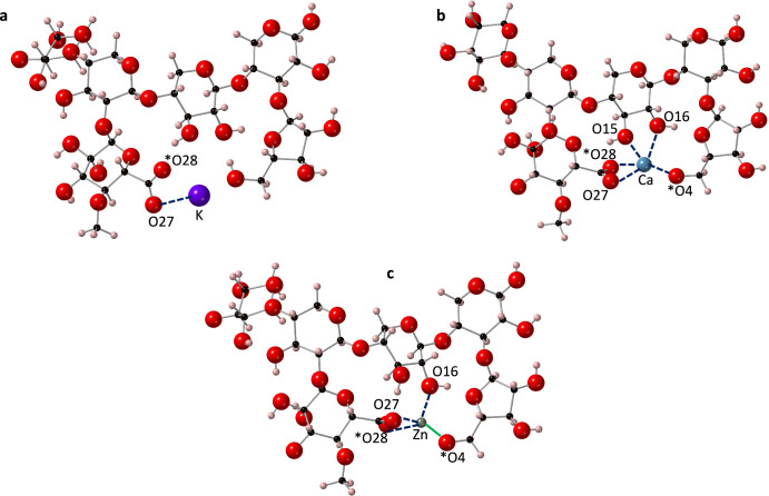
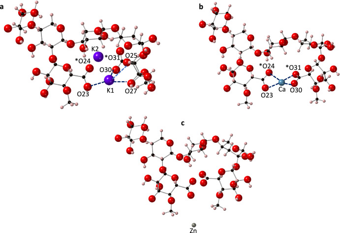
Finger millet, a cereal grain widely consumed in India and Africa, has gained more attention in recent years due to its high dietary fibre (arabinoxylan) and trace mineral content, and its climate resilience. The aim of this study was to understand the interactions between potassium (K+), calcium (Ca2+) and zinc (Zn2+) ions and the arabinoxylan structure and determine its ion-binding capacity. Three variations of a proposed model of the arabinoxylan structure were constructed and first principles Density Functional Theory calculations were carried out to determine the cation-binding capacity of the arabinoxylan complexes. Zn2+-arabinoxylan complexes were highly unstable and thermodynamically unfavourable in all three models. Ca2+ and K+ ions, however, form thermodynamically stable complexes, particularly involving two glucuronic acid residues as a binding pocket. Glucuronic acid residues are found to play a key role in stabilising the cation-arabinoxylan complex, and steric effects are more important to the stability than charge density. Our results highlight the most important structural features of the millet fibre regarding ion-storage capacity, and provide valuable preliminary data for confirmatory experimental studies and for the planning of clinical trials where the bioavailability of bound ions following digestion may be tested.


Conflict of interest statement

The authors declare no potential conflicts of interest with respect to the research, authorship, and/or publication.

Figures

Fig. 1
Fig. 1. The arabinoxylan models.
a PolyXA arabinoxylan consisting of the xylan backbone with one arabinose residue. b PolyXGA arabinoxylan consisting of the xylan backbone with one arabinose and one glucuronic acid residue. c PolyXGG arabinoxylan consisting of the xylan backbone and two glucuronic acid residues. Carbon atoms are shown in black, hydrogen in pink and oxygen in red. Residues (arabinose (A) and glucuronic acid (G)) are also labelled.
Fig. 2
Fig. 2. Optimised PolyXA structures with three different cations.
a K+, b Ca2+ and c Zn2+. Ionic bonds to the cations are shown with dotted lines and populations are marked. Atoms with a formal charge are marked with an asterisk (*). Calcium is shown in blue, potassium in purple, zinc in grey, oxygen in red, carbon in black and hydrogen in pink.
Fig. 3
Fig. 3. Optimised PolyXGA structures with three different cations.
a K+, b Ca2+ and c Zn2+. Ionic bonds to the cations are shown with dotted lines and the one covalent bond from Zn2+ to O4 is shown as a solid green line. Atoms with a formal charge are marked with an asterisk (*). Note that due to resonance effects, the formal charge has become equally split between O27 and O28 upon optimisation. Bond lengths and populations are provided in Table 2. Calcium is shown in blue, potassium in purple, zinc in grey, oxygen in red, carbon in black and hydrogen in pink.
Fig. 4
Fig. 4. Optimised PolyXGG structures with three different cations.
a K+, b Ca2+ and c Zn2+. Ionic bonds to the cations are shown with dotted lines. Atoms with a formal charge are marked with an asterisk (*). Note that due to resonance effects, upon optimisation, the formal charge has become equally split between O23 and O24, and O30 and O31. Bond lengths and populations are provided in Supplementary Table 2. Calcium is shown in blue, potassium in purple, zinc in grey, oxygen in red, carbon in black and hydrogen in pink.
Fig. 5
Fig. 5. A close-up view of the two K+ ions in the PolyXGG complex, illustrating the similar binding pocket environments.
The Mulliken-predicted bonds to K1 are shown as blue dotted line, and bond distances (not predicted as bonding) to K2 are shown with red dotted lines.

References

    1. Kumssa DB, et al. Dietary calcium and zinc deficiency risks are decreasing but remain prevalent. Sci. Rep. 2015;5:10974. doi: 10.1038/srep10974. - DOI - PMC - PubMed
    1. Zimmermann MB, Hurrell RF. Nutritional iron deficiency. Lancet. 2007;370:511–520. doi: 10.1016/S0140-6736(07)61235-5. - DOI - PubMed
    1. Osendarp SJM, et al. Large-scale food fortification and biofortification in low- and middle-income countries: a review of programs, trends, challenges, and evidence gaps. Food Nutr. Bull. 2018;39:315–331. doi: 10.1177/0379572118774229. - DOI - PMC - PubMed
    1. Baye K, Guyot JP, Mouquet-Rivier C. The unresolved role of dietary fibers on mineral absorption. Crit. Rev. Food Sci. Nutr. 2017;57:949–957. doi: 10.1080/10408398.2014.953030. - DOI - PubMed
    1. Rousseau S, Kyomugasho C, Celus M, Hendrickx MEG, Grauwet T. Barriers impairing mineral bioaccessibility and bioavailability in plant-based foods and the perspectives for food processing. Crit. Rev. Food Sci. Nutr. 2020;60:826–843. doi: 10.1080/10408398.2018.1552243. - DOI - PubMed

LinkOut - more resources