. 2024 Dec;20(12):1577-1585.
doi: 10.1038/s41589-024-01620-6. Epub 2024 May 14.

Antibodies expand the scope of angiotensin receptor pharmacology

Affiliations

Antibodies expand the scope of angiotensin receptor pharmacology

Meredith A Skiba et al. Nat Chem Biol. 2024 Dec.

Abstract

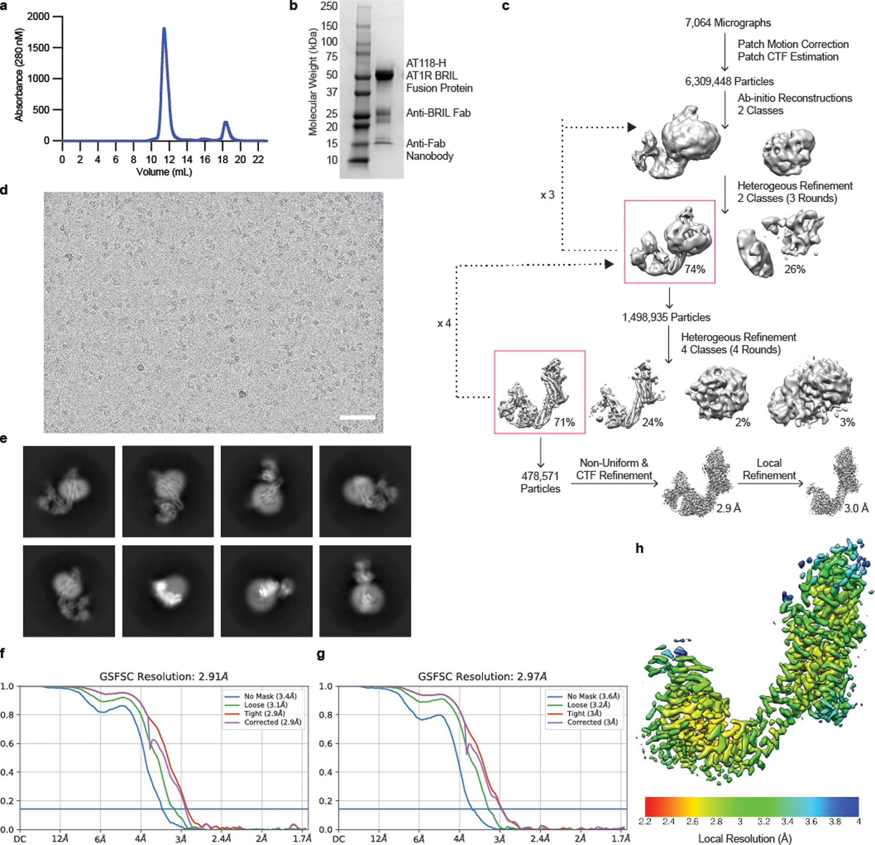
G-protein-coupled receptors (GPCRs) are key regulators of human physiology and are the targets of many small-molecule research compounds and therapeutic drugs. While most of these ligands bind to their target GPCR with high affinity, selectivity is often limited at the receptor, tissue and cellular levels. Antibodies have the potential to address these limitations but their properties as GPCR ligands remain poorly characterized. Here, using protein engineering, pharmacological assays and structural studies, we develop maternally selective heavy-chain-only antibody ('nanobody') antagonists against the angiotensin II type I receptor and uncover the unusual molecular basis of their receptor antagonism. We further show that our nanobodies can simultaneously bind to angiotensin II type I receptor with specific small-molecule antagonists and demonstrate that ligand selectivity can be readily tuned. Our work illustrates that antibody fragments can exhibit rich and evolvable pharmacology, attesting to their potential as next-generation GPCR modulators.


Conflict of interest statement

Competing interests: A.C.K., C.M., L.M.W., D.P.S. and M.A.S. are co-inventors on a patent application for AT1R blocking nanobodies. A.C.K. is a cofounder and consultant for biotechnology companies Tectonic Therapeutic and Seismic Therapeutic, and also for the Institute for Protein Innovation, a nonprofit research institute. L.M.W. is a scientific advisor for Septerna. D.P.S. is a Septerna employee. C.M. is a Sanofi employee. P.B. holds patents and provisional patent applications in the field of engineered T-cell therapies and protein design. The other authors declare no competing interests.

Figures

Extended Data Figure 1.
Extended Data Figure 1.. Engineering of high affinity AT1R nanobody antagonist with low non-specific binding.
a) Flowchart of nanobody selection. AT1R binders were enriched through two rounds of magnetic-activated cell sorting (MACS). Fluorescence-activated cell sorting (FACS) was used to isolate clone with low polyreactivity. A final FACS step enriched high-affinity AT1R binders. b) FACS round 1 plot. 1.38% of the population containing high-affinity AT1R binders with reduced polyspecificity were collected. c) FACS round 2 plot 0.7% of the population was collected containing high affinity AT1R binders. d) Binding of yeast-display library to FLAG-AT1R throughout each selection round. e) Distribution of AT1R-binding and polyreactive nanobodies in the yeast-display library throughout the selection process.
Extended Data Figure 2.
Extended Data Figure 2.. Effects of AT118-L and AT118-L-Fc fusion proteins on AT1R binding and signaling.
a) Binding of AT118-H variants displayed on yeast to detergent solubilized AT1R. Data are presented as mean ± SEM from three experiments. b) Non-specific binding of AT118-H variants displayed on yeast to biotinylated insect cell membrane polyspecificity reagent. Data are presented as mean ± SEM from three experiments. c-e) 3H-olmesartan competition experiments in Expi293 cell membranes containing AT1R with purified AT118-H variants. Variants containing D103N and S102G fail to displace olmesartan. The addition of V101D to D103N rescues the loss in pharmacological function. Data are presented as mean ± SEM from three experiments. Error is too small to be displayed if no bar is present. f) Accumulation of AT118-L F47T Y98F Fc fusion protein, that does not bind AT1R, in fetal serum. Data are presented as mean ± SEM from nine embryos from three separate litters for the control Fc (pMAS512, Supplementary Table 1) and eight embryos from two litters for the engineered Non-FcRn binding Fc (pMAS513, Supplementary Table 1).
Extended Data Figure 3.
Extended Data Figure 3.. Cryo-EM Construct Screening.
a) Fusion of AT118-H to the N-terminus of AT1R enhances total receptor expression. Data are presented as mean ± SEM from three experiments. b-f) Constructs screened for structure determination and representative 2-D class averages. b) AT118-AT1R fusion protein, c) Anti-nanobody Fab fragment bound to free AT118, but not AT118 in complex with AT1R. d) MBP-AT118 in complex with AT1R. e) AT118-AT1R-kappa opioid receptor (κOR) ICL3 fusion protein in complex nanobody 6 with an engineered alpaca framework, which binds κOR ICL3, anti-nanobody Fab, and anti-Fab nanobody,. f) AT118-AT1R-BRIL fusion protein in complex with anti-BRIL Fab and an anti-Fab nanobody.
Extended Data Figure 4.
Extended Data Figure 4.. AT118-H AT1R Data Processing.
a) Size-exclusion chromatography trace b) SDS-PAGE gel under reducing conditions. c) cryo-EM data processing scheme, d) representative micrograph (n = 7,064, scale bar = 50 nm), and e) representative 2D class averages of AT118-H-AT1R-BRIL, anti-BRIL Fab, anti-Fab nanobody complex. Two independent purifications of this complex yielded similar size-exclusion and SDS-PAGE results. Fourier shell correlation (FSC) used to determine the f) global map and g) locally refined map resolutions. h) Local resolution estimate after local refinement.
Extended Data Figure 5.
Extended Data Figure 5.. Molecular Dynamics Simulations.
Distance landscape of a) AT118-H AT1R-BRIL and b) AT118-H AT1R between TM3 (residue 3.50) and TM6 (residue 6.34) and TM3 (residue 3.50) and TM7 (residue 7.53) displayed as individual runs (colored in purple, orange, blue, and yellow) and overall density. Active state (PDB ID: 6OS0) and inactive state (PDB ID: 4YAY) are provided for reference. Neither construct, AT1R or AT1R-BRIL in complex with AT118-H, visits an active like state. c) Examination of dihedral distributions of allosteric activation network within AT1R’s core for AT1R and AT1R-BRIL in complex with AT118-H plotted by Chi 1 angles. KL divergence between the two constructs is zero, indicating that the BRIL fusion does not induce substantial conformational change in the activation network.
Extended Data Figure 6.
Extended Data Figure 6.. Binding of AT118-H and AT118-L with a broad panel of small molecule AT1R antagonists.
a) Molecular structures of small-molecule AT1R antagonists. b) Binding of AT118-H (orange) and AT118-L (purple) with a series of small-molecule AT1R antagonists. Error bars represent mean ± SEM from three independent experiments.
Extended Data Figure 7.
Extended Data Figure 7.. Allosteric effects of AT118-H and AT118-L.
a) Binding of AT118-H with modeled olmesartan. D103CDR3 of AT118-H would clash with olmesartan. Weak density for W842.60 is observed in the antagonist binding site in the orthosteric pocket of the AT118-H AT1R fusion protein structure. b) Binding of AT118-L with modeled ZD7155 (pink sticks, PDB 4YAY). c) Allosteric effect of AT118-L on small molecule inhibition of AT1R activation. AT118-L potentiates the inhibitory effects of losartan (gray), but has a non-significant effect on olmesartan (green). Log EC50 data are expressed as mean ± SEM from three independent experiments. *p = 0.026 was determined with one-way repeated-measures ANOVA with Dunnett’s correction for multiple comparison.
Extended Data Figure 8.
Extended Data Figure 8.. AT118-L AT1R Data Processing.
a) Size exclusion trace, b) SDS-PAGE gel under reducing conditions, and c) cryo-EM data processing scheme of AT118-L AT1R-BRIL, anti-BRIL Fab, anti-Fab nanobody complex. One purification of this complex was performed and similar size-exclusion and SDS-PAGE results are in agreement with the analogous complex prepared in Extended Data Fig. 4. d) Fourier shell correlation (FSC) used to determine the global map resolution. e) FSC used to determine locally refined map resolution. f) Experimental density of losartan within the orthosteric binding pocket from locally refined map. g) Comparison of orthosteric binding pocket in AT118-L, Losartan, AT1R-BRIL complex (blue) and olmesartan bound AT1R (purple, PDB 4ZUD).
Extended Data Figure 9.
Extended Data Figure 9.. Antibody GPCR binding model.
Nanobodies and other antibody fragments can adopt modular binding modes where one region mediates GPCR binding and another influences pharmacological function. The GPCR binding moiety can be formatted into a conventional antibody increasing avidity for the target GPCR or combined with secondary antibodies that recognize a tissue specific marker in a bispecific format. Pharmacokinetics, effector function, and tissue localization and delivery, can be further tuned by the antibodies constant Fc region.
Figure 1.
Figure 1.. Evolution of AT118 family of nanobody antagonists.
a) Structure of AT118-H (top, PDB 7T83) and CDR sequences of nanobodies described in this study colored by CDR (bottom). Shaded positions vary between nanobodies. Full nanobody sequences are provided in Supplementary Table 1. b) Sites substituted in AT118-H library. Variant enrichment was tracked through each selection round via deep sequencing. c) AT118-H and evolved variant AT118-L displace olmesartan from cell membranes containing AT1R. Data are presented as mean ± SEM from three experiments. d) AT118-L and AT118-L Fc fusion proteins (pMAS493, Supplementary Table 1) suppress G⍺q signaling (IP1 accumulation), whereas AT118-L F47T Y98F (pMAS513, Supplementary Table 1), the non-AT1R binding control, Fc fusion protein has no effect. Data are presented as mean ± SEM from three experiments. e) AT118-H and AT118-L suppress the recruitment of β-arrestin2 to AT1R upon AngII stimulation. Data are presented as mean ± SEM from three experiments. f) The AT118-L Fc fusion protein (pMAS493, Supplementary Table 1), suppresses the increase in mouse mean arterial blood pressure in vivo in response to AngII, compared to the non-AT1R binding control AT118-L F47T Y98F (pMAS513, Supplementary Table 1). Data are expressed as mean ± SEM, N=8 each group. 10 ng AngII, *p = 0.0011; 20 ng AngII, **p = 0.0006, 50 ng AngII, *p = 0.0022 by two-way repeated-measures ANOVA with Sidak correction, p<0.05 for interaction. g) AT118-L Fc fusion proteins lacking the ability to interact with FcRn (pMAS493, Supplementary Table 1) do not accumulate in fetal serum of mice compared to AT118-L Fc fusion proteins with a control WT Fc (pMAS430, Supplementary Table 1). Data are presented as mean ± SEM from twelve embryos from three separate litters.
Fig 2.
Fig 2.. Structure of AT118-H bound to AT1R.
a) Cryo-EM map colored by component: AT118-H (gray), AT1R (light blue), BRIL (purple), anti-BRIL Fab (light pink), anti-Fab nanobody (dark pink) b) Model of the AT118-H AT1R complex from local refinement maps. Image is colored as in a) with extracellular loops and AT118-H CDRs colored as follows: ECL1 (yellow), ECL2 (light pink), ECL3 (red), CDR1 (dark blue), CDR2 (green), CDR3 (orange).
Fig 3.
Fig 3.. AT118-H mimics peptide agonist binding to AT1R.
a) Surface rendering of AT118-H bound to AT1R as colored in Fig 2b. CDR3 (orange) engages with the orthosteric pocket of AT1R. b) surface rendering of active-state AT1R bound to the endogenous peptide agonist AngII (gray) (PDB 6OS0). AngII engages deeply in the orthosteric pocket. c) AT118-H binding is driven by engagement of CDRs 2 (green) and 3 (orange) with ECL2 (pink). Dashed lines indicate hydrogen bonds. d) AT118-H CDR3 (orange) forms β-strand and hydrophobic interactions with ECL2 (pink). The hydrophobic network is further supported by residues on ECL1 and TM2. e) AngII (gray sticks) and CDR3 share similar hydrophobic interactions with ECL2 (pink). f) In peptide agonist bound structures, the N-terminus of AT1R stabilizes the position of ECL2 through β-strand interactions.
Fig 4.
Fig 4.. AT118-H binding to AT1R and AT2R variants.
Binding of AT118-H to Expi293F TetR cells expressing AT1R variants or AT2R. Data are presented as mean ± SEM from at least three experiments. In the AT2R N-terminus chimera residues 2–16 of AT1R are replaced with residues of 3–33 of human AT2R. The AT2R ECL1 chimera contains M90Y, E91R, R93D, and P95L substitution and ECL3 chimera contains I270V, R272N, D273S substitutions.
Fig 5.
Fig 5.. AT118-H stabilizes a hybrid active-inactive state of AT1R.
a-b) R31CDR1 (a, blue sticks) and R99CDR3 (a, orange sticks) of AT118-H mimic R2 of AngII (b, gray sticks) positioning the extracellular side of Asp rich TMs 6 and 7. c) TM6, ECL3, and TM7 are displaced outward in AT118-H (light blue) and AngII (green) bound AT1R compared to small-molecule antagonist bound AT1R (purple) (PDB 4ZUD). d) AT118-H partially engages the allosteric activation network between AT1R’s orthosteric pocket and the intracellular side of AT1R. Switches indicated with the following arrows are activated (black), partially activated (dashed lines), unengaged (gray). e) AT118-H induced movements of TM5 and TM6 (light blue) induces inward movement of K1195.42 and downward rotation of W2536.48 compared to the inactive state (purple). f) W2536.48 does not fully rotate into its active-state position (green) and cannot displace N1113.35 and L1123.36 g) Partial movement of W2536.48 repositions Y2927.43 and F2085.51 from the canonical inactive state (purple) to h) active state positions (green), but is not sufficient to rotate the F2496.44 and F2506.45 rachet and displace TM6 to open up the intracellular effector binding site.
Fig 6.
Fig 6.. AT118 family members exhibit probe dependence with small molecules.
a) AT118-H and the evolved AT118-L variant accommodate binding of the small-molecule antagonist losartan (gray sticks). AT118-L CDR3 (purple) engages deeper within the orthosteric pocket compared to AT118-H (orange). b) S102CDR3 of AT118-H and c) D101CDR3 of AT118-L controls the depth of CDR3 within the orthosteric pocket through interactions with TM7. d) The backbone carbonyl of S102CDR3 of AT118-L is within hydrogen binding distance of the alcohol substituent (R1 in e) of losartan (gray sticks). e) Olmesartan and losartan derivatives vary in substituents on the imidazole ring. f) Probe dependent binding of AT118-H (orange) and AT118-L (purple) with losartan and olmesartan derivatives. Data are presented as mean ± SEM from three experiments. g) CDR3 of AT118-L clashes with the carboxylic acid (R1 in e) of modeled olmesartan (green sticks, modeled based upon PDB: 4ZUD).

Update of

References

    1. Mullard A 2022 FDA approvals. Nat Rev Drug Discov 22, 83–88 (2023). 10.1038/d41573-023-00001-3 - DOI - PubMed
    1. Hauser AS, Attwood MM, Rask-Andersen M, Schioth HB & Gloriam DE Trends in GPCR drug discovery: new agents, targets and indications. Nat Rev Drug Discov 16, 829–842 (2017). 10.1038/nrd.2017.178 - DOI - PMC - PubMed
    1. Peterson SM et al. Discovery and design of G protein-coupled receptor targeting antibodies. Expert Opin Drug Discov 18, 417–428 (2023). 10.1080/17460441.2023.2193389 - DOI - PubMed
    1. Niwa R et al. Defucosylated chimeric anti-CC chemokine receptor 4 IgG1 with enhanced antibody-dependent cellular cytotoxicity shows potent therapeutic activity to T-cell leukemia and lymphoma. Cancer Res 64, 2127–2133 (2004). 10.1158/0008-5472.can-03-2068 - DOI - PubMed
    1. Smith EL et al. GPRC5D is a target for the immunotherapy of multiple myeloma with rationally designed CAR T cells. Sci Transl Med 11 (2019). 10.1126/scitranslmed.aau7746 - DOI - PMC - PubMed

LinkOut - more resources