[Preprint]. 2025 Apr 5:2024.04.26.591113.
doi: 10.1101/2024.04.26.591113.

Mimicking opioid analgesia in cortical pain circuits

Affiliations

Mimicking opioid analgesia in cortical pain circuits

Corinna S Oswell et al. bioRxiv. .

Abstract

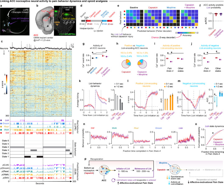
The anterior cingulate cortex is a key brain region involved in the affective and motivational dimensions of pain, yet how opioid analgesics modulate this cortical circuit remains unclear. Uncovering how opioids alter nociceptive neural dynamics to produce pain relief is essential for developing safer and more targeted treatments for chronic pain. Here we show that a population of cingulate neurons encodes spontaneous pain-related behaviors and is selectively modulated by morphine. Using deep-learning behavioral analyses combined with longitudinal neural recordings in mice, we identified a persistent shift in cortical activity patterns following nerve injury that reflects the emergence of an unpleasant, affective chronic pain state. Morphine reversed these neuropathic neural dynamics and reduced affective-motivational behaviors without altering sensory detection or reflexive responses, mirroring how opioids alleviate pain unpleasantness in humans. Leveraging these findings, we built a biologically inspired gene therapy that targets opioid-sensitive neurons in the cingulate using a synthetic mu-opioid receptor promoter to drive chemogenetic inhibition. This opioid-mimetic gene therapy recapitulated the analgesic effects of morphine during chronic neuropathic pain, thereby offering a new strategy for precision pain management targeting a key nociceptive cortical opioid circuit with safe, on-demand analgesia.


Conflict of interest statement

Competing interests. G.C, K.D., C.R. and G.J.S. are inventors on a provisional patent application through the University of Pennsylvania and Stanford University regarding the custom sequences used to develop, and the applications of synthetic opioid promoters (patent application number: 63/383,462 462 ‘Human and Murine Oprm1 Promotes and Uses Thereof’).

Figures

Fig. 1 │
Fig. 1 │. Deep learning analysis of natural behavior reveals how pain and opioids shape internal affective-motivational states.
(a) Schematic of the standardized LUPE platform and chamber for high-speed infrared videography. (b) A 20-body point DeepLabCut pose-tracking model was built from a bottom-up camera for male and female mouse pain behavior. (c) Behavior segmentation models trained iteratively on supervised annotations of holistic behaviors (e.g., licking vs. grooming vs. walking vs. rearing etc.) with A-SOiD, followed by further unsupervised sub-clustering with B-SOiD. (d) Motion energy heat maps illustrating spatial trajectories and intensity distributions for each of the six primary behavioral repertoires. (e) Temporal probability plots for each of the six primary behavior repertoires in 1-min bins, comparing mice in uninjured conditions to those injured by left hindpaw injections of 1% formalin, 5% formalin, or capsaicin. (f) Raster plots of rapid behavioral transitions within a 30-sec window from uninjured, formalin, and capsaicin injuries. (g) Procedure for behavioral state inference from statistical structure of spontaneous behavior. (h) Left panels: Model centroid transition matrices characterizing each of six inferred states. Right panels: Comparing fraction occupancy of mice in each state between uninjured (gray), formalin (magenta), and capsaicin (Cyan) pain models (n=20/group; 1-way ANOVA + Tukey: pState 1 = 0.0007, pState 3 = 0.0078, pState 4 < 0.0001). (i) Two-dimensional visualization of PCA of state occupancies across pain models. Dots are individual animals. (j) Magnitude of coefficients of each state in each PCA. (k) Scores of each animal along PC1 (top) and PC2 (bottom) across pain models (n=20/group; One-way ANOVA, Tukey correction: pPC1 = 0.0027, pPC2 = 0.0082). (l) Dose-response of morphine on PC1 (top) and PC2 (bottom) scores in uninjured, formalin, and capsaicin administered mice (n=20/group and dose; 1-way ANOVA + Tukey: pPC1, uninjured < 0.0001, pPC1 formalin < 0.0001, pPC1 capsaicin < 0.0001, pPC2, uninjured < 0.0001, pPC2, formalin < 0.0001, pPC2, capsaicin < 0.0001). ★= P < 0.05. Bars, lines, or dots are mean; error bars and shaded areas are SEM. See Supplementary Table 1 for statistics.
Fig. 2:
Fig. 2:. Neural dynamics in ACC track acute pain and analgesia.
(a) Microendoscope calcium imaging synced with LUPE behavior tracking. (b) GRIN lens implant and hSyn-GCaMP8m expression in ACC Cg1. Yellow FOV bar = 1.0-mm. (c) From top to bottom panels: Average and single cell neural activity (z-score) from a representative mouse, aligned with behaviors, states inferred by our behavioral state model, and probability of behaviors given states and behavior history (binomial GLM). (d) Imaging protocol during capsaicin injury (i.pl, 2%, left hindpaw) and morphine (i.p., 0.5 mg/kg; n=5 mice). (e) Fisher decoder accuracies predicting behaviors from neural activity, averaged over mice. (Permutation test, Fig. S10g). (f) auROC of GLMs predicting pLick from (e) in each animal (n=5). (g) Calcium events per second of all ACC neurons in all sessions. (h) Mean ± SEM fraction of positive and negative pLick neurons during capsaicin (red outline) and capsaicin+morphine (purple outline) sessions. (i,j) Calcium events per second of positive (i) and negative (j) pLick neurons in capsaicin and capsaicin+morphine sessions. (l,m) Left: average activity in positive (l) and negative (m) pLick neurons at lick bout onset, pooled across animals. Right: AUC of Lick probability from 0–1 seconds post-initiation and 1–2 seconds post-initiation (Unpaired t-test: pNegative, 0–1s = 0.0003). (n) Behavior probability as a function of fraction time in Pain State-4 in all capsaicin injured mice administered 0.0 and 0.5 mg/kg morphine (n=19–20/group). (o) Cumulative lick probability over fraction of time remaining in Pain State-4 between groups (Kologomorov-Smirnov test, p = 1.1e-23). (p) Summary of results. Morphine inhibits ACC neurons encoding affective-motivational pain behaviors, disrupting the pain-recuperation loop and reducing pain state expression.★ = P < 0.05. Bars, lines, or dots are mean; error bars and shaded areas are SEM. See Supplementary Table 2 for statistics.
Fig. 3:
Fig. 3:. Morphine targets functionally compromised ACC neurons to relieve chronic pain.
(a) SNI protocol for chronic neuropathic pain. (b) Log-transformed rate of spontaneous licking at the injury limb at −1, 1, 7, 14, and 21 days post-SNI (red, n=9) vs. uninjured controls (gray, n=9; 2-Way RM ANOVA + Tukey: pinteraction = 0.0036). (c) Heatmap of average state occupancy. (d) Occupancy of pain (top) and non-pain (bottom) states in SNI and uninjured control mice (2-way RM ANOVA +Tukey: pinteraction < 0.0001). (e) AMPS score (state PC2) score in SNI and uninjured control mice before and after SNI or anesthesia (2-way RM ANOVA + Tukey: pinteraction = 0.0089). (f) AMPS score (state PC2) score in SNI and uninjured control mice three weeks post-SNI or no-injury, before and after morphine (0.5 mg/kg, i.p; 2-way RM ANOVA +Tukey: pinjury = 0.0085, ptreatment = 0.0041). (g,h) Left: Lick-evoked activity in positive (g) and negative (h) pLick neurons, respectively, before (black) and after SNI (warm color gradient, yellow = 1 day post-SNI, red = 3 weeks post-SNI). Right: Area under the curve of lick-evoked activity (0–1s post-onset) in SNI (red) and uninjured (gray) control mice (2-way ANOVA + Tukey: pPositive, interaction < 0.0001, pNegative, interaction = 0.021). (i,j) Left: Same as (g,h left) visualizing lick-evoked activity in baseline (black), three weeks post-SNI (red), and three weeks post-SNI + morphine (blue) in SNI mice. Right: Same as (g,h) right comparing lick-evoked activity three weeks post-SNI vs. uninjured controls (2-way ANOVA + Tukey: pPositive, interaction = 0.035, pNegative, treatment < 0.0001, pNegative, injury < 0.0001). (k,l) Spontaneous calcium event rate in positive (k) and negative (l) pLick neurons, before and after morphine (2-Way ANOVA + Tukey: pPositive, treatment = 0.0023, pNegative, treatment = 0.0124). (m) Linear regression predicting log-transformed lick rate (purple) and pain state occupancy (gray) from the average magnitude of lick-evoked activity three weeks post-SNI (top), after morphine (middle), and change between sessions (bottom; Bonferroni-corrected p-values displayed). ★ = P < 0.05. Bars, lines, or dots = mean; error bars and shaded areas = SEM. See Supplementary Table 3 for statistics.
Fig. 4 │
Fig. 4 │. Precision neuromodulation of affective chronic pain via an ACC opioid cell-type specific gene therapy.
(a) Strategies to deliver inhibitory chemogenetic actuators to ACC MOR+ neurons (Red: MORp-hM4) or nociceptive/MOR+ cell-types (Blue: MORp-FlpO + CreON/FlpON-hM4 or MORp-DIO-iC++ in painTRAP mice). (b) Co-expression of MORp-eYfp mRNA and endogenous Oprm1 mRNA. (c) MORp-driven fluorophore or hM4 transgene expression across ACC layers. (d) Timeline of chronic neuropathic pain and DCZ exposure (0.3 mg/kg, i.p., once daily. Gray triangles) with LUPE and sensory testing (N=19 (10 male and 9 female) MORp-eYFP mice, N=30 (equal sexes) MORp-hM4 mice). (e-h) Effect of nerve injury and chemogenetic inhibition on spontaneous licking of the injured limb (e, pInteraction = 0.0012), occupancy of behavioral states (f), fraction of time spent in pain versus non-pain states (g, pain states pInteraction<0.0001, non-pain states pInteraction<0.0001), and AMPS scores (h, pInteraction = 0.0003) in MORp-hM4 vs. MORp-eYFP mice. 2-way RM-ANOVA + Tukey for all panels. (i,j) Brain-wide projections of ACC MOR+ axons expressing hM4 or eYFP (i) to assess neuropathic activity (touchFOS) within connected brain areas with or without ACC inhibition (j). t-Test, 2-tailed per region. (k) Analgesic efficacy for morphine (0.5 mg/kg, i.p) vs. MORp-eYFP, MORp-hM4, or nociceptive-MORp-hM4 + DCZ (0.3 mg/kg, i.p.) in uninjured and chronic neuropathic pain conditions. Effects on % Maximum Possible Analgesia on evoked mechanical thresholds (left, pInteraction = 0.0061), and evoked affective-motivational behaviors to acetone (middle, pInteraction <0.0001), and 55°C hot water (right, pInteraction P<0.0001). 1-way ANOVA + Tukey. (l) AMPS scores for spontaneous pain behaviors to assess analgesic efficacy of morphine vs. acute or chronic MORp-hM4 ACC inhibition in uninjured or chronic pain conditions. (Neuropathic left: 1-way ANOVA + Tukey, pTreatment =0.0085. Neuropathic right: 2-way ANOVA + Tukey, pInteraction =0.0003). (m) Overview of advancements: using deep-learning behavior tracking software to classify behavior states in pain, paired with calcium imaging we uncovered new cortical mechanisms of opioid analgesia, which informed our creation of an opioid cell-type gene therapy approach for chronic pain that mimics morphine analgesia with circuit-targeted precision. For detailed statistics, see Supplemental Table 4 rows 1–10. ★ = P < 0.05. Errors bars = s.e.m.

References

    1. Kimmey B. A., McCall N. M., Wooldridge L. M., Satterthwaite T. D. & Corder G. Engaging endogenous opioid circuits in pain affective processes. J Neurosci Res 1–33 (2020) doi: 10.1002/jnr.24762. - DOI - PMC - PubMed
    1. Wiech K. Deconstructing the sensation of pain: The influence of cognitive processes on pain perception. Science (1979) 354, 584–587 (2016). - PubMed
    1. Tan L. L. & Kuner R. Neocortical circuits in pain and pain relief. Nature Reviews Neuroscience vol. 22 458–471 Preprint at 10.1038/s41583-021-00468-2 (2021). - DOI - PubMed
    1. Corder G., Castro D. C., Bruchas M. R. & Scherrer G. Endogenous and Exogenous Opioids in Pain. Annu Rev Neurosci 453–73 (2018) doi:/ 10.1146/annurev-neuro-080317-061522. - DOI - PMC - PubMed
    1. Woolf C. J. Capturing Novel Non-opioid Pain Targets. Biol Psychiatry 87, 74–81 (2020). - PMC - PubMed

Publication types