[Preprint]. 2024 Apr 30:2024.04.29.591704.
doi: 10.1101/2024.04.29.591704.

P66 is a bacterial mimic of CD47 that binds the anti-phagocytic receptor SIRPα and facilitates macrophage evasion by Borrelia burgdorferi

Affiliations

P66 is a bacterial mimic of CD47 that binds the anti-phagocytic receptor SIRPα and facilitates macrophage evasion by Borrelia burgdorferi

Michal Caspi Tal et al. bioRxiv. .

Abstract

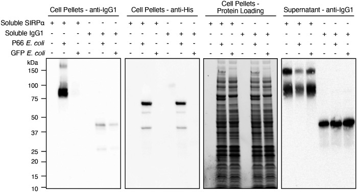
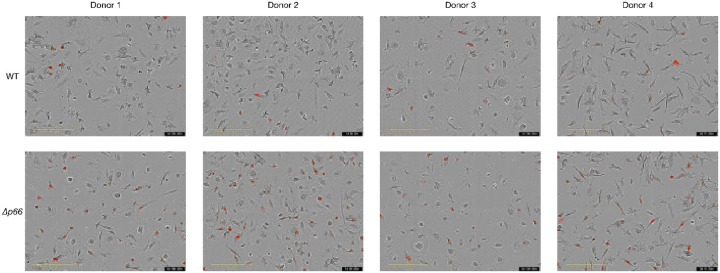
Innate immunity, the first line of defense against pathogens, relies on efficient elimination of invading agents by phagocytes. In the co-evolution of host and pathogen, pathogens developed mechanisms to dampen and evade phagocytic clearance. Here, we report that bacterial pathogens can evade clearance by macrophages through mimicry at the mammalian anti-phagocytic "don't eat me" signaling axis between CD47 (ligand) and SIRPα (receptor). We identified a protein, P66, on the surface of Borrelia burgdorferi that, like CD47, is necessary and sufficient to bind the macrophage receptor SIRPα. Expression of the gene encoding the protein is required for bacteria to bind SIRPα or a high-affinity CD47 reagent. Genetic deletion of p66 increases phagocytosis by macrophages. Blockade of P66 during infection promotes clearance of the bacteria. This study demonstrates that mimicry of the mammalian anti-phagocytic protein CD47 by B. burgdorferi inhibits macrophage-mediated bacterial clearance. Such a mechanism has broad implications for understanding of host-pathogen interactions and expands the function of the established innate immune checkpoint receptor SIRPα. Moreover, this report reveals P66 as a novel therapeutic target in the treatment of Lyme Disease.


Figures

Extended Data Figure 1.
Recombinant P66 is enriched by CV1-G4 or SIRPα but not isotype control as determined by in vitro binding assay. Representative full gel and Western blotting images of all 4 replicates of binding assay quantified in Figure 1D.
Extended Data Figure 2.
P66 and CD47 are structurally distinct proteins. A. Structure of P66 as predicted by Alphafold2. B. The human CD47-SIRPα binding interface. The crystal structure of the IgSF domain of CD47 bound to the N-terminal IgV domain SIRPα (PDB: 2JJT) was aligned with the cryo-EM structure of full-length human CD47 (PDB: 7MYZ).
Extended Data Figure 3.
Detergent partitioning of P66-expressing E. coli reveals P66 is selectively detected in the membrane (detergent) fraction by anti-His Western blot.
Extended Data Figure 4.
Full Western blot and gels from pelleting assay described in Figure 1C. P66- or GFP-expressing E. coli were incubated with soluble SIRPa-Fc or isotype control, prior to Western blot analysis.
Extended Data Figure 5.
Full Western blot and gels from cell pelleting assays described in Figure 1D. Soluble SIRPa-Fc, SIRPa-His or isotype control was incubated with E. coli expressing P66 or GFP prior to Western blot analysis.
Extended Data Figure 6.
Peak phagocytosis images with macrophages from 4 donors (denoted by arrows in Figure 3D). Red events indicate lysosomal localization of wild-type (WT) or p66 knockout (Δp66) B. burgdorferi.
Figure 1.
Figure 1.
The surface protein P66 binds to a high-affinity CD47 binding reagent and the CD47 receptor SIRPα. A. B. burgdorferi stain for the high-affinity CD47 blocking reagent CV1-G4 by flow cytometry analysis. **** p-value < 0.0001, df = 4, N = 4; Unpaired 2-tailed t-test. B. B. burgdorferi was cultured under conditions to stain either positively or negatively for CV1-G4 as confirmed by flow cytometry. Bacteria were lysed under non-denaturing conditions, and lysate was subjected to enrichment with CV1-G4 or IgG4 prior to SDS-PAGE. Gel bands of interest were excised and subjected to in-gel trypsin digestion followed by mass spectrometry analysis. Arrow denotes putative P66 band. C. Data filtration parameters identified P66 as a putative CV1-G4 binding protein. D. Recombinant his-tagged P66 is enriched by CV1-G4 or SIRPα but not isotype control as determined by in vitro binding assay. A representative blot is shown as is quantification from 4 replicates. ** p-value = 0.0065, N = 4, df = 3. Paired two-tailed t-test; *** p-value < 0.0008, N = 4, df = 3;. Error bars calculated as SEM.
Figure 2.
Figure 2.
P66 is necessary and sufficient for binding to a CD47 affinity reagent and SIRPα. A. Wild-type (WT), p66 deficient (Δp66) B. burgdorferi, Δp66 B. burgdorferi reconstituted with WT p66, Δp66 B. burgdorferi reconstituted with the p66D184A D186A mutant, or Δp66 B. burgdorferi reconstituted with the p66Δ181–187 loop deletion mutant were stained with CV1-G4, IgG4 or MIAP410 and analyzed by flow cytometry. B. Predicted structure of P66 by Alphafold2 with mutated residues or loop deletions highlighted in orange. C. P66-His- or GFP-expressing E. coli were incubated with soluble SIRPa-Fc or IgG1 isotype control, prior to Western blot analysis. D. Soluble SIRPa-Fc, SIRPa-His or IgG1 isotype control was incubated with E. coli expressing P66-His or GFP prior to Western blot analysis.
Figure 3.
Figure 3.
Δp66 B. burgdorferi are more-readily phagocytosed by human macrophages compared to WT as measured by pHrodo-positive phagocytic events. A. Phagocytosis assays using human monocyte derived macrophages averaged across 4 donors reveal that Δp66 bacteria are more-readily taken up by macrophages throughout the course of the experiment. 2 technical replicates per donor with 3 fields of view per technical replicate, allowing for 6 fields of view measured per donor. B. Δp66 B. burgdorferi have a greater rate of increase in phagocytic events during the first 10 hours. 95% confidence intervals for the event rate are shown as dashed lines. C. Fold-change in rates of phagocytic events between Δp66 and WT bacteria were calculated for each donor over the first 10 hours. D. Individual macrophage phagocytosis data from 4 human donors, 2 technical replicates per donor with 3 fields of view per technical replicate for a total of 6 measurements per donor. Arrows denote maximum for each donor with each strain. E. Fold-increase in total number of phagocytic events with Δp66 B. burgdorferi compared to WT across co-incubation time. Error bars calculated as SEM.
Figure 4.
Figure 4.
P66 is a therapeutic target in Lyme Disease. A. C3H Female mice were infected with 105 luciferase-expressing B. burgdorferi, strain ML23-pBBE22luc, that were pretreated with CV1-G4 or isotype control (6 μg). Mice were imaged at Day 0, Day 7, and Day 14 following injection with D-luciferin for in vivo imaging. Error bars calculated as SEM. ** p-value = 0.00478; N = 5; * p-value = 0.04541; N = 5; N.S. = not significant. Unpaired Welch’s t-test. B. C3H Female mice were infected with 106 luciferase-expressing B. burgdorferi, strain ML23-pBBE22luc, that were pretreated with CV1-G4 or isotype control (6 μg). Mice were imaged at Day 0 and Day 9 following injection with D-luciferin for in vivo imaging. Error bars calculated as SEM. Ratio of average radiance for Day 9/Day 0 was calculated for each mouse. ** p-value = 0.0075; N = 5; Unpaired Welch’s t-test.

References

    1. Mantovani A., Allavena P., Marchesi F. & Garlanda C. Macrophages as tools and targets in cancer therapy. Nat. Rev. Drug Discov. 21, 799–820 (2022). - PMC - PubMed
    1. Jaiswal S. et al. CD47 is upregulated on circulating hematopoietic stem cells and leukemia cells to avoid phagocytosis. Cell 138, 271–285 (2009). - PMC - PubMed
    1. Majeti R. et al. CD47 is an adverse prognostic factor and therapeutic antibody target on human acute myeloid leukemia stem cells. Cell 138, 286–299 (2009). - PMC - PubMed
    1. Oldenborg P.-A. et al. Role of CD47 as a Marker of Self on Red Blood Cells. Science 288, 2051–2054 (2000). - PubMed
    1. Tal M. C. et al. Upregulation of CD47 Is a Host Checkpoint Response to Pathogen Recognition. Mbio 11, e01293–20 (2020). - PMC - PubMed

Publication types