[Preprint]. 2024 Apr 30:2024.04.30.591319.
doi: 10.1101/2024.04.30.591319.

Functional selection in SH3-mediated activation of the PI3 kinase

Affiliations

Functional selection in SH3-mediated activation of the PI3 kinase

Safia S Aljedani et al. bioRxiv. .

Abstract

The phosphoinositide-3 kinase (PI3K), a heterodimeric enzyme, plays a pivotal role in cellular metabolism and survival. Its deregulation is associated with major human diseases, particularly cancer. The p85 regulatory subunit of PI3K binds to the catalytic p110 subunit via its C-terminal domains, stabilising it in an inhibited state. Certain Src homology 3 (SH3) domains can activate p110 by binding to the proline-rich (PR) 1 motif located at the N-terminus of p85. However, the mechanism by which this N-terminal interaction activates the C-terminally bound p110 remains elusive. Moreover, the intrinsically poor ligand selectivity of SH3 domains raises the question of how they can control PI3K. Combining structural, biophysical, and functional methods, we demonstrate that the answers to both these unknown issues are linked: PI3K-activating SH3 domains engage in additional "tertiary" interactions with the C-terminal domains of p85, thereby relieving their inhibition of p110. SH3 domains lacking these tertiary interactions may still bind to p85 but cannot activate PI3K. Thus, p85 uses a functional selection mechanism that precludes nonspecific activation rather than nonspecific binding. This separation of binding and activation may provide a general mechanism for how biological activities can be controlled by promiscuous protein-protein interaction domains.

Keywords: Grb2; cancer; kinase; selectivity; signalling; tertiary interactions.


Figures
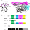
Figure 1.
Figure 1.. Structure and interactions of p85, p110 and SH3.
A) Example of a canonical interaction between the Fyn SH3 domain (black ribbon) and a PXXP containing peptide (stick model with carbon atoms in magenta). The PR motif inserts its two key prolines (pink) into hydrophobic grooves on the SH3 domain (residues shown stick models). In the PR motif, the position of an arginine with respect to the prolines determines the binding orientation by engaging a salt bridge with an aspartic acid from the SH3 RT loop (Feng et al., 1994) Shown is a class II peptide (RXXPXXP) bound to Fyn SH3 (extracted from the HIV-1 Nef:Fyn SH3 complex, PDB 1AVZ). R96 of Fyn SH3 is shown as stick model. B) Top: Schematic drawing of full-length p85 and relevant fragments thereof used in this work. Bottom: schematic drawing of p110. The colours are maintained throughout the manuscript. C) Structure of p110 (black, with catalytic domain in grey) kept in an inhibited conformation through interactions with p85 iSH2 (pink) and nSH2 (cyan). Structure taken from PDB 8DCP.
Figure 2.
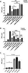
Figure 2.. Lipid kinase assays.
Immunoprecipitated p110α protein from EFE-184 cells was incubated with A) doubly phosphorylated PDGFR peptide (pY) and/or recombinant purified wild-type Fyn (R96) or SH3 mutant (R96I or R96W), B) pY and/or recombinant purified wild-type Fyn (R96) or SH3 mutant L112A or L112N (with wild-type R96 background), or C) wild-type SH3 domain of the indicated proteins (100 μm), prior to lipid kinase assay and absorbance measurement. Reaction without incubation with any peptide served as control. Immunoprecipitation using normal IgG instead of anti-p110α antibody in the control sample was performed in parallel for background signal. Data in graphs are levels of PIP3 after subtracting the absorbance of IgG samples and are presented as the mean of triplicates ± S.D. P-values were obtained using unpaired t-test. ns, not statistically significant.
Figure 3.
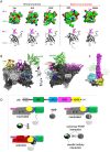
Figure 3.. NMR interaction mapping onto Fyn SH3 variants.
The crystal structure of Fyn R96 (PDB accession 1AVZ), Fyn I96 (6IPY), and Fyn W96 (6IPZ) were used to map the chemical shifts. The PR sequence from the HIV-1 Nef protein (PDB 1AVZ; shown as green stick model) has been taken as a guide for ‘canonical’ binding of a PR to Fyn SH3. Four different orientations are shown (I-IV). A) Binding of the PR1 motif to Fyn SH3 wild type (R96) and the I96 and W96 mutants, as mapped by chemical shift analysis. B) Binding of 15N labelled Fyn SH3 R96, I96 and W96 to p85ΔSH3. Shown are the residues that reappear early during the titration (at p85ΔSH3: SH3 ratios of 1:4, 1:3 and 1:6 for WT, RI and WR, respectively) or later during the titration (at p85ΔSH3:SH3 ratios of 1:6, 1:6 and 1:8 for WT, RI and WR, respectively).
Figure 4.
Figure 4.. Molecular mechanism for the activation of p85 by SH3 domains.
A) Classification of SH3 domains that either bind and activate p85 (left) or just bind to p85 without activating it (right). Top: SH3 domain surfaces, colour coded according to atoms being hydrophobic: green; sulphur: yellow; acidic: red; basic: blue; polar nitrogen: cyan; polar-oxygen: salmon. The PR sequence from the HIV-1 Nef protein (PDB 1AVZ; shown as magenta stick model) has been taken as a guide for ‘canonical’ binding of a PR to SH3 domains. The position corresponding to Fyn L112 is circled in ‘activating’ SH3 domains, and roughly indicated by an arrow in ‘non-activating’ SH3 domains, where the structure at this position is different. Bottom: Ribbon model corresponding to the surface view. The PR motif is in magenta. L112 position and nature of corresponding residue is given. B) Two 90° views XLMS-mapped lysines. p110 is shown as a surface model with the catalytic domain in light grey, and other domains in dark grey. Domains of p85ΔSH3 are colour-coded. Lysines identified in XLMS and being compatible with a membrane-associated p110:p85 complex are shown as sphere models, coloured in blue (crosslinked only in presence of Fyn SH3), purple (only Grb2 cSH3), or red (crosslinked with both Fyn and Grb2 SH3). The p110:p85ΔSH3 model was produced by AlphaFold. C) Speculative 3D model to illustrate how binding of an activating SH3 domain may contact and rearrange both SH2 domains of p85. Model produced by AlphaFold. D) Speculative model for PI3K activation through SH3 domains. The schematic p85 drawing recapitulates the domain colours. p110 is shown in dark grey. Top left: In absence of stimulating ligands, p85 binds p110 through the iSH2 domain. Interactions between the p85 SH2 domains and p110 keep p110 in the catalytically inactive conformation. Top right: SH3 domains that lack the tertiary interfaces may still bind to p85 PR, but cannot activate PI3K. Bottom left: When an SH3 domain with correct tertiary surface binds (green; only one such surface is shown), then the resulting interactions with the p85 nSH2-iSH2-cSH2 fragment remove the inhibitory SH2 interactions, activating p110 (red). Bottom right: Schematic drawing of SH3 domains with or without tertiary surfaces.

Similar articles

References

    1. Aldehaiman A., Momin A.A., Restouin A., Wang L., Shi X., Aljedani S., Opi S., Lugari A., Shahul Hameed U.F., Ponchon L., Morelli X., Huang M., Dumas C., Collette Y., Arold S.T., 2021. Erratum: Synergy and allostery in ligand binding by HIV-1 Nef (Biochemical Journal 478:8 (1525–1545) DOI: 10.1042/BCJ20201002). Biochem. J. 478. 10.1042/BCJ20201002_COR - DOI - PMC - PubMed
    1. Arold S., Franken P., Strub M.P., Hoh F., Benichou S., Benarous R., Dumas C., 1997. The crystal structure of HIV-1 Nef protein bound to the Fyn kinase SH3 domain suggests a role for this complex in altered T cell receptor signaling. Structure 5. 10.1016/S0969-2126(97)00286-4 - DOI - PubMed
    1. Arold S., O’Brien R., Franken P., Strub M.P., Hoh F., Dumas C., Ladbury J.E., 1998. RT loop flexibility enhances the specificity of Src family SH3 domains for HIV-1 Nef. Biochemistry 37. 10.1021/bi980989q - DOI - PubMed
    1. Axelsson L., Hellberg C., Melander F., Smith D., Zheng L., Andersson T., 2000. Clustering of β2-integrins on human neutrophils activates dual signaling pathways to Ptdlns 3-kinase. Exp. Cell Res. 256. 10.1006/excr.2000.4816 - DOI - PubMed
    1. Backer J.M., 2010. The regulation of class IA PI 3-kinases by inter-subunit interactions. Curr. Top. Microbiol. Immunol. 346. 10.1007/82-2010-52 - DOI - PMC - PubMed

Publication types