[Preprint]. 2024 May 5:2024.05.02.592258.
doi: 10.1101/2024.05.02.592258.

In vitro heterochronic parabiosis identifies pigment epithelium-derived factor as a systemic mediator of rejuvenation by young blood

Affiliations

In vitro heterochronic parabiosis identifies pigment epithelium-derived factor as a systemic mediator of rejuvenation by young blood

Xizhe Wang et al. bioRxiv. .

Abstract

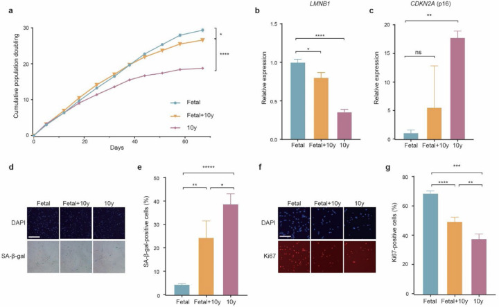
Several decades of heterochronic parabiosis (HCPB) studies have demonstrated the restorative impact of young blood, and deleterious influence of aged blood, on physiological function and homeostasis across tissues, although few of the factors responsible for these observations have been identified. Here we develop an in vitro HCPB system to identify these circulating factors, using replicative lifespan (RLS) of primary human fibroblasts as an endpoint of cellular health. We find that RLS is inversely correlated with serum donor age and sensitive to the presence or absence of specific serum components. Through in vitro HCPB, we identify the secreted protein pigment epithelium-derived factor (PEDF) as a circulating factor that extends RLS of primary human fibroblasts and declines with age in mammals. Systemic administration of PEDF to aged mice reverses age-related functional decline and pathology across several tissues, improving cognitive function and reducing hepatic fibrosis and renal lipid accumulation. Together, our data supports PEDF as a systemic mediator of the effect of young blood on organismal health and homeostasis and establishes our in vitro HCPB system as a valuable screening platform for the identification of candidate circulating factors involved in aging and rejuvenation.


Conflict of interest statement

Competing interests The authors declare no competing interests.

Figures

Extended Data Figure 1.
a, Replicative lifespan curves of IMR90 cultured in fetal (FBS), aged (10y), and heterochronic (50% fetal/50% aged) bovine serum. b, qPCR analysis of LMNB1 in mid-late passage IMR90 cultured under different serum conditions, normalized to fetal serum condition. c, qPCR analysis of CDKN2A in mid-late passage IMR90 cultured under different serum conditions, normalized to fetal serum condition. d-e, Representative images (d) and quantification (e) of senescence-associated β-galactosidase (SA-β-Gal) staining of IMR90 at mid-late passage from different serum age conditions. f-g, Representative images (f) and quantification (g) of Ki67 staining of IMR90 at mid-late passage from different serum age conditions. Statistical analysis was performed using two-tailed unpaired t-tests (a-c,e and g). Data represented as mean ±SEM from three technical replicates, *p<0.05, **p<0.01, ***p<0.001, ****p<0.0001, ns=non-significant. Scale bars, 500 µm (d) and 200 µm (f).
Extended Data Figure 2.
a, Principal component analysis (PCA) of protein signatures of different aged bovine serum.
Extended Data Figure 3.
a, Expression of SERPINF1 (PEDF) across human tissue from GTEx RNA-seq analysis. b, Expression of SERPINF1 (PEDF) across human central nervous system tissue from GTEx RNA-seq analysis.
Extended Data Figure 4.
a-c, Significant DEGs (p<0.05) after RNA-seq analysis of IMR90 24hr post-serum change into either male young or aged serum, or aged serum supplemented with PEDF (a), and associated pathways (b-c). d-f, Significant DEGs (p<0.05) after RNA-seq analysis of IMR90 24hr post-serum change into either female young or aged serum, or aged serum supplemented with PEDF (d), and associated pathways (e-f). Statistical analysis was performed using Wald tests (a and d), and Fisher’s exact test (b-c, and e-f).
Extended Data Figure 5.
a, Quantification of Oil Red O staining in liver of aged mice (n=14 (saline), n=15 (PEDF) mice). b, Quantification of Trichrome staining in kidney of aged mice (n=15 (saline), n=15 (PEDF) mice). Statistical analysis was performed using two-tailed unpaired t-tests. Data represented as mean ±SEM, ns=non-significant.
Figure 1.
Figure 1.. Aged serum reduces replicative lifespan and induces early senescence of human primary fibroblasts.
a, Replicative lifespan curves of human primary fibroblast, IMR90, under different bovine serum conditions from different ages (fetal, 1.5y, 3y, 6y, and 10y). b, qPCR analysis of LMNB1 in mid-late passage IMR90 cultured under different serum age conditions, relative to fetal serum. c, qPCR analysis of CDKN2A in mid-late passage IMR90 cultured under different serum age conditions, relative to fetal serum. d-e, Representative images (d) and quantification (e) of senescence-associated β-galactosidase (SA-β-Gal) staining of IMR90 at mid-late passage from different serum age conditions. f-g, Representative images (f) and quantification (g) of Ki67 staining of IMR90 at mid-late passage from different serum age conditions. h-k, Significant DEGs (FDR<0.05) after RNA-seq analysis of IMR90 24hr post-serum change from fetal to 10-year-old serum (h), associated pathways (i-j), and expression of CDKN2A and SenMayo senescence genes among significant DEGs (k). Statistical analysis was performed using two-tailed unpaired t-tests (a-c,e and g), Wald tests (h and k), and Fisher’s exact test (i-j). Data represented as mean ±SEM from three technical replicates, *p<0.05, **p<0.01, ***p<0.001, ****p<0.0001. Scale bars, 500 µm (d) and 200 µm (f).
Figure 2.
Figure 2.. Young serum counteracts deleterious impact of aged serum in heterochronic culture system.
a, Replicative lifespan curves of IMR90 cultured in young (1.5y), aged (10y), and heterochronic (50% young/50% aged) bovine serum. b, qPCR analysis of LMNB1 in mid-late passage IMR90 cultured under different serum conditions, normalized to young serum condition (1.5 years). c, qPCR analysis of CDKN2A in mid-late passage IMR90 cultured under different serum conditions, normalized to young serum condition (1.5 years). d-e, Representative images (d) and quantification (e) of senescence-associated β-galactosidase (SA-β-Gal) staining of IMR90 at mid-late passage from different serum age conditions. f-g, Representative images (f) and quantification (g) of Ki67 staining of IMR90 at mid-late passage from different serum age conditions. h-i, Replicative lifespan curves of IMR90 cultured in young (22–29y), aged (58–68y), and heterochronic (50% young/50% aged) male (h) and female (i) human serum. Statistical analysis was performed using two-tailed unpaired t-tests (a-c,e and g-i). Data represented as mean ±SEM from three technical replicates, *p<0.05, **p<0.01, ***p<0.001, ****p<0.0001. Scale bars, 500 µm (d) and 200 µm (f).
Figure 3.
Figure 3.. Serum proteins have the strongest impact on replicative lifespan.
a, Replicative lifespan curves of IMR90 cultured in fetal serum subjected to different fraction depletion/inactivation manipulations. b, qPCR analysis of LMNB1 in mid-late passage IMR90 cultured in different serum conditions, normalized to unmanipulated fetal serum. c, qPCR analysis of CDKN2A in mid-late passage IMR90 cultured in different serum conditions, normalized to unmanipulated fetal serum. d-e, Representative images (d) and quantification (e) of senescence-associated β-galactosidase (SA-β-Gal) staining of IMR90 at mid-late passage in different serum conditions. f-g, Representative images (f) and quantification (g) of Ki67 staining of IMR90 at mid-late passage in different serum age conditions. Statistical analysis was performed using two-tailed unpaired t-tests (a-c,e and g). Data represented as mean ±SEM from three technical replicates, *p<0.05, **p<0.01, ***p<0.001, ****p<0.0001. Scale bars, 200 µm (d and f). ED=exosome depleted; CS=charcoal stripped; HI=heat inactivated
Figure 4.
Figure 4.. Quantitative bovine serum proteomics identifies conserved protein aging trajectories.
a, Quantitative DIA mass spectrometry detected 647 bovine serum proteins across ages. Heatmap displays average abundance across technical replicates for each sample age. b, Serum protein aging trajectories. Protein levels were z scored and trajectories were estimated by LOESS. c, Hierarchical clustering dendrogram of protein trajectories estimated by LOESS regression. d, Protein trajectories for the 10 identified clusters. Clusters are grouped by similarity of trajectory, with the thick colored lines representing the average trajectory across proteins in that cluster. The number of proteins in each cluster and the top enriched Gene ontology: biological process pathways determined by Fisher’s exact test are indicated. e, Conserved serum proteins with significant differential abundance with age in both our bovine serum proteomic analysis and across 3 public large-scale human plasma/serum studies. Proteins ranked by statistical significance as determined by a linear model from top to bottom. Arrow direction/color indicates correspondence of abundance change between bovine and human. Blue/down = decreased with age, red/up = increased with age.
Figure 5.
Figure 5.. PEDF increases cell viability and replicative lifespan of human primary fibroblasts exposed to aged serum.
a, ELISA of PEDF in young (23–29y, n=6) and aged (58–85y, n=4) human serum used for replicative lifespan assays. b-c, Relative cell viability (b) and replicative lifespan curves (c) of IMR90 cultured in very aged (85y, male) human serum with and without PEDF supplementation (4 µg ml−1), as compared to young human serum (29y, male). d-e, Replicative lifespan curves of IMR90, cultured in young (23–29y), aged (58–68y), and aged + PEDF (4 µg ml−1) male (d) and female (e) human serum. f-g, Replicative lifespan curves of IMR90 cultured in young (22–29y) and young + PEDF neutralizing antibody male (f) and female (g) human serum. Statistical analysis was performed using two-tailed unpaired t-tests (a-e). Data represented as mean ±SEM from three technical replicates, *p<0.05, **p<0.01, ***p<0.001, ****p<0.0001, ns=non-significant.
Figure 6.
Figure 6.. Systemic PEDF improves age-related phenotypes across several tissues in mice.
a, Schematic of treatment administration timeline to aged (20 months) male mice. s.c., subcutaneous. b, Object recognition memory was assessed by Novel Object Recognition (NOR) test as the preference index for the novel object (n = 10 (saline), and 12 (PEDF) mice). c-e, Significant DEGs (p<0.05) after RNA-seq analysis (c), associated GO:BP terms (d), and the fold change in expression (DEseq2 normalized counts) of example genes among significant DEGs (e) in hippocampus from aged mice (n=3 (saline) and 4 (PEDF) mice). f-g, Representative images (f) and quantification (g) of Picrosirius Red staining in liver of aged mice (n=14 (saline), n=15 (PEDF) mice). h-j, Significant DEGs (p<0.05) after RNA-seq analysis (h), associated GO:BP terms (i), and the fold change in expression (DEseq2 normalized counts) of example genes among significant DEGs (j) in liver from aged mice (n=3 (saline) and 4 (PEDF) mice). k-l, Representative images (k) and quantification (l) of Oil Red O staining in kidney of aged mice (n=15 (saline), n=15 (PEDF) mice). m-o, Significant DEGs (p<0.05) after RNA-seq analysis (m), associated GO:BP terms (n), and the fold change in expression (DEseq2 normalized counts) of example genes among significant DEGs (o) in kidney from aged mice (n=3 (saline) and 4 (PEDF) mice). Statistical analysis was performed using two-tailed one-sample t-test compared to theoretical mean of 0 (b), Wald tests (e,j, and o), Fisher’s exact test (d,i and n), and two-tailed unpaired t-tests (g and i). Data represented as mean ±SEM, *p<0.05, **p<0.01, ***p<0.001, ****p<0.0001, ns=non-significant. Scale bars, 500 µm (g and i).

Similar articles

References

    1. Conboy I.M. et al. Rejuvenation of aged progenitor cells by exposure to a young systemic environment. Nature 433, 760–4 (2005). - PubMed
    1. Baht G.S. et al. Exposure to a youthful circulaton rejuvenates bone repair through modulation of beta-catenin. Nat Commun 6, 7131 (2015). - PMC - PubMed
    1. Sousa-Victor P. et al. MANF regulates metabolic and immune homeostasis in ageing and protects against liver damage. Nat Metab 1, 276–290 (2019). - PMC - PubMed
    1. Villeda S.A. et al. The ageing systemic milieu negatively regulates neurogenesis and cognitive function. Nature 477, 90–4 (2011). - PMC - PubMed
    1. Villeda S.A. et al. Young blood reverses age-related impairments in cognitive function and synaptic plasticity in mice. Nat Med 20, 659–63 (2014). - PMC - PubMed

Publication types