. 2024 Dec;8(12):e2400210.
doi: 10.1002/smtd.202400210. Epub 2024 May 15.

Biomimetic Approach of Brain Vasculature Rapidly Characterizes Inter- and Intra-Patient Migratory Diversity of Glioblastoma

Affiliations

Biomimetic Approach of Brain Vasculature Rapidly Characterizes Inter- and Intra-Patient Migratory Diversity of Glioblastoma

Michele Crestani et al. Small Methods. 2024 Dec.

Abstract

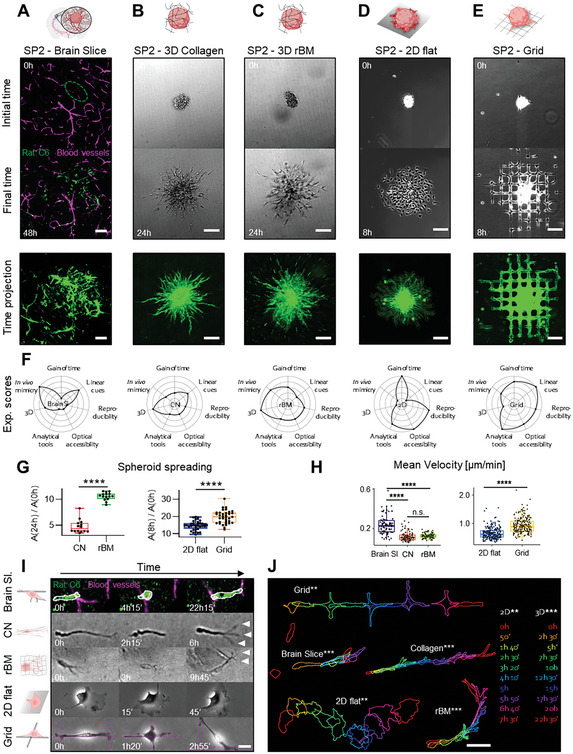
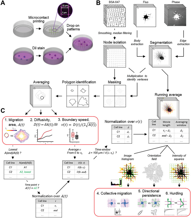
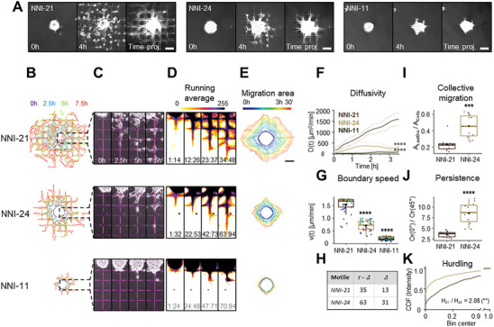
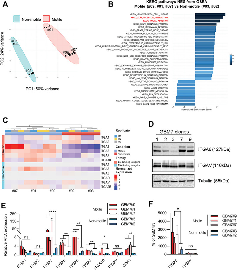
Glioblastomas exhibit remarkable heterogeneity at various levels, including motility modes and mechanoproperties that contribute to tumor resistance and recurrence. In a recent study using gridded micropatterns mimicking the brain vasculature, glioblastoma cell motility modes, mechanical properties, formin content, and substrate chemistry are linked. Now is presented, SP2G (SPheroid SPreading on Grids), an analytic platform designed to identify the migratory modes of patient-derived glioblastoma cells and rapidly pinpoint the most invasive sub-populations. Tumorspheres are imaged as they spread on gridded micropatterns and analyzed by this semi-automated, open-source, Fiji macro suite that characterizes migration modes accurately. SP2G can reveal intra-patient motility heterogeneity with molecular correlations to specific integrins and EMT markers. This system presents a versatile and potentially pan-cancer workflow to detect diverse invasive tumor sub-populations in patient-derived specimens and offers a valuable tool for therapeutic evaluations at the individual patient level.

Keywords: cell migration; cytoskeleton; glioblastoma; micropatterning.


Conflict of interest statement

The authors declare no conflict of interest.

Figures

Figure 1
Figure 1
Spheroid spreading on grids (SP2G) mimics glioblastoma invasion on brain blood vessels. A–E) Rat C6 glioma cells cultured as spheroids were stained (green, DiOC6 dye) and seeded in brain slices (A), in collagen gel (B), in reconstituted basement membrane (rBM) (C), on laminin‐coated dishes (D), on gridded micropatterns (E) and imaged for 48 h (A), 24 h (B, C) or 8 h (D, E). The first and last images of the movies (upper panels) and time projections (lower panels) are shown. The dashed oval in A corresponds to the initial area of the spheroid in the brain slice. F) Radar plots summarizing experimental scores (1 to 5) of gain of time, presence of linear cues, experimental reproducibility, optical accessibility, possibility to develop analytical tools, 3D and in vivo mimicry for each setting. G) Quantification of spheroid spreading in collagen gel, rBM, 2D flat, and grid (n = 14, 15, 35, 35 spheroids respectively). H) Mean velocities of single cells migrating in brain slices, collagen gel, rBM, 2D flat and grid (n = 80, 95, 90, 215, 215 tracks respectively, 5 to 7 tracks per spheroid, each dot is a cell). I) Snapshots of single cells moving away from the spheroid in each set, extracted from Video S1 (Supporting Information). J) Panel summarizing cell shapes for C6 cell motility in each setting. Time is color‐coded as indicated. Bars are 100 µm (A–E), 20 µm (I), and 50 µm (J).
Figure 2
Figure 2
SP2G experimental setup and image analysis workflow. A) Spheroids are stained (red, Dil), seeded on fluorescent gridded micro‐patterns (coated with laminin mixed with BSA 647), and imaged for 8 h. B) Cells and grids images are segmented in binary images that are multiplied to isolate the grid nodes covered by the invasive boundary. Polygons tracking the spheroid spreading are reconstructed and averaged. The time trend is projected and color‐coded to visualize migration area A(t) (output #1). Diffusivity D(t) is obtained by differentiating A(t) (output #2). Spheroid boundary speed v(t) is obtained from D(t) and A(t)(output #3). Normalization of A(t) and v(t): τ, corresponding to the frame at which each cell line has migrated the same distance as the slowest cell line (C2) at the end of the acquisition (8 h or more), and Δ, corresponding to the time window to complete 100 µm, are identified and all the movies are cut at τ. Running average (RA) movies are created by shifting Δ in the interval 1:τ and cell motility modes are characterized by extrapolating features from the RA movies: collective migration (output #4) is obtained by thresholding the area (outlined in green) of pixels belonging to the last bin of the histogram; directional persistence (output #5) is obtained by evaluating image orientation; hurdling (output #6) is obtained by sampling the intensities of the grid squares.
Figure 3
Figure 3
SP2G quantifies cell migratory tactics. Patient‐derived glioblastoma spheroids were seeded on fluorescent gridded micropatterns, imaged for 8 h, and analyzed as indicated in Figure 2 (n = 10, 11, 13 spheroids, n = 2, 2, 2 independent experiments for NNI‐21, NNI‐24 and NNI‐11 respectively). A) Snapshots of the movies at 0 h, 4 h, and corresponding time projections. B–D) Cellular edges (B) and corresponding overlays of the phase contrast and the fluorescent grid images (C) at 4‐time points (0, 2.5, 5, 7.5 h) and corresponding running average (RA) (D). The time window Δ constituting the corresponding RA frame is indicated at the bottom of each panel: for the non‐motile τ = 94, Δ = 24. E) Averaged polygons visualizing migration areas. F) Diffusivity over 3.5 h; dashed lines are standard deviations. G) Mean boundary speed over 3.5 h. Each dot represents a time point and is color‐coded as in (E). H) Δ (number of frames needed to travel 100 µm) and τ – Δ (number of frames in the RA movie) in motile cell lines. I) Collective migration quantification. Each dot represents a spheroid. J) Directional persistence is visualized as the ratio between the orientation along 0° and along 45° (Or(0°) / Or(45°), see methods). Each dot represents a spheroid. K) Hurdling is visualized as the Cumulative Distribution Function (CDF) of the normalized mean intensity of the grid squares (image intensity is sampled in each square). The ratio indicates the relationship between the average mean intensities (the sum of the mean intensity from all the squares divided by the total number of squares) of the 2 cell lines. Scale bars are 100 µm. Time and image intensity are color‐coded as indicated. Statistical analyses are shown for NNI‐21 compared to the other cell lines (see Supporting Information for complete analysis).
Figure 4
Figure 4
SP2G reveals migration heterogeneity in glioblastoma sub‐populations isolated from patient‐derived tumor‐spheres. Spheroids from the GBM7 original cell line (bulk) and isolated subpopulations (clones #01, #02, #03, #07, #09) were seeded on fluorescent gridded micropatterns, imaged for 8 h, and analyzed as indicated in Figure 2. A) Snapshots of the movies of the original population (bulk) at the indicated time points. B) Phase‐contrast pictures of the GBM7 sub‐populations cultured on laminin (top) and after SP2G at 8 hours. C) Spheroid spreading of GBM7 sub‐populations (green, DiOC6 dye) in brain slices at 0 h and 40 h. D–G) SP2G analysis of the clones #01, #02, #03, #07, #09 (n = 22, 23, 20, 22, 22 spheroids respectively; n = 3 independent experiments): Cellular edges (D), corresponding overlays of the phase contrast and the fluorescent grid images at 4‐time points (E), and corresponding running average (RA) (F). The time window Δ constituting the corresponding RA frame is indicated at the bottom of each panel: for the non‐motile τ = 94, Δ = 24. (G) Averaged polygons visualizing migration areas. H) Δ and τ – Δ of the motile subpopulations. I) Diffusivity over 3 h 30′. Dashed lines are the standard deviation. J) Mean boundary speed over 3 h 30′. Each dot represents a time point and is color‐coded as in (G). K) Collective migration for the motile cells. Each dot represents a spheroid. L) Directional persistence for the motile cells is visualized as the ratio between the orientation along 0° and 45° (see Experimental Section). Each dot represents a spheroid. M) Hurdling, visualized as the Cumulative Distribution Function (CDF) of the normalized mean intensity of the grid squares. The ratio indicates the relationship between the average mean intensities of the most hurdling (#01) against the others. N) Mean velocities of single cells migrating in brain slices n = 20, 13 15, 15, 22 tracks for clones #1, 2, 3, 7, 9 respectively, each dot is a cell). Bars are 100 µm (A, B bottom panel, C) and 50 µm (B, top panel). Time and image intensity are color‐coded as indicated. Statistical analyses are shown for clone #7 compared to all the other clones (see Supporting Information for a complete analysis).
Figure 5
Figure 5
Intra‐patient heterogeneity in migration ability is correlated with specific molecular signatures. A) Principal component analysis showing segregation of the 5 GBM7 sub‐populations in motile and non‐motile groups. B) Gene set enrichment analysis (GSEA) of differentially expressed genes in the motile versus non‐motile group. GSEA was performed using the Kyoto Encyclopedia of Genes and Genomes (KEGG) gene set in the GSEA molecular signatures database. Moderated t‐statistic was used to rank the genes. Reported are Normalized Enrichment Scores (NES) of enriched pathways (with the fill color of the bar corresponding to the p‐value). P‐value was calculated as the number of random genes with the same or more extreme ES value divided by the total number of generated gene sets. C) Heatmap representing z‐score of expression levels of integrins. D) Expression of ITGA6, ITGAV, and tubulin, in total cell extracts of the 5 GBM7 sub‐populations growing on laminin. E) Relative mRNA expression levels of ITGA1, ITGA2, ITGA3, ITGA5, ITGA6, ITGA7, ITGA10, ITGAV, and CD44 in the 5 GBM7 sub‐populations. Each integrin expression is reported to its expression in clone #3. GAPDH and B2M were used as housekeeping genes. n = 3 independent experiments. Error bars are S.E.M. F) Quantification of the expression of ITGA6 and ITGAV in each condition reported to their expression in clone #3. 2 independent western blots were quantified.
Figure 6
Figure 6
Intra‐patient heterogeneity in motility modes is correlated with specific molecular signatures. A) Principal component analysis showing segregation of the 3 motile sub‐populations in 2 groups. B) Heatmap representing row‐normalized expression levels of the 20 most variable genes between the 2 motile groups. Genes belonging to the EMT pathway are upregulated in clone #01 compared to clones #07 and #09. C) Gene set enrichment analysis (GSEA) of differentially expressed genes in the motile groups (clones #07 and #09 versus clone #01). GSEA was performed using the KEGG gene set in the GSEA molecular signatures database as in Figure. 5.

References

    1. Ostrom Q. T., Price M., Neff C., Cioffi G., Waite K. A., Kruchko C., Barnholtz‐Sloan J. S., Neuro Oncol 2022, 24, v1. - PMC - PubMed
    1. Louis D. N., Perry A., Wesseling P., Brat D. J., Cree I. A., Figarella‐Branger D., Hawkins C., Ng H. K., Pfister S. M., Reifenberger G., Soffietti R., von Deimling A., Ellison D. W., Neuro Oncol 2021, 23, 1231. - PMC - PubMed
    1. Weller M., Le Rhun E., Van den Bent M., Chang S. M., Cloughesy T. F., Goldbrunner R., Hong Y.‐K., Jalali R., Jenkinson M. D., Minniti G., Nagane M., Razis E., Roth P., Rudà R., Tabatabai G., Wen P. Y., Short S. C., Preusser M., Neuro Oncol 2023, 25, 1200. - PMC - PubMed
    1. Xie Q., Mittal S., Berens M. E., Neuro Oncol 2014, 16, 1575. - PMC - PubMed
    1. Brennan C. W., Verhaak R. G. W., McKenna A., Campos B., Noushmehr H., Salama S. R., Zheng S., Chakravarty D., Sanborn J. Z., Berman S. H., Beroukhim R., Bernard B., Wu C.‐J., Genovese G., Shmulevich I., Barnholtz‐Sloan J., Zou L., Vegesna R., Shukla S. A., Ciriello G., Yung W. K., Zhang W., Sougnez C., Mikkelsen T., Aldape K., Bigner D. D., Meir E. G. V., Prados M., Sloan A., Black K. L., et al., Cell 2013, 155, 462. - PubMed

Publication types

LinkOut - more resources