. 2024 Mar 26;134(10):e159884.
doi: 10.1172/JCI159884.

Noncanonical WNT5A controls the activation of latent TGF-β to drive fibroblast activation and tissue fibrosis

Affiliations

Noncanonical WNT5A controls the activation of latent TGF-β to drive fibroblast activation and tissue fibrosis

Thuong Trinh-Minh et al. J Clin Invest. .

Abstract

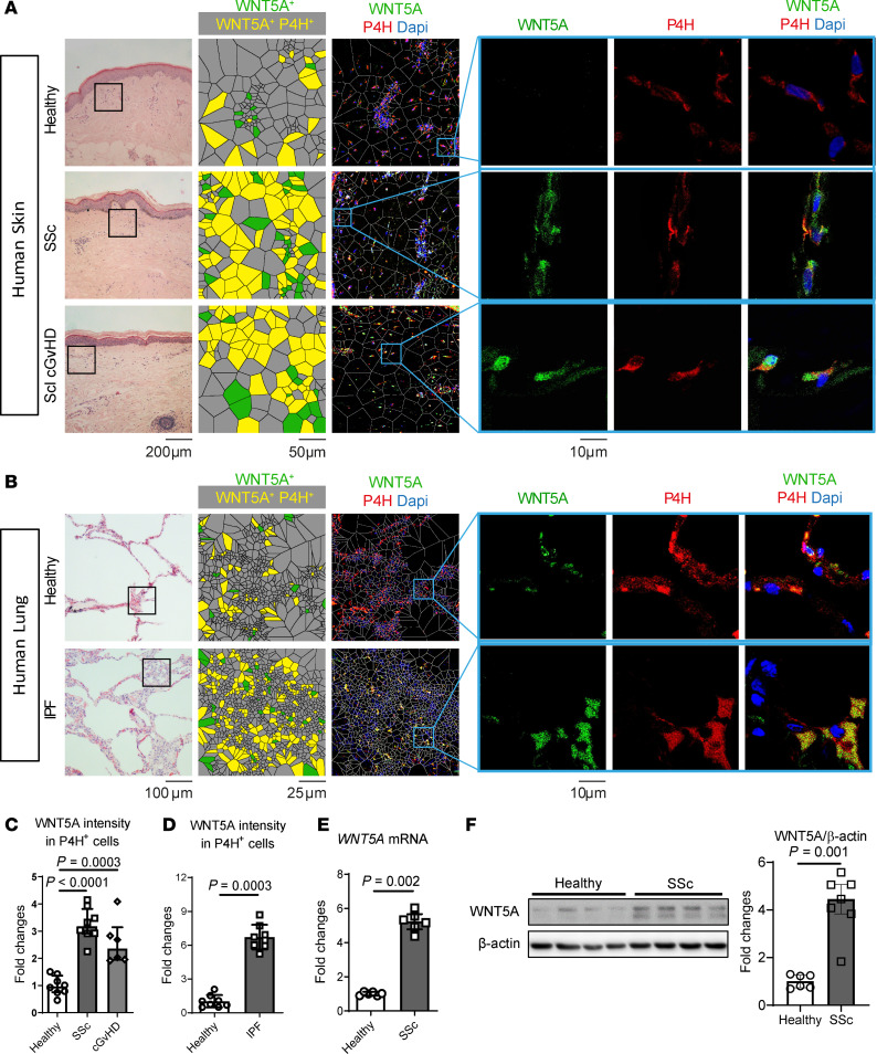
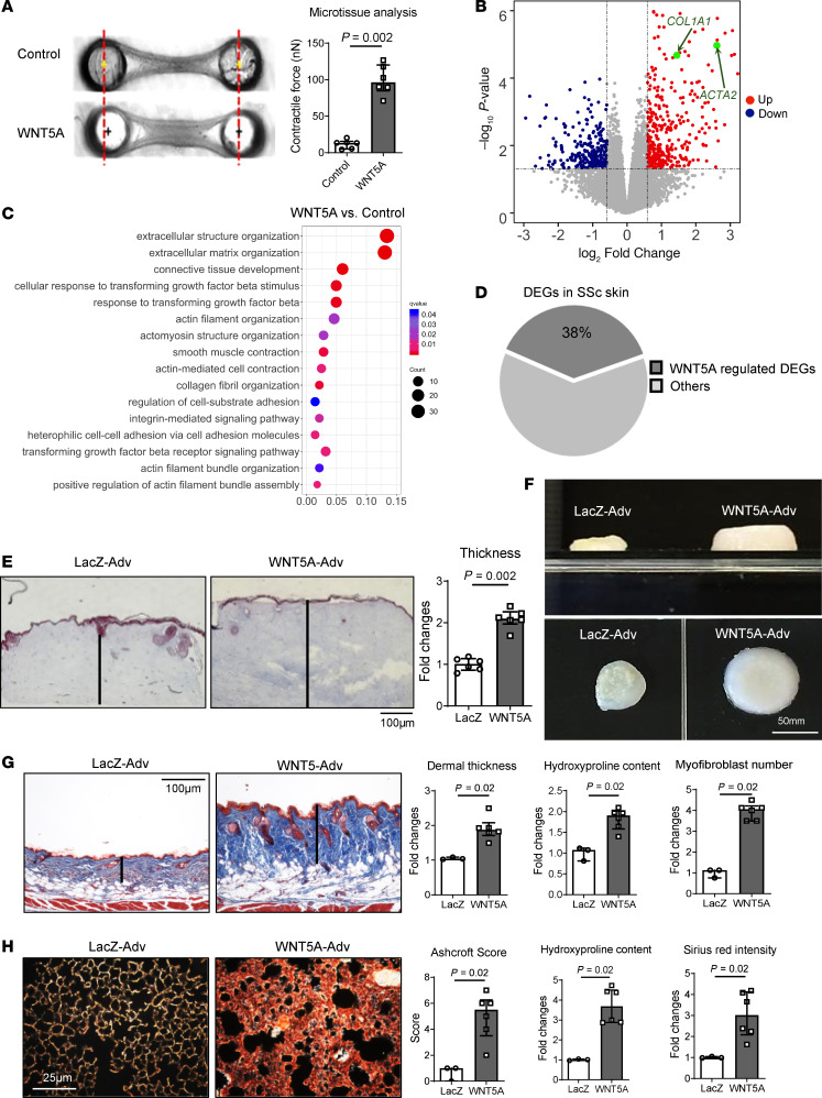
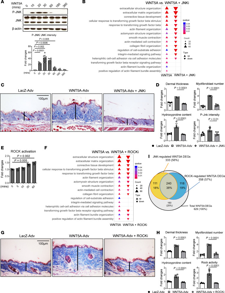
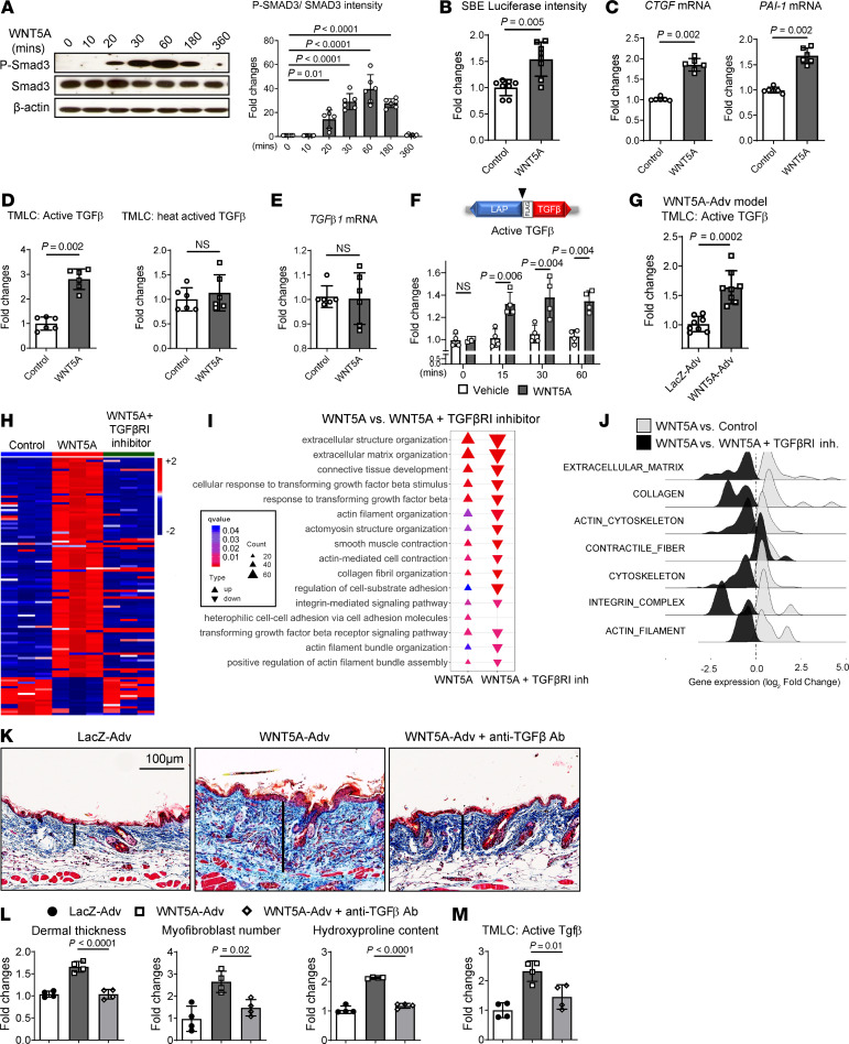
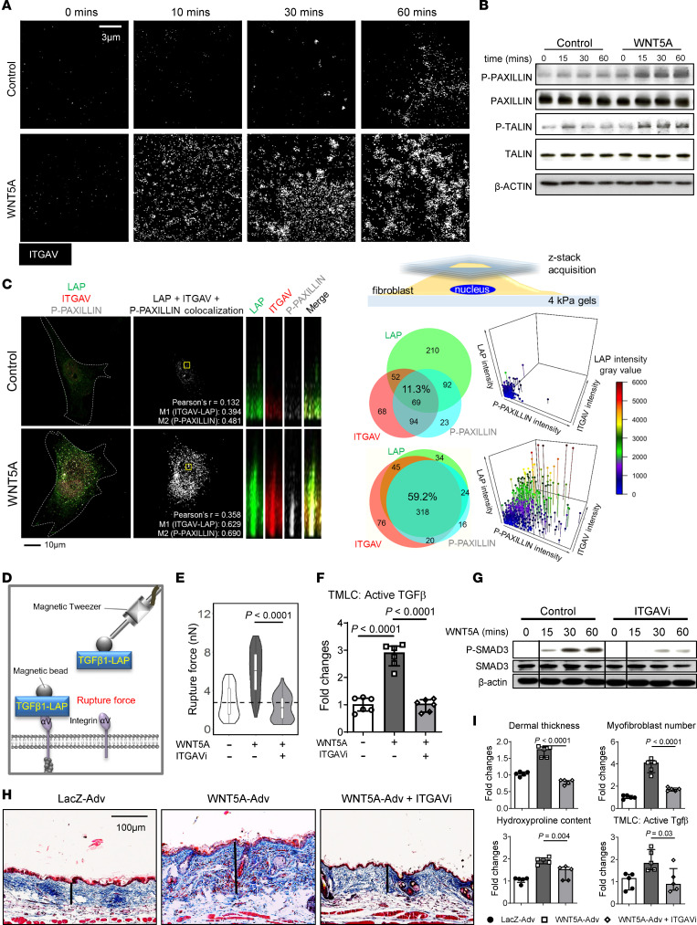
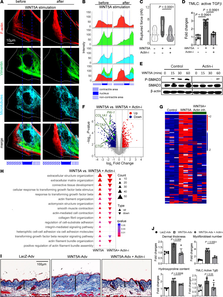
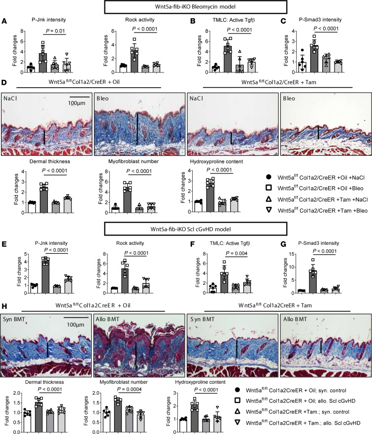
Transforming growth factor β (TGF-β) signaling is a core pathway of fibrosis, but the molecular regulation of the activation of latent TGF-β remains incompletely understood. Here, we demonstrate a crucial role of WNT5A/JNK/ROCK signaling that rapidly coordinates the activation of latent TGF-β in fibrotic diseases. WNT5A was identified as a predominant noncanonical WNT ligand in fibrotic diseases such as systemic sclerosis, sclerodermatous chronic graft-versus-host disease, and idiopathic pulmonary fibrosis, stimulating fibroblast-to-myofibroblast transition and tissue fibrosis by activation of latent TGF-β. The activation of latent TGF-β requires rapid JNK- and ROCK-dependent cytoskeletal rearrangements and integrin αV (ITGAV). Conditional ablation of WNT5A or its downstream targets prevented activation of latent TGF-β, rebalanced TGF-β signaling, and ameliorated experimental fibrosis. We thus uncovered what we believe to be a novel mechanism for the aberrant activation of latent TGF-β in fibrotic diseases and provided evidence for targeting WNT5A/JNK/ROCK signaling in fibrotic diseases as a new therapeutic approach.

Keywords: Dermatology; Fibrosis; Pulmonology.


Conflict of interest statement

Conflict of interest: Although none of the authors has any direct conflict of interest related to noncanonical WNT signaling, OD and JHWD are involved in the development of new targeted therapies for fibrotic diseases such as SSc. OD has consultancy relationships and/or has received research funding from Actelion, Pfizer, Ergonex, BMS, Sanofi-Aventis, United BioSource Corporation, Roche/Genentech, Medac, Biovitrium, Boehringer Ingelheim, Novartis, 4D Science, Active Biotech, Bayer, Sinoxa, Serodapharm, EpiPharm, GSK, Pharmacyclics, and Biogen. JHWD has consultancy relationships with Actelion, Active Biotech, Anamar, ARXX, Bayer Pharma, Boehringer Ingelheim, Celgene, Galapagos, GSK, Inventiva, JB Therapeutics, Medac, Pfizer, RuiYi, and UCB. JHWD has received research funding from Anamar, Active Biotech, Array Biopharma, aTyr, BMS, Bayer Pharma, Boehringer Ingelheim, Celgene, Galapagos, GSK, Inventiva, Novartis, Sanofi-Aventis, RedX, and UCB. JHWD is stock owner of 4D Science and Scientific head of FibroCure.

Figures

Figure 1
Figure 1. WNT5A is expressed at increased levels in human fibrotic diseases such as SSc, Scl cGvHD, and IPF.
(A) Representative H&E staining, IF stainings for WNT5A (green) in combination with the human fibroblast marker P4H (red) and DAPI (blue) and results of Voronoi tessellation in skin sections of patients with SSc, in skin sections of patients with sclerodermatous (Scl) cGvHD (n = 8 for healthy and SSc patients, n = 6 for Scl cGvHD patients) and (B) in lung sections of patients with IPF (n = 8 for each group), all with control sections from nonfibrotic skin or lungs, respectively. (C and D) Quantification of the WNT5A staining in each fibrotic disease. (E) Fold changes of WNT5A mRNA in fibroblasts isolated from SSc skin or from healthy skin (n = 6 for each group). (F) Protein levels of WNT5A in fibroblasts (average passage 5–7) analyzed by representative Western blots and quantification (n = 6 for healthy fibroblasts and n = 7 for SSc fibroblasts). Results are shown as median ± IQR with data representing individual data points. The statistical significance was determined by 2-tailed Mann-Whitney U test if 2 groups were compared or 1-way ANOVA with Tukey’s multiple comparison test in case of more than 2 comparisons.
Figure 2
Figure 2. WNT5A promotes fibroblast-to-myofibroblast transition and induces dermal and pulmonary fibrosis.
(A) Microtissue assay. Representative microtissue images and quantification of the contractile force exerted by fibroblasts (n = 6 for each group). (B and C) RNA-Seq of human dermal fibroblasts stimulated with WNT5A compared with control fibroblasts (n = 3 for each group). (B) Volcano plot of DEGs. The expression of each gene is plotted as the log-fold change of expression compared with controls; the 1.5-fold change threshold is marked by dotted lines. (C) Bubble plots displaying significant enrichment of GO biological processes. The color of the bubble represents the q value, and the size of the bubble represents the number of DEGs in the data sets associated with the GO processes. (D) Pie chart showing the percentage of DEGs in SSc skin (29) that overlap with WNT5A target genes in fibroblasts. (E and F) Full-thickness skin organoids. (E) Trichrome stainings and quantification, and (F) representative microscopic images of dermal thickness in skin organoids (n = 6 for each group). (G) Forced overexpression of Wnt5a in the skin of mice. Representative Trichrome stainings, quantification of the dermal thickness, the collagen content, and myofibroblast counts (n = 3 for LacZ-Adv group and n = 6 for WNT5A-Adv group). (H) Forced overexpression of Wnt5a in murine lungs. Representative sirius red stainings, ashcroft scores, hydroxyproline content, and quantifications of sirius red staining (n = 3 for LacZ-Adv group and n = 6 for WNT5A-Adv group). Results are shown as median ± IQR with data representing individual data points. The statistical significance was determined by 2-tailed Mann-Whitney U test. Adv, Adenovirus.
Figure 3
Figure 3. WNT5A-induced fibrosis requires JNK and ROCK.
(A-D) JNK signaling. (A) Representative Western blot and quantification of pJNK in WNT5A stimulated human dermal fibroblasts (n = 6 for each group). (B) Negative enrichment scores (deenrichment) of GO biological processes related to fibroblast-to-myofibroblast transition and fibrosis in WNT5A-stimulated fibroblasts. (C and D) Effects of Jnk inhibition on Wnt5a-induced skin fibrosis in mice. (C) Representative trichrome stainings. (D) Quantification of the dermal thickness, the collagen content, myofibroblast counts, and P-Jnk immunofluorescence staining levels in tissue sections (n = 5 for each group). (EH) ROCK signaling. (E) Quantification of ROCK activity in WNT5A-stimulated human dermal fibroblasts as analyzed by ROCK activity assays (n = 4 for each group). (F) Negative enrichment scores (deenrichment) of GO biological processes related to fibroblast-to-myofibroblast transition and fibrosis in WNT5A-stimulated fibroblasts treated with ROCKi compared with vehicle-treated WNT5A-stimulated fibroblasts (n = 3 for each group). (G and H) Effects of Rock inhibition on Wnt5a-induced skin fibrosis in mice. (G) Representative trichrome stainings. (H) Quantification of the dermal thickness, the collagen content, myofibroblast counts, and the Rock activity measured in tissue lysates (n = 5 for each group). (I) Venn diagram showing the number and percentage of JNK- and ROCK-regulated DEGs among all WNT5A-regulated DEGs. Results are shown as median ± IQR with data representing individual data points. The statistical significance was determined 1-way ANOVA with Tukey’s multiple comparison test. Adv, Adenovirus.
Figure 4
Figure 4. WNT5A induces activation of latent TGF-β in vitro and in vivo.
(A) Representative Western blots and quantification of the levels of P-SMAD3 in dermal fibroblasts stimulated with WNT5A (n = 6 for each group). (B) Changes in the activity of a SBE-reporter construct (n = 8 for each group). (C) mRNA levels of the CTGF and PAI-1 genes (n = 6 for each group). (D) Levels of active TGF-β and of total, heat-activated TGF-β in the supernatant of WNT5A-stimulated fibroblasts as measured by TMLC assays (n = 6 for each group). (E) Absence of changes in TGF-β1 mRNA (n = 6 for each group). (F) Quantification of active TGF-β1 in supernatants from fibroblasts expressing LAP-TGF-β1 with a cleavage exposed FLAG-tag (▼) between the LAP and the TGF-β1 coding region (n = 4 for each group). (G) Active TGF-β in the skin of mice overexpressing WNT5A (n = 8 for each group). (H) Heatmap illustration of DEGs in WNT5A-stimulated fibroblasts with or without-βRIi (n = 3 for each group). (I) Negative enrichment scores for GO biological processes related to fibroblast-to-myofibroblast transition and fibrosis. (J) Ridgeline plot highlighting the deenrichment of GSEA-gene sets related to fibroblast-to-myofibroblast transition and tissue fibrosis in WNT5A-stimulated fibroblasts treated with or without TGF-βRIi. (K) Representative trichrome stainings and (L) quantification of dermal thickness, hydroxyproline content, myofibroblast counts, and (M) active TGF-β in skin lysate from mice with WNT5A-induced fibrosis with or without TGF-βRIi (n = 4 for each group). Results are shown as median ± IQR. The statistical significance was determined by 2-tailed Mann-Whitney U-test if 2 groups were compared or 1-way ANOVA with Tukey’s multiple comparison test in figure A, L, and M, or 2-way ANOVA in figure F. Adv, Adenovirus.
Figure 5
Figure 5. WNT5A -induced activation of latent TGF-β requires Integrin αV.
(A) Representative confocal IF images of ITGAV clusters in dermal fibroblasts incubated with or without WNT5A (n = 4 for each group). (B) Representative Western blots of P-PAXILLIN, PAXILLIN, P-TALIN and TALIN (n = 3 for each group). (C) Representative confocal images, z-stack reconstructions, and Venn diagrams showing colocalization of LAP with ITGAV and P-PAXILLIN, and 3 -dimensional scatter plots showing the distribution of each marker at each localization (n ≥ 10 for each group); schematic overview of the experimental conditions and areas of assessment. Pearson’s r is the Pearson’s correlation coefficient between LAP-ITGAV and P-PAXILLIN voxel intensities. M1 and M2 representing for Manders’ split coefficients. (D) Schematic illustration of the experiment for measuring the rupture force with magnetic tweezers. (E) Violin plots showing the force required to rupture magnetic beads coupled with peptides containing the RGD domain of LAP-TGF-β1 (RRGDLATISPASSKGGGGSRLLLLLLR) from the cell surface of dermal fibroblasts incubated with WNT5A with or without ITGAV inhibitor (ITGAVi) (n ≥ 50 for each group). (F) Levels of active TGF-β in the cell culture supernatant of dermal fibroblasts incubated with WNT5A with or without ITGAVi (n = 6 for each group). (G) Representative Western blots of P-SMAD3 in dermal fibroblasts stimulated by WNT5A with and without the ITGAVi (n = 3 for each group). The black thin vertical lines were drawn to separate noncontiguous lanes. (H) Representative Trichrome stainings and (I) Quantification of the dermal thickness, the hydroxyproline content, myofibroblast counts, and active TGF-β in the skin tissue of WNT5A-induced skin fibrosis mice treated with or without ITGAVi (n = 5 for each group). Results are shown as median ± IQR. The statistical significance was determined by 1-way ANOVA with Tukey’s multiple comparison test. Adv, Adenovirus.
Figure 6
Figure 6. WNT5A induces coordinated cytoskeletal changes to promote activation of latent TGF-β.
(A) Representative images showing WNT5A-induced changes in F-actin, Vimentin filaments, and microtubules with (B) quantification of each cytoskeletal component in relation to its subcellular localization. (C) Violin plots showing the force required to rupture magnetic beads coupled with LAP-TGF-β1 peptides from dermal fibroblasts incubated with WNT5A with or without the actin inhibitor cytochalasin D (n ≥ 50 for each group). (D) Levels of active TGF-β in the supernatant of dermal fibroblast incubated with WNT5A in the presence or absence of cytochalasin D measured by TMLC assays (n = 6 for each group). (E) Representative Western blots showing the levels of P-SMAD3 in dermal fibroblasts stimulated with WNT5A and cytochalasin D (n = 3 for each group). The black thin vertical lines were drawn to separate noncontiguous lanes. (F) Volcano plot and (G) heatmap illustration of DEGs from RNA-Seq of WNT5A-stimulated human dermal fibroblasts treated with Cytochalasin D or with vehicle. (H) Deenrichment of GO biological processes related to fibroblast-to-myofibroblast transition and fibrosis (n = 3 for each group). (I) Representative Trichrome stainings, and (J) quantification of the dermal thickness, hydroxyproline content, myofibroblast counts, and active TGF-β in skin lysates from mice with WNT5A-induced skin fibrosis with or without ITGAV inhibitor (n = 5 for each group). Results are shown as median ± IQR with data representing individual data points. The statistical significance was determined by 1-way ANOVA with Tukey’s multiple comparison test. Adv, Adenovirus; Actin-i, Actin inhibitor.
Figure 7
Figure 7. Inactivation of Wnt5a signaling ameliorates experimental skin fibrosis.
(AD) Fibroblast-specific knockout of Wnt5a in bleomycin-induced skin fibrosis. (A) Quantification of the IF staining of P-Jnk and of the Rock activity. (B) Levels of active TGF-β measured by TMLC. (C) Quantification of the IF staining for P-Smad3. (D) Representative Trichrome stainings and quantification of dermal thickness, myofibroblast counts, and hydroxyproline content. (EH) Fibroblast-specific knockout of Wnt5a in murine scl cGvHD. (E) quantification of the IF staining for P-Jnk and of Rock activity. (F) Level of active TGF-β measured by TMLC. (G) Quantification of the IF staining for P-Smad3. (H) Representative trichrome stainings and quantification of the dermal thickness, myofibroblast counts, and hydroxyproline content (n = 6 for each group). Results are shown as median ± IQR with data representing individual data points. The statistical significance was determined by 1-way ANOVA with Tukey’s multiple comparison test. Bleo, Bleomycin; Tam, Tamoxifen; Syn, Syngenic; Allo, Allogenic; BMT, Bone marrow transplantation.

References

    1. Wynn TA. Cellular and molecular mechanisms of fibrosis. J Pathol. 2008;214(2):199–210. doi: 10.1002/path.2277. - DOI - PMC - PubMed
    1. Distler JH, et al. Review: Frontiers of antifibrotic therapy in systemic sclerosis. Arthritis Rheumatol. 2017;69(2):257–267. doi: 10.1002/art.39865. - DOI - PubMed
    1. Thannickal VJ, et al. Fibrosis: ultimate and proximate causes. J Clin Invest. 2014;124(11):4673–4677. doi: 10.1172/JCI74368. - DOI - PMC - PubMed
    1. Yoon YJ, et al. Antifibrotic therapies: where are we now? Semin Liver Dis. 2016;36(1):87–98. doi: 10.1055/s-0036-1571295. - DOI - PubMed
    1. Strieter RM, Mehrad B. New mechanisms of pulmonary fibrosis. Chest. 2009;136(5):1364–1370. doi: 10.1378/chest.09-0510. - DOI - PMC - PubMed

MeSH terms