. 2024 May 15;134(10):e176577.
doi: 10.1172/JCI176577.

The mechanosensory channel PIEZO1 functions upstream of angiopoietin/TIE/FOXO1 signaling in lymphatic development

Affiliations

The mechanosensory channel PIEZO1 functions upstream of angiopoietin/TIE/FOXO1 signaling in lymphatic development

Jing Du et al. J Clin Invest. .

Abstract

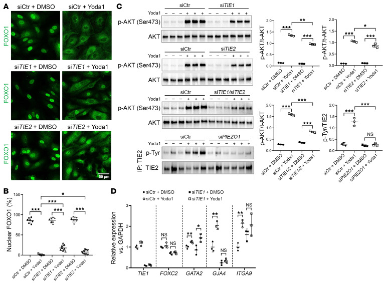
Lymphedema is a debilitating disease with no effective cure and affects an estimated 250 million individuals worldwide. Prior studies have identified mutations in piezo-type mechanosensitive ion channel component 1 (PIEZO1), angiopoietin 2 (ANGPT2), and tyrosine kinase with Ig-like and EGF-like domains 1 (TIE1) in patients with primary lymphedema. Here, we identified crosstalk between these molecules and showed that activation of the mechanosensory channel PIEZO1 in lymphatic endothelial cells (LECs) caused rapid exocytosis of the TIE ligand ANGPT2, ectodomain shedding of TIE1 by disintegrin and metalloproteinase domain-containing protein 17 (ADAM17), and increased TIE/PI3K/AKT signaling, followed by nuclear export of the transcription factor FOXO1. These data establish a functional network between lymphedema-associated genes and provide what we believe to be the first molecular mechanism bridging channel function with vascular signaling and intracellular events culminating in transcriptional regulation of genes expressed in LECs. Our study provides insights into the regulation of lymphatic function and molecular pathways involved in human disease.

Keywords: Development; Embryonic development; Endothelial cells; Ion channels; Vascular biology.


Figures

Figure 1
Figure 1. Bulk RNA-Seq analysis of dermal LECs reveals transcriptomic changes driven by FOXO1 overactivation in the Tie1–/– group.
(A) Generation of Tie1 whole-body inducible or lymphatic endothelium–specific Tie1-KO mice. Tie1 whole-body inducible KO (Tie1WB–/–) was generated by crossing Tie1fl mice with Rosa26rtTA TetOCre mice and timed induction with Dox water. Tie1 lymphatic-specific Tie1 KO (Tie1LEC–/–) was generated by crossing Tie1fl mice with PdpnCre mice. (B) Gross phenotypes of Tie1WB–/– embryos that were induced and examined at different time points (first number indicates the induction time point; second number indicates the harvest time point). White asterisk shows blood-filled lymphatics at E15.5 (5 of 7 of the Tie1WB–/– embryos); arrow shows edema at E18.5 (7 of 9 of the Tie1WB–/– embryos); and arrowhead shows chylous ascites at P2 (6 of 6 of the Tie1WB–/– pups). (C) LEC-specific Tie1 KO resulted in a phenotype similar to that of whole-body KO. (D) Workflow of dermal LEC bulk RNA-Seq. E18.5 embryos induced at E13.5 were euthanized. The skin from each embryo was removed and placed in an Eppendorf tube for enzymatic digestion. The single-cell suspension was labeled with antibodies against CD45, CD31, and Lyve1. CD31+LYVE1+ cells were sorted into lysis buffer for RNA extraction. Bulk RNA-Seq was performed on the Illumina HiSeq 4000 system. (E) Number of differentially expressed genes using a P value of less than 0.01 as the cutoff. Down, downregulated; Up, upregulated. (F) Volcano plot shows some of the most differentially expressed genes including Ccl21a, valve genes and tip cell–enriched genes. (G) Heatmaps of manually selected vascular-relevant genes categorized as labeled. (H) Overlap between our data set (blue) and a data set from HUVECs with transcriptionally active FOXO1 (orange). Some commonly regulated genes are listed in the square, including tip cell genes (red), polarization genes (blue), ion channel genes (green), and valve genes (black).
Figure 2
Figure 2. TIE-mediated regulation of FOXO1 subcellular localization in vivo and in vitro.
(A) Whole mounts of E16.5 skin with lymphatic vessels stained for PROX1 and FOXO1. Nuclei within areas of LECs from WT control, Tie1WB–/–E13.5, and Tie2WB–/–E13.5 mice are indicated by arrowheads (branching points) and arrows (nonbranching points). Scale bars: 20 μm. (B) Quantification of FOXO1 localizations in PROX1+ LECs. Averaged values calculated from 3 mice in each group are presented. (C) P1 Tie1WB–/–E18.5 pups and their littermate controls were intraperitoneally injected with 1 μg/g BW Hepta-ANG1 or PBS. After a 30-minute period, pups were euthanized, and mesentery specimens were harvested and stained for PROX1 and FOXO1. Scale bars: 50 μm. (D) Quantification of FOXO1 localizations from 4 mice in each group. (E) FOXO1 staining of HDLECs transfected with siCtr, siTIE1, or siTIE2 for 48 hours and subsequently treated with Hepta-ANG1 (1 μg/mL) or PBS for 30 minutes. This experiment was replicated 3 times. Scale bar: 50 μm. (F) Quantification of FOXO1 localization in 3 fields of view selected from each group. (G) Western blot analysis of p-AKT levels in HDLECs transfected with siCtr, siTIE1, or siTIE2 and treated with either vehicle or Hepta-ANG1. Each band represents a biological replicate sample (n = 3). Data are expressed as the mean ± SD. **P < 0.01 and ***P < 0.001, by 2-tailed, unpaired Student’s t test (D) and 2-way ANOVA followed by Tukey’s multiple-comparison test (F and G).
Figure 3
Figure 3. Activation of PIEZO1 signaling promotes nuclear exclusion of FOXO1 and activates the AKT pathway.
(A) Mesenteries isolated from P1 Piezo1WB–/–E18.5 pups or their littermate controls were subjected to a 30-minute incubation at 37°C with either 250 nM Yoda1 or vehicle. After fixation in 2% PFA for 30 minutes, the mesenteries were stained for PROX1 and FOXO1. Scale bars: 50 μm. (B) Quantification of mouse LECs with nuclear FOXO1 localization (n = 4 mice in each group). (C) HDLECs were transfected with siCtr or siPIEZO1 for 48 hours and subsequently treated with either vehicle or 250 nM Yoda1 for 30 minutes. Following fixation, cells were stained for FOXO1. Scale bar: 50 μm. (D) Quantification of cells exhibiting nuclear FOXO1 staining. (E) Western blot analysis of p-AKT levels in HDLECs transfected with siCtr or siPIEZO1 and treated with either vehicle or Yoda1. Each band represents a biological replicate sample (n = 3). Data are expressed as the mean ± SD. *P < 0.05, **P < 0.01, and ***P < 0.001, by 2-tailed, unpaired Student’s t test (B) and 2-way ANOVA followed by Tukey’s multiple-comparison test (D and E).
Figure 4
Figure 4. TIE signaling partially mediates AKT/FOXO1 activation triggered by PIEZO1 signaling.
(A) HDLECs were transfected with siCtr, siTIE1, siTIE2, or siTIE1/siTIE2 for 48 hours and then exposed to either vehicle or 250 nM Yoda1 for 30 minutes. After fixation, cells were stained for FOXO1. This experiment was carried out concurrently with the one depicted in Figure 3C. The samples used in the siCtr plus DMSO and siCtr plus Yoda1 groups were identical to those in Figure 3C. Scale bar: 50 μm. (B) Quantification of cells exhibiting nuclear FOXO1 staining. This experiment was repeated 3 times. (C) HDLECs were transfected with the specified siRNAs and treated with vehicle or Yoda1 as described above. Cell lysates were subjected to Western blot analysis to assess AKT phosphorylation. TIE2 was isolated from cell lysates via immunoprecipitation and subsequently analyzed by Western blotting to evaluate its phosphorylation status. Each band represents a biological replicate sample (n = 3). (D) qPCR analysis of HDLECs transfected with siCtr or siTIE1 and subsequently treated with Yoda1 (250 nM, 24 hours) or vehicle. Expression levels of TIE1, FOXC2, GATA2, GJA4, and ITGA9 genes were measured. Data are expressed as the mean ± SD. *P < 0.05, **P < 0.01, and ***P < 0.001, by 2-way ANOVA followed by Tukey’s multiple-comparison test (B and C) and 2-tailed, unpaired Student’s t test (D).
Figure 5
Figure 5. PIEZO1 activation induces ANGPT2 exocytosis in LECs.
(A) Immunostaining for ANGPT2 and FOXO1 in HDLECs transfected with siCtr or siANGPT2 and treated with DMSO or Yoda1 (250 nM, 30 minutes). Scale bar: 50 μm. (B) Western blot analysis of ANGPT2 expression in lysates from HDLECs treated with Yoda1 or DMSO. ANGPT2 concentration in the HDLEC culture medium was measured by ELISA (lower right panel). Each band represents a biological replicate sample (n = 3). (C) Quantification of cells exhibiting nuclear FOXO1 staining in A. This experiment was repeated 3 times, and 3 fields were counted in each group. (D) Western blot analysis of p-AKT levels in lysates from HDLECs treated with siCtr or siANGPT2 and DMSO or Yoda1. Each band represents a biological replicate sample (n = 3). (E) Skins isolated from P1 Piezo1WB–/–E18.5 pups and their littermate controls were stained for PROX1 and ANGPT2. Scale bar: 50 μm. (F) Quantification of the ANGPT2+ areas in lymphatic vessels from 3 mice in each group. (G) Western blot analysis of AKT activation following treatment with rANGPT2 or rANGPT1 at the indicated concentrations for 30 minutes. Each band represents a biological replicate sample (n = 3). (H) FOXO1 immunostaining of HDLECs treated with vehicle or rANGPT2 (600 ng/mL, 30 minutes) and quantification of cells displaying nuclear FOXO1 staining. Scale bar: 50 μm. (I) Western blot analysis of p-AKT levels under the indicated conditions, with rANGPT2 administered at 600 ng/mL and Yoda1 at 250 nM. Each band represents a biological replicate sample (n = 3). Data are expressed as the mean ± SD. *P < 0.05, **P < 0.01, and ***P < 0.001, by 2-way ANOVA followed by Tukey’s multiple-comparison test (C, D, and I) and 2-tailed, unpaired Student’s t test (B, F, and H).
Figure 6
Figure 6. PIEZO1 activation promotes ADAM17-mediated TIE1 shedding in HDLECs.
(A) Following a 30-minute treatment with Yoda1, HDLECs displayed a reduced distribution of TIE1 at cell-cell junctions (indicated by arrows), as observed in TIE1 and tight-junction protein 1 (ZO-1) immunostaining. Scale bar: 20 μm. (B) Yoda1-treated HDLECs exhibited increased TIE1 shedding, as confirmed by Western blot analysis (left panel: cell lysate protein samples; right panel: protein samples obtained from TCA precipitation of the culture medium). Each band represents a biological replicate sample (n = 3). (C) HDLECs treated with either siCtr or siADAM17 were stimulated with Yoda1 or vehicle control, followed by staining for TIE1 and ZO-1. Arrows highlight the areas at cell-cell junctions where TIE1 shedding occurred. Scale bars: 50 μm. (D) TIE1 shedding was assessed by Western blot analysis using medium samples subjected to TCA precipitation from siCtr-, siADAM17-, or siADAM10-treated HDLECs after vehicle or Yoda1 treatment. sTIE1, soluble TIE1. Each band represents a biological replicate sample (n = 3). Data are expressed as the mean ± SD. *P < 0.05, **P < 0.01, and ***P < 0.001, by 2-tailed, unpaired Student’s t test (B) or 2-way ANOVA followed by Tukey’s multiple-comparison test (D).
Figure 7
Figure 7. Yoda1 induces TIE1 shedding and ANGPT2 exocytosis via extracellular calcium influx.
(A) Intracellular calcium levels in HDLECs treated with Yoda1 or vehicle were visualized using confocal microscopy with the cell-permeable Ca2+ indicator Fluo-8 AM. Scale bar: 50 μm. (B) Quantification of Yoda1-induced calcium influx in HDLECs pretreated with varying concentrations of the calcium chelator BAPTA. RFU, relative fluorescence units; T–T0, difference between the value measured at time point (T) and the value measured immediately prior to the treatment (T0). (C) Immunostaining for FOXO1, ANGPT2, and TIE1 in HDLECs treated with either vehicle or the calcium ionophore A23187 for 30 minutes. Arrows indicate the areas at cell-cell junctions where TIE1 shedding occurred. Scale bar: 50 μm. (D) Western blot analysis of TIE1 shedding and ANGPT2 exocytosis in HDLECs treated with vehicle or A23187. Each band represents a biological replicate sample (n = 3). (E) Western blot analysis of Yoda1-triggered TIE1 shedding and ANGPT2 exocytosis in the presence or absence of BAPTA. Each band represents a biological replicate sample (n = 3). Data are expressed as the mean ± SD. **P < 0.01 and ***P < 0.001, by 2-tailed, unpaired Student’s t test (D) and 2-way ANOVA followed by Tukey’s multiple-comparison test (E).
Figure 8
Figure 8. Model of the PIEZO1/ANGPT/TIE/FOXO1 axis in the regulation of lymphatic development.
Activation of the mechanosensory cation channel PIEZO1 initiates a cascade of events crucial for lymphatic development. This activation leads to an increase in intracellular calcium levels, subsequently triggering the release of ANGPT2 from intracellular vesicles and activation of the protease ADAM17. ADAM17 cleaves cell membrane–anchored TIE1, facilitating the binding and activation of TIE2 by the released ANGPT2. This activation, in turn, initiates downstream signaling through the PI3K/AKT/FOXO1 pathways. The translocation of FOXO1 from the nucleus to the cytoplasm alleviates its repression of lymphatic valve– and other lymphatic-associated genes that are crucial for lymphatic development. This finely orchestrated axis plays a pivotal role in governing the intricate processes involved in the formation and maturation of the lymphatic system.

References

    1. Schwartz MA, Simons M. Lymphatics thrive on stress: mechanical force in lymphatic development. EMBO J. 2012;31(4):781–782. doi: 10.1038/emboj.2011.484. - DOI - PMC - PubMed
    1. Choi D, et al. Piezo1-regulated mechanotransduction controls flow-activated lymphatic expansion. Circ Res. 2022;131(2):e2–e21. doi: 10.1161/CIRCRESAHA.121.320565. - DOI - PMC - PubMed
    1. Choi D, et al. Piezo1 incorporates mechanical force signals into the genetic program that governs lymphatic valve development and maintenance. JCI Insight. 2019;4(5):e125068125068. doi: 10.1172/jci.insight.125068. - DOI - PMC - PubMed
    1. Nonomura K, et al. Mechanically activated ion channel PIEZO1 is required for lymphatic valve formation. Proc Natl Acad Sci U S A. 2018;115(50):12817–12822. doi: 10.1073/pnas.1817070115. - DOI - PMC - PubMed
    1. Han S, et al. A novel homozygous missense mutation of PIEZO1 leading to lymphatic malformation-6 identified in a family with three adverse pregnancy outcomes due to nonimmune fetal hydrops. Front Genet. 2022;13:856046. doi: 10.3389/fgene.2022.856046. - DOI - PMC - PubMed

MeSH terms