. 2024 Jul 1;5(4):258-266.
doi: 10.1158/2643-3230.BCD-23-0263.

Long-term Remissions Following CD20-Directed Chimeric Antigen Receptor-Adoptive T-cell Therapy

Affiliations

Long-term Remissions Following CD20-Directed Chimeric Antigen Receptor-Adoptive T-cell Therapy

George Mo et al. Blood Cancer Discov. .

Abstract

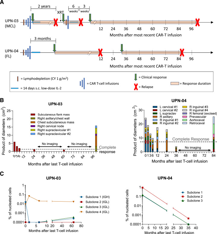
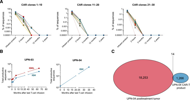
Chimeric antigen receptor (CAR) T-cell therapy produces high response rates in refractory B-cell non-Hodgkin lymphoma, but long-term data are minimal to date. In this study, we present long-term follow-up of a pilot trial testing a CD20-targeting third-generation CAR in patients with relapsed B-cell lymphomas following cyclophosphamide-only lymphodepletion. Two of the three patients in the trial, with mantle cell lymphoma and follicular lymphoma, had remissions lasting more than 7 years, though they ultimately relapsed. The absence of B-cell aplasia in both patients suggested a lack of functional CAR T-cell persistence, leading to the hypothesis that endogenous immune responses were responsible for these long-term remissions. Correlative immunologic analyses supported this hypothesis, with evidence of new humoral and cellular antitumor immune responses proximal to clinical response time points. Collectively, our results suggest that CAR T-cell therapy may facilitate epitope spreading and endogenous immune response formation in lymphomas. Significance: Two of three patients treated with CD20-targeted CAR T-cell therapy had long-term remissions, with evidence of endogenous antitumor immune response formation. Further investigation is warranted to develop conditions that promote epitope spreading in lymphomas.


Conflict of interest statement

I.R. Kirsch reports personal fees and other support from Adaptive Biotechnologies during the conduct of the study, as well as personal fees and other support from Adaptive Biotechnologies outside the submitted work. R. Gottardo reports other support from Ozette Technologies, Takeda, Sanofi, and Arcellx and grants from 10X Genomics and Owkin outside the submitted work, as well as a patent for markers, methods and systems for identifying cell populations, and diagnosing, monitoring, predicting, and treating conditions issued. K.S. Smythe reports other support from Exicure and X4 Pharmaceuticals and personal fees and other support from Sensei Bio outside the submitted work. A. Greenbaum reports grants from ASH during the conduct of the study. D.J. Green reports grants and other support from Juno Therapeutics, personal fees from GlaxoSmithKline and Ensoma Therapeutics, grants and personal fees from Janssen Biotech, and grants from SpringWorks Therapeutics, Sanofi, Seattle Genetics, Cellectar Biosciences, and Celgene outside the submitted work, as well as a patent for 62/582,270 issued to the Fred Hutchinson Cancer Center and a patent for 62/582,308 issued to Juno Therapeutics. D.G. Maloney reports personal fees from Genentech during the conduct of the study; grants and personal fees from Bristol Myers Squibb, Celgene, Juno Therapeutics, and Kite Pharma, grants from Legend Biotech, personal fees from Caribou Biosciences, Janssen, Chimeric Therapeutics, Bristol Myers Squibb, Genentech, Gilead, and Novartis, and personal fees and other support from A2 Biotherapeutics and NAVAN Technologies outside the submitted work; and a patent for Juno Therapeutics and Bristol Myers Squibb licensed and with royalties paid. B.G. Till reports grants from the Damon Runyon Cancer Research Foundation, Fred Hutchinson Cancer Center, David and Patricia Giuliani Family Foundation, and NIH during the conduct of the study; grants and personal fees from Mustang Bio, grants from Bristol Myers Squibb and Juno, and personal fees from Proteios Technology outside the submitted work; and a patent for Mustang Bio issued, licensed, and with royalties paid. No disclosures were reported by the other authors.

Figures

Figure 1.
Figure 1.
Clinical response summary. A, Timeline of UPN-03 (MCL) and UPN-04 (FL) disease course relative to CAR-T infusions. UPN-03 received a fourth infusion 2+ years after his first series of infusions. B, Products of the largest diameters of the indicated tumor sites at various time points after the last CAR T-cell infusion. C, MRD testing was performed on peripheral blood samples at the indicated time points. Deep sequencing of the BCR locus (IGH and IGL for UPN-03 and IGH for UPN-04) was performed, and the frequency of the dominant clones present in the tumor biopsy at each time point is represented as a percentage of nucleated cells. A value of “0.0001” indicates an undetectable sample, as 0 cannot be plotted on a log scale. XRT, radiotherapy.
Figure 2.
Figure 2.
Lack of CAR-T persistence with B-cell recovery over time and minimal CAR T-cell presence in tumor. A, The top 30 most abundant TCR sequences within the CAR T-cell infusion product for patient UPN-04 (representing 94.3% of all the TCR clonotypes within the CAR product), as determined by NGS TCRβ analysis. Clonotype frequency of these sequences over time is shown. The value of “0.00001” represents an undetectable sample. B, B-cell recovery over time after the most recent CAR T-cell infusion for UPN-03 and UPN-04. Patient PBMCs at the time points shown were analyzed for clonal IGH or IGK/IGL analysis using the immunoSEQ NGS assay (Adaptive Biotechnologies). The numbers of total productive sequences identified, representing the BCR repertoire, are shown. C, Overlapping TCRβ sequences between the CAR T-cell infusion product and post–CAR T-cell infusion lymph node biopsy in patient UPN-04 were determined by immunoSEQ. The 14 common sequences shown in the Venn diagram represent 53.8% of the productive TCRβ sequence reads in the CAR-T infusion product and 0.086% of productive TCRβ sequence reads in the tumor biopsy.
Figure 3.
Figure 3.
Development of antitumor immune responses. A, IFNγ ELISpot assays using patient PBMCs from serial time points, incubated with either autologous tumor cells or media, were performed to assess antitumor T-cell responses. Red arrows denote timing of clinical responses. Error bars represent the SEM of triplicate wells. Comparisons were made using an unpaired two-tailed t test. B, Immunoblotting analysis to detect humoral antitumor immune responses was performed using lysates from patient tumor cells, incubated with 1:1,000 diluted patient serum obtained at the indicated time points relative to the last infusion of CD20 CAR T cells, and probed with a secondary antihuman IgG antibody. C, NGS TCRβ analysis to identify differentially expressed TCR sequences compared with the apheresis sample (FDR <0.05) for patient UPN-04. Clonotype frequency of these sequences is represented as log2FC from the apheresis sample. D, Multiparameter flow cytometry was performed on a single-cell suspension of a tumor biopsy obtained 24 hours after the final CAR T-cell infusion for UPN-04, with the identification of intratumoral immune cell subsets. A total of 499,885 viable singlet events were collected. The gating strategy is shown in Supplementary Fig. S3. E, mIHC of UPN-04 posttreatment tumor biopsy using a 6-plex panel containing the antibodies shown at right plus DAPI nuclear stain. Panel 1: ×5 magnification demonstrating FL with the pink/purple nodules surrounded by a mixed population of CD4+ and CD8+ T cells. Panel 2: ×20 magnification of the interfollicular region adjacent to a neoplastic follicle from the yellow box gate in the first panel, showing the complex interactions between T cells, monocytes, and DCs. Panel 3: ×40 magnification of the interfollicular region highlighted in the yellow box in the second panel but restricted to CD8 and CLEC9A markers, showing the interactions between CD8+ T cells and CLEC9A+ DCs. Panel 4: ×40 magnification of the region highlighted in the second panel, restricted to CD4 and CLEC9A markers. Note the relatively increased percentages of CD4+ cells and that both CD4+ and CD8+ T cells are seen in close contact with CLEC9A+ DCs. The figure has been enhanced (brightness/contrast) for easier viewing. log2FC, log2-transformed fold change; Treg, regulatory T cells. MDSC, myeloid-derived suppressor cells; mDC, myeloid dendritic cells; TFH, T-follicular helper cells; TFR, T-follicular regulatory cells.

References

    1. Cappell KM, Kochenderfer JN. Long-term outcomes following CAR T cell therapy: what we know so far. Nat Rev Clin Oncol 2023;20:359–71. - PMC - PubMed
    1. Wang M, Munoz J, Goy A, Locke FL, Jacobson CA, Hill BT, et al. . Three-year follow-up of KTE-X19 in patients with relapsed/refractory mantle cell lymphoma, including high-risk subgroups, in the ZUMA-2 study. J Clin Oncol 2023;41:555–67. - PMC - PubMed
    1. Wang Y, Jain P, Locke FL, Maurer MJ, Frank MJ, Munoz JL, et al. . Brexucabtagene autoleucel for relapsed or refractory mantle cell lymphoma in standard-of-care practice: results from the US lymphoma CAR T Consortium. J Clin Oncol 2023;41:2594–606. - PMC - PubMed
    1. Jacobson CA, Chavez JC, Sehgal AR, William BM, Munoz J, Salles G, et al. . Axicabtagene ciloleucel in relapsed or refractory indolent non-Hodgkin lymphoma (ZUMA-5): a single-arm, multicentre, phase 2 trial. Lancet Oncol 2022;23:91–103. - PubMed
    1. Fowler NH, Dickinson M, Dreyling M, Martinez-Lopez J, Kolstad A, Butler J, et al. . Tisagenlecleucel in adult relapsed or refractory follicular lymphoma: the phase 2 ELARA trial. Nat Med 2022;28:325–32. - PubMed

LinkOut - more resources