. 2024 May 30;128(21):4288-4296.
doi: 10.1021/acs.jpca.4c01866. Epub 2024 May 15.

1H/17O Chemical Shift Waves in Carboxyl-Bridged Hydrogen Bond Networks in Organic Solids

Affiliations

1H/17O Chemical Shift Waves in Carboxyl-Bridged Hydrogen Bond Networks in Organic Solids

Gang Wu et al. J Phys Chem A. .

Abstract

We report solid-state 1H and 17O NMR results for four 17O-labeled organic compounds each containing an extensive carboxyl-bridged hydrogen bond (CBHB) network in the crystal lattice: tetrabutylammonium hydrogen di-[17O2]salicylate (1), [17O4]quinolinic acid (2), [17O4]dinicotinic acid (3), and [17O2]Gly/[17O2]Gly·HCl cocrystal (4). The 1H isotropic chemical shifts found for protons involved in different CBHB networks are between 8.2 and 20.5 ppm, which reflect very different hydrogen-bonding environments. Similarly, the 17O isotropic chemical shifts found for the carboxylate oxygen atoms in CBHB networks, spanning a large range between 166 and 341 ppm, are also remarkably sensitive to the hydrogen-bonding environments. We introduced a simple graphical representation in which 1H and 17O chemical shifts are displayed along the H and O atomic chains that form the CBHB network. In such a depiction, because wavy patterns are often observed, we refer to these wavy patterns as 1H/17O chemical shift waves. Typical patterns of 1H/17O chemical shift waves in CBHB networks are discussed. The reported 1H and 17O NMR parameters for the CBHB network models examined in this study can serve as benchmarks to aid in spectral interpretation for CBHB networks in proteins.


Figures
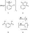
Figure 1.
Figure 1.
Molecular structures of (a) 1, (b) 2, (c) 3, and (d) 4.
Figure 2.
Figure 2.
1H MAS NMR spectra of (a) 1, (b) 2, (c) 3, and (d) 4. All spectra were obtained at 16.4 T with a sample spinning frequency of 30 kHz. In each case, a total of 16 transients were collected with a recycle delay of 60 s.
Figure 3.
Figure 3.
Experimental (black trace) and simulated (red trace) 17O MAS NMR spectra of (a) 1, (b) 2, (c) 3, and (d) 4. For easy comparison, sub-spectra (green, purple, turquoise, and brown traces) from individual sites are also shown. The spectra shown in (a) and (d) were obtained at 18.8 T whereas those in (b) and (c) were acquired at 21.1 T. The sample spinning frequencies were (a) 17.00, (b) 31.25, (c) 22.00, and (d) 16.00 kHz, respectively. Other acquisition parameters are: (a) 4096 transients, 0.5 s recycle delay; (b) 3072 transients, 30 s recycle delay; (c) 3072 transients, 30 s recycle delay; (d) 4096 transients, 1 s recycle delay.
Figure 4.
Figure 4.
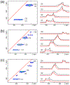
Left panel: Experimental 2D 17O 3QMAS NMR spectra of (a) 2, (b) 3, and (c) 4. Right panel: Corresponding experimental (black trace) and simulated (red trace) slice spectra. The 2D spectra shown in (a) and (b) were obtained at 18.8 T whereas the 2D spectrum in (c) was acquired at 21.1 T. The sample spinning frequency was 16 kHz in all three cases. The signals marked as * are spinning sidebands. The red line shown in each 2D spectrum corresponds to the “chemical shift axis” where the f1/f2 slope is 1.
Figure 5.
Figure 5.
Partial crystal structures of (a) 1, (b) 2, (c) 3, and (d) 4 to illustrate the CBHB networks in these compounds. Color coding for atoms: H (white), C (grey), N (light blue), O (red). Relevant HB lengths are shown.
Figure 6.
Figure 6.
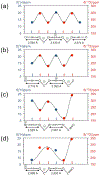
Illustration of the “1H/17O chemical shift waves” observed in (a) 1, (b) 2, (c) 3, and (d) 4. The horizontal axis corresponds to the atomic positions of H (blue) and O (red) atoms along the CBHB network for each compound.
Scheme 1.
Scheme 1.
Several CBHB networks postulated by Huggins for carboxylic acids in the solid state.
Scheme 2.
Scheme 2.
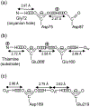
Three examples of CBHB networks found in proteins: (a) 1k7c, (b) 4kxw, (c) 5mop.

Similar articles

References

    1. Huggins ML Hydron bridges in organic compounds. J. Org. Chem 1936, 1, 407–456.
    1. Hendricks SB The orientation of the oxalate group in oxalic acid and some of its salts. Z. Kristallogr 1935, 91, 48–64.
    1. Derissen JL; Smith PH Refinement of the crystal structures of anhydrous [alpha]- and [beta]-oxalic acids. Acta Crystallogr., Sect. B 1974, 30, 2240–2242.
    1. Leiserowitz L Molecular packing modes. Carboxylic acids. Acta Cryst. 1976, B32, 775–802.
    1. D'Ascenzo L; Auffinger P A comprehensive classification and nomenclature of carboxyl-carboxyl(ate) supramolecular motifs and related catemers: implications for biomolecular systems. Acta Crystallogr., Sect. B 2015, 71, 164–175. - PMC - PubMed

LinkOut - more resources