. 2024 Aug 1;200(2):299-311.
doi: 10.1093/toxsci/kfae062.

Effects of ozone exposure on lung injury, inflammation, and oxidative stress in a murine model of nonpneumonic endotoxemia

Affiliations

Effects of ozone exposure on lung injury, inflammation, and oxidative stress in a murine model of nonpneumonic endotoxemia

Jared Radbel et al. Toxicol Sci. .

Abstract

Recent studies have identified exposure to environmental levels of ozone as a risk factor for the development of acute respiratory distress syndrome (ARDS), a severe form of acute lung injury (ALI) that can develop in humans with sepsis. The aim of this study was to develop a murine model of ALI to mechanistically explore the impact of ozone exposure on ARDS development. Mice were exposed to ozone (0.8 ppm, 3 h) or air control followed 24 h later by intravenous administration of 3 mg/kg lipopolysaccharide (LPS) or PBS. Exposure of mice to ozone + LPS caused alveolar hyperplasia; increased BAL levels of albumin, IgM, phospholipids, and proinflammatory mediators including surfactant protein D and soluble receptor for advanced glycation end products were also detected in BAL, along with markers of oxidative and nitrosative stress. Administration of ozone + LPS resulted in an increase in neutrophils and anti-inflammatory macrophages in the lung, with no effects on proinflammatory macrophages. Conversely, the numbers of resident alveolar macrophages decreased after ozone + LPS; however, expression of Nos2, Arg1, Cxcl1, Cxcl2, Ccl2 by these cells increased, indicating that they are activated. These findings demonstrate that ozone sensitizes the lung to respond to endotoxin, resulting in ALI, oxidative stress, and exacerbated pulmonary inflammation, and provide support for the epidemiologic association between ozone exposure and ARDS incidence.

Keywords: acute lung injury; inflammation; oxidative stress; ozone; sepsis.


Figures

Figure 1.
Figure 1.
Effects of ozone and LPS on terminal bronchiolar-alveolar hyperplasia. Lung sections, prepared from mice exposed to air + PBS, air + LPS, ozone + PBS, or ozone + LPS, were stained with H&E. Images were acquired using the Olympus VS120 Virtual Microscopy system and visualized with QuPath software. Fifteen random 30× fields of bronchiolar-alveolar junctions were examined for the presence of bronchiolar-alveolar epithelial hyperplasia. One representative field from 7 mice/treatment group is shown. Arrows indicate areas of alveolar parenchymal thickening. Abbreviations: A, alveolar space; TB terminal bronchiole. Original magnification, 10× (center panels), 40× (peripheral panels).
Figure 2.
Figure 2.
Effects of ozone and LPS on alveolar-capillary barrier function, cells and lung phospholipids. BAL, collected from mice exposed to air + PBS, air + LPS, ozone + PBS, or ozone + LPS, was centrifuged and cell-free supernatants analyzed for IgM, albumin and phospholipid content. Top left panel: IgM was measured by ELISA (n = 8–11 mice/treatment group). Top right panel: albumin was measured by ELISA (n = 13–15 mice/treatment group). Bottom left panel: Total phospholipids were quantified in large aggregate fractions of cell-free BAL as described in Materials and Methods (n = 5–12 mice/treatment group). Bottom right panel: Cells were enumerated by trypan blue dye exclusion using a hemocytometer (n = 11–12 mice/treatment group). Bars, mean ± SD; *Significant difference (p .05) between groups as indicated.
Figure 3.
Figure 3.
Effects of ozone and LPS on inflammatory proteins in BAL. Equal volumes of cell-free BAL, prepared from mice exposed to air + PBS, air + LPS, ozone + PBS, or ozone + LPS, were analyzed by western blotting for sRAGE (Left panel) and SP-D (Right panels). Bands were visualized using an ECL Prime detection system (GE Health Care). Blots are representative of 4 experiments; each lane is one animal. Band densities were quantified by ImageJ, normalized to air + PBS controls, and presented as fold change relative to control. Bars (sRAGE), mean ± SD (n = 10–12 mice/treatment group). Bars (SP-D), mean ± SD (n = 13–15 mice/treatment group). *Significant difference (p .05) between groups as indicated. Complete Western blots are shown in Supplementary Figure 3.
Figure 4.
Figure 4.
Effects of ozone and LPS on lung oxidative stress. Tissue was collected from mice exposed to air + PBS, air + LPS, ozone + PBS, or ozone + LPS, and immediately frozen at −80°C. Left panel: Lung homogenates were separated into microsomal and cytosolic (S9) fractions and analyzed for hydrogen peroxide (H2O2) using an Amplex Red assay. Right panel: Lung homogenates were analyzed for oxidized (GSSG) and reduced (GSH) glutathione by ELISA. Bars, mean ± SD (n = 5–6 mice/treatment group). *Significant difference (p .05) between groups as indicated.
Figure 5.
Figure 5.
Effects of ozone and LPS on HO-1 expression. Lung sections, prepared from mice exposed to air + PBS, air + LPS, ozone + PBS, or ozone + LPS, were immunostained with antibody to HO-1 (StressGen Biotechnologies; 1:250) or rabbit anti-mouse IgG antibody control. Binding was visualized using a 3,3′-Diaminobenzidine (DAB) peroxidase substrate kit (Vector Laboratories). One representative section from 3 to 4 mice/treatment group is shown (original magnification, 40×). Positively staining AMs were enumerated from 20 random fields (20× magnification/lung). Inset: mean ± SD (n = 3–4). aSignificantly different (p .05) from air + PBS. Lower magnifications of lung histology are shown in Supplementary Figure 4.
Figure 6.
Figure 6.
Effects of ozone and LPS on markers of nitrosative stress. BAL was collected from mice exposed to air + PBS, air + LPS, ozone + PBS, or ozone + LPS, centrifuged and cell-free supernatants analyzed for NOx levels by vanadium chloride reduction (upper left panel). Bars, mean ± SD, n = 7–12 mice/treatment group. Equal volumes of BAL were acetone precipitated to enrich for protein and lipid (organic fraction) and analyzed for organic NOx levels (lower left panel). Bars, mean ± SD, n = 4–5 mice/treatment group. A biotin switch assay followed by Western blotting was used to detect SNO-SP-D in BAL (right panels). Blots are representative of 2 experiments; each lane is 1 mouse. Band densities were quantified by ImageJ, normalized to air + PBS controls and presented as fold change. Bars, mean + SD (n = 5–6 mice/treatment group). *Significant difference (p .05) between groups as indicated. Complete Western blots are shown in Supplementary Figure 3.
Figure 7.
Figure 7.
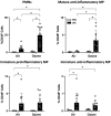
Effects of ozone and LPS on lung inflammatory cells. Cells collected from mice exposed to air + PBS, air + LPS, ozone + PBS, or ozone + LPS by BAL + lung massage were immunostained with antibodies to CD45, CD11b, Siglec F, CD11c, Ly6G, F4/80, and Ly6C and analyzed by flow cytometry. PMNs were defined as CD45+CD11b+Siglec F-Ly6G+Cd11c, immature proinflammatory macrophages as CD11b+ Siglec FLy6GCd11cF4/80+Ly6Chi, mature anti-inflammatory macrophages as CD11b+ Siglec FLy6GCd11c+F4/80+Ly6Clo, immature anti-inflammatory macrophages as CD11b+ Siglec FLy6GCd11c-F4/80+Ly6Clo. Mature proinflammatory macrophages (CD11b+ Siglec FLy6GCd11c+F4/80+Ly6Chi cells) were not detected. Data are representative of at least 3 experiments and presented as % CD45+ cells (leukocytes). Bars, mean ± SD (n = 11–14 mice/treatment group). *Significant difference (p .05) between groups as indicated. MP, macrophage.
Figure 8.
Figure 8.
Effects of ozone and LPS on neutrophil infiltration into the lungs. Sections, prepared from mice exposed to air + PBS, air + LPS, ozone + PBS, or ozone + LPS, were stained with H&E. Images were acquired using the VS120 Virtual Microscopy system and visualized with QuPath software. Neutrophils were enumerated in ten random 40× fields from 5 lobes (2 fields/lobe) in the interstitial (I) and alveolar (A) space. One representative field from 7 mice/treatment group is shown. Arrows indicate alveolar neutrophils; arrowheads indicate interstitial neutrophils. Original magnification, 40×. aSignificantly different (p .05) from air + PBS. bSignificantly different from air + LPS. cSignificantly different from ozone + PBS.
Figure 9.
Figure 9.
Effects of ozone and LPS on resident alveolar macrophages (AMs). Lung cells from mice exposed to air + PBS, air + LPS, ozone + PBS, or ozone + LPS were immunostained with antibodies to CD45, CD11b, Siglec F, CD11c, Ly6G, F4/80, and Ly6C and analyzed by flow cytometry. Upper left panel: Resident AMs were identified as CD45+CD11bSiglec F+. Data are presented as % CD45+ cells (n = 11–14 mice/treatment group). Separate groups of lung cells were immunostained with metallic bead-conjugated antibodies to CD11b; CD11b AMs were collected by magnetic separation and analyzed by qPCR using the 2–ΔΔCt method with Actb as a control housekeeping gene. Arg1 (n = 5–6 mice/group), Cxcl1 (n = 5–6 mice/group), Cxcl2 (n = 5–6 mice/group), and Ccl2 (n = 3 mice/group); data are presented as fold change relative to air + PBS controls; Nos2 (n = 6–7 mice/group); data are presented as fold change relative to air+ LPS-treated mice, as no expression was detected in the air + PBS and ozone + PBS controls. Ct values above 36 were considered undetectable. Bars, mean ± SE. *Significant difference (p .05) between groups as indicated. ND, not detected.
Figure 10.
Figure 10.
Nos2 and Arg1 expression in resident alveolar macrophages (AMs). Histological sections were prepared from mice exposed to ozone + LPS. Tissue was hybridized using probes to Arg1 or Nos2 mRNA and then immunostained with antibody to Siglec F (Abcam). Nos2 and Arg1 mRNA were visualized using the RNAScope® assay (Bio-Techne). Siglec F binding was visualized using a DAB peroxidase substrate kit. One representative section from 2 mice is shown. Original magnification, 40×; brown staining, Siglec F; red dots, Arg1 (upper panel) or Nos2 (lower panel). Insets: Enlarged Siglec F and mRNA-positive macrophages (100×). Lower magnifications are shown in Supplementary Figure 5.
Figure 11.
Figure 11.
Two-hit model of ozone + LPS-induced ARDS. Ozone inhalation results in damage to the alveolar epithelium (bottom left panel) while endotoxemia activates resident AMs which release chemokines, such as Cxcl2, attracting circulating neutrophils (PMNs) into the airspace (top right panel). ROS and RNS generated following ozone + LPS exposure further damage epithelial cells, disrupt the alveolar-capillary barrier, and activate resident AMs. Activated AMs release chemokines and inflammatory mediators exacerbating PMN recruitment and ALI. Created with BioRender.com.

References

    1. Atochina E. N., Beers M. F., Hawgood S., Poulain F., Davis C., Fusaro T., Gow A. J. (2004). Surfactant protein-D, a mediator of innate lung immunity, alters the products of nitric oxide metabolism. Am. J. Respir. Cell Mol. Biol. 30, 271–279. - PubMed
    1. Atochina-Vasserman E. N., Beers M. F., Kadire H., Tomer Y., Inch A., Scott P., Guo C. J., Gow A. J. (2007). Selective inhibition of inducible NO synthase activity in vivo reverses inflammatory abnormalities in surfactant protein D-deficient mice. J. Immunol. 179, 8090–8097. - PMC - PubMed
    1. Atochina-Vasserman E. N., Winkler C., Abramova H., Schaumann F., Krug N., Gow A. J., Beers M. F., Hohlfeld J. M. (2011). Segmental allergen challenge alters multimeric structure and function of surfactant protein D in humans. Am. J. Respir. Crit. Care Med. 183, 856–864. - PMC - PubMed
    1. Bankhead P., Loughrey M. B., Fernández J. A., Dombrowski Y., McArt D. G., Dunne P. D., McQuaid S., Gray R. T., Murray L. J., Coleman H. G., et al. (2017). QuPath: Open source software for digital pathology image analysis. Sci. Rep. 7, 16878. - PMC - PubMed
    1. Bellani G., Laffey J. G., Pham T., Fan E., Brochard L., Esteban A., Gattinoni L., van Haren F., Larsson A., McAuley D. F., et al.; ESICM Trials Group. (2016). Epidemiology, patterns of care, and mortality for patients with acute respiratory distress syndrome in intensive care units in 50 countries. JAMA 315, 788–800. - PubMed

MeSH terms