

Structural neuroimaging changes associated with subjective cognitive decline from a clinical sample
- PMID: 38749146
- PMCID: PMC11109886
- DOI: 10.1016/j.nicl.2024.103615
Structural neuroimaging changes associated with subjective cognitive decline from a clinical sample
Abstract
Background: Alzheimer's disease (AD) is characterized by progressive deterioration of cognitive functions. Some individuals with subjective cognitive decline (SCD) are in the early phase of the disease and subsequently progress through the AD continuum. Although neuroimaging biomarkers could be used for the accurate and early diagnosis of preclinical AD, the findings in SCD samples have been heterogeneous. This study established the morphological differences in brain magnetic resonance imaging (MRI) findings between individuals with SCD and those without cognitive impairment based on a clinical sample of patients defined according to SCD-Initiative recommendations. Moreover, we investigated baseline structural changes in the brains of participants who remained stable or progressed to mild cognitive impairment or dementia.
Methods: This study included 309 participants with SCD and 43 healthy controls (HCs) with high-quality brain MRI at baseline. Among the 99 subjects in the SCD group who were followed clinically, 32 progressed (SCDp) and 67 remained stable (SCDnp). A voxel-wise statistical comparison of gray and white matter (WM) volume was performed between the HC and SCD groups and between the HC, SCDp, and SCDnp groups. XTRACT ATLAS was used to define the anatomical location of WM tract damage. Region-of-interest (ROI) analyses were performed to determine brain volumetric differences. White matter lesion (WML) burden was established in each group.
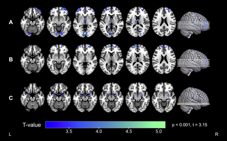
Results: Voxel-based morphometry (VBM) analysis revealed that the SCD group exhibited gray matter atrophy in the middle frontal gyri, superior orbital gyri, superior frontal gyri, right rectal gyrus, whole occipital lobule, and both thalami and precunei. Meanwhile, ROI analysis revealed decreased volume in the left rectal gyrus, bilateral medial orbital gyri, middle frontal gyri, superior frontal gyri, calcarine fissure, and left thalamus. The SCDp group exhibited greater hippocampal atrophy (p < 0.001) than the SCDnp and HC groups on ROI analyses. On VBM analysis, however, the SCDp group exhibited increased hippocampal atrophy only when compared to the SCDnp group (p < 0.001). The SCD group demonstrated lower WM volume in the uncinate fasciculus, cingulum, inferior fronto-occipital fasciculus, anterior thalamic radiation, and callosum forceps than the HC group. However, no significant differences in WML number (p = 0.345) or volume (p = 0.156) were observed between the SCD and HC groups.
Conclusions: The SCD group showed brain atrophy mainly in the frontal and occipital lobes. However, only the SCDp group demonstrated atrophy in the medial temporal lobe at baseline. Structural damage in the brain regions was anatomically connected, which may contribute to early memory decline.
Keywords: Alzheimer’s disease; Magnetic resonance imaging; Region-of-interest; Subjective cognitive decline; Voxel-based morphometry; White matter lesions.
Copyright © 2024 The Author(s). Published by Elsevier Inc. All rights reserved.
Conflict of interest statement
Declaration of competing interest The authors declare that they have no known competing financial interests or personal relationships that could have appeared to influence the work reported in this paper.
Figures


References
-
- Albert M.S., DeKosky S.T., Dickson D., et al. The diagnosis of mild cognitive impairment due to Alzheimer’s disease: recommendations from the National Institute on Aging-Alzheimer’s Association workgroups on diagnostic guidelines for Alzheimer’s disease. Alzheimers Dement. 2011;7:270–279. doi: 10.1016/j.jalz.2011.03.008. - DOI - PMC - PubMed
MeSH terms
LinkOut - more resources
Full Text Sources
Medical
