. 2024 May 15;15(1):4128.
doi: 10.1038/s41467-024-48530-8.

Post-translational modification-dependent oligomerization switch in regulation of global transcription and DNA damage repair during genotoxic stress

Affiliations

Post-translational modification-dependent oligomerization switch in regulation of global transcription and DNA damage repair during genotoxic stress

Prathama Talukdar et al. Nat Commun. .

Abstract

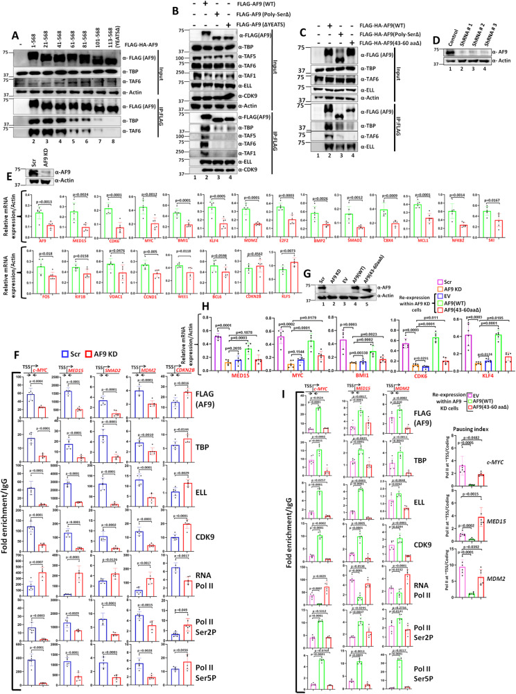
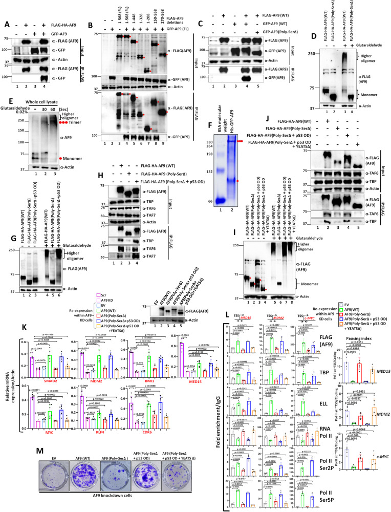
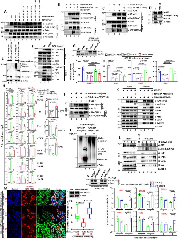
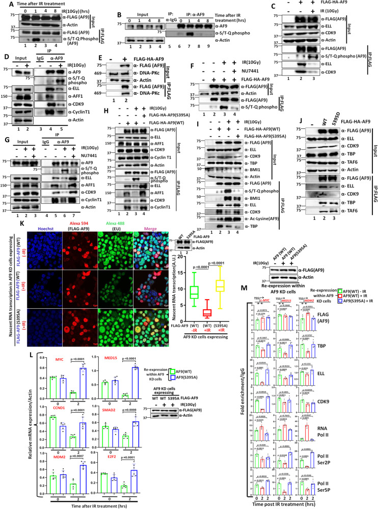
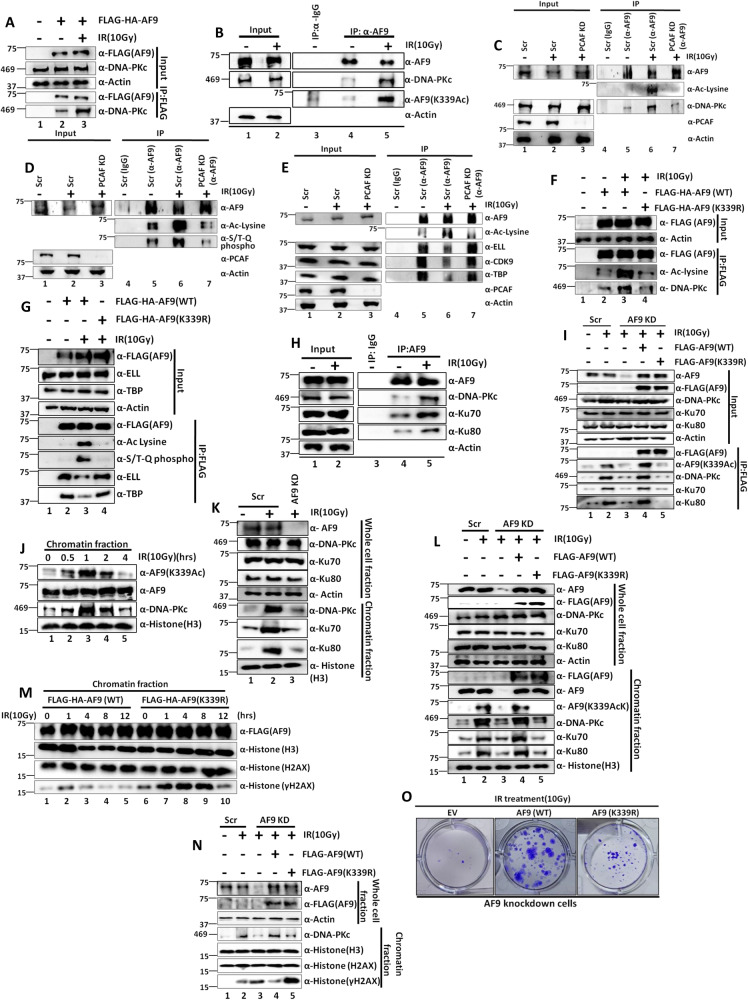
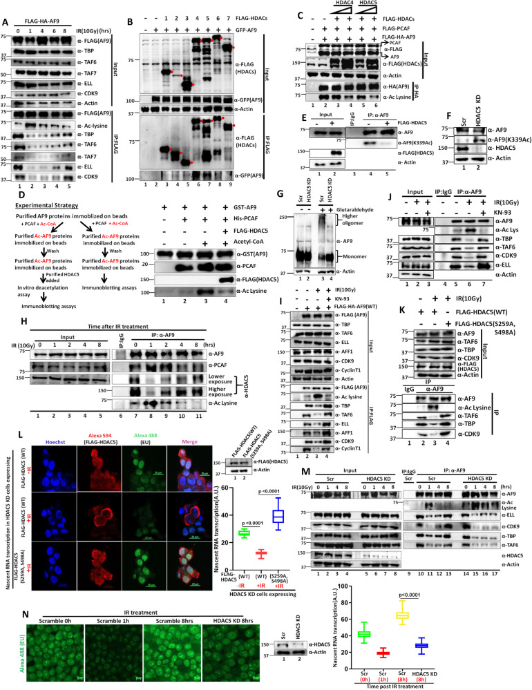
Mechanisms of functional cross-talk between global transcriptional repression and efficient DNA damage repair during genotoxic stress are poorly known. In this study, using human AF9 as representative of Super Elongation Complex (SEC) components, we delineate detailed mechanisms of these processes. Mechanistically, we describe that Poly-Serine domain-mediated oligomerization is pre-requisite for AF9 YEATS domain-mediated TFIID interaction-dependent SEC recruitment at the promoter-proximal region for release of paused RNA polymerase II. Interestingly, during genotoxic stress, CaMKII-mediated phosphorylation-dependent nuclear export of AF9-specific deacetylase HDAC5 enhances concomitant PCAF-mediated acetylation of K339 residue. This causes monomerization of AF9 and reduces TFIID interaction for transcriptional downregulation. Furthermore, the K339 acetylation-dependent enhanced AF9-DNA-PKc interaction leads to phosphorylation at S395 residue which reduces AF9-SEC interaction resulting in transcriptional downregulation and efficient repair of DNA damage. After repair, nuclear re-entry of HDAC5 reduces AF9 acetylation and restores its TFIID and SEC interaction to restart transcription.


Conflict of interest statement

The authors declare no competing interests.

Figures

Fig. 1
Fig. 1. Besides Poly-Ser, AF9 also requires YEATS domain for TFIID interaction and transcriptional activation within 293 T cells.
A Immunoblotting analysis showing interaction of ectopically expressed N-terminal-deleted AF9 fragments with TFIID components as indicated (n = 3 replicates). B Immunoblotting analysis showing the effect of AF9 YEATS and Poly-Ser domain deletion on its interaction with TFIID and SEC components as indicated (n = 3 replicates). C Immunoblotting analysis showing the requirement of N-terminal minimal region (43–60 aa) of AF9 for its exclusive interaction with TFIID (n = 2 replicates). D Immunoblotting analysis showing stable knockdown of endogenous AF9 protein by multiple shRNAs using actin as a loading control (n = 3 replicates). E qRT-PCR analysis showing effect of AF9 stable knockdown (KD) on mRNA expression of indicated AF9-target genes. The inset immunoblot panel shows a knockdown of AF9 in the cells that were used for the RNA analysis (n = 2 replicates). F ChIP analysis showing the effect of AF9 stable knockdown on recruitment of indicated target factors at the promoter-proximal region of selected AF9-target genes (from (E)) as mentioned (n = 2 replicates). G Immunoblotting analysis showing re-expression of AF9(WT) and AF9(43–60 aaΔ) proteins in AF9 KD cells through their ectopic expression (n = 2 replicates). H qRT-PCR analysis showing effect of re-expression of AF9(WT) and AF9(43–60 aaΔ) proteins in stable AF9 KD cells on mRNA expression of indicated AF9-target genes (n = 2 replicates). I ChIP analysis showing effect of re-expression of AF9(WT) and AF9(43–60 aaΔ) proteins in stable AF9 KD cells on recruitment of indicated target factors at the promoter-proximal region of selected AF9-target genes (left panel). Parallel use of empty vector (EV) was used as a control in our experimental setup. Statistical significance was calculated as Scr vs AF9 KD, and AF9 KD vs EV, AF9(WT), AF9(43–60 aaΔ). Pausing index analysis of Pol II on the selected AF9-target genes as indicated (right panel) (n = 2 replicates). In this figure, for data showing RNA and ChIP analyses by qRT-PCR, the error bar represents mean ± SD and statistical analyses were performed using one-tailed Student’s t test. p values for each experimental data is indicated on the bar diagram.
Fig. 2
Fig. 2. Poly-Ser domain-mediated oligomerization of AF9 protein is essential for its interaction with TFIID for transcriptional activation of target genes within 293 T cells.
AC Immunoblotting analysis showing self-association between ectopically expressed FLAG-HA-AF9 and GFP-AF9 (A), n = 4 replicates), different AF9 domains (B), n = 2 replicates), and between ectopically expressed FLAG-HA-AF9(WT) and GFP-AF9 (WT and Poly-SerΔ) (C), n = 3 replicates) within mammalian cells. D Immunoblotting assay showing oligomerization of ectopically expressed FLAG-HA-AF9(WT) and FLAG-HA-AF9(Poly-SerΔ) proteins (n = 2 replicates). E Immunoblotting analysis showing oligomerization of endogenous AF9 protein with indicated period of incubation time (n = 3 replicates). F Native PAGE Coomassie staining showing formation of trimer and monomer by purified recombinant His-GFP-AF9 protein. Filled red single dot indicates monomer, and triple dot indicates trimer formation (n = 3 replicates). G Immunoblotting assay showing restoration of oligomerization capacity of AF9(Poly-SerΔ) protein upon introduction of oligomerization domain (OD) of p53 protein (n = 2 replicates). H Immunoblotting analysis showing restoration of interaction of TFIID components with AF9(Poly-SerΔ) protein upon addition of p53 OD within this fragment ((AF9(Poly-SerΔ + p53 OD)) (n = 2 replicates). I Immunoblotting assay using cell lysates containing indicated AF9 proteins showing YEATS domain-independent oligomerization of AF9 (n = 2 replicates). J Immunoblotting analysis showing absolute requirement of YEATS domain for AF9 interaction with TFIID components (n = 1 replicate). K qRT-PCR analysis showing effect of re-expression of AF9(WT), AF9(Poly-SerΔ), AF9(Poly-SerΔ + p53 OD) and AF9(Poly-SerΔ + p53 OD + YEATSΔ) proteins in stable AF9 KD cells on mRNA expression of indicated AF9-target genes (n = 2 replicates). L ChIP analysis showing effect of re-expression of indicated AF9 proteins in stable AF9 KD cells on recruitment of indicated target factors at the promoter-proximal region of indicated AF9-target genes (n = 2 replicates). M Colony formation assay showing restoration of colony formation ability of stable AF9 KD cells that re-express indicated AF9 proteins (n = 2 replicates). For this figure, the data showing RNA and ChIP analyses by qRT-PCR, the error bar represents mean ± SD and statistical analyses were performed using one-tailed Student’s t test. p values for each experimental data is mentioned on the bar diagram.
Fig. 3
Fig. 3. PCAF-mediated acetylation of AF9 reduces its oligomerization potential and TFIID interaction during exposure to genotoxic stress within 293 T cells.
A Reduced oligomerization of endogenous AF9 within mammalian cells upon IR(10 Gy) treatment(n = 2 replicates). B Immunoblotting analysis showing effect of IR(10 Gy) treatment on interaction of TFIID and SEC components with endogenous AF9 protein (n = 3 replicates). C Nascent RNA transcription analysis showing dynamic regulation of global transcription within mammalian cells upon IR(10 Gy) treatment. Error bar represents mean ± SD (n = 100 cells). This experiment was performed once. D Effect of IR-induced genotoxic stress on dynamic acetylation of ectopically expressed AF9 within mammalian cells (n = 4 replicates). E Immunoblotting analysis showing effect of IR(10 Gy) treatment on acetylation of endogenous AF9 and its concomitant reduced interaction with TFIID and SEC components (n = 3 replicates). F Analysis of specificity of AF9 acetylation by various acetyl transferases as indicated (n = 1 replicate). G SDS Coomassie staining showing purification of recombinant HAT domain of PCAF (n = 2 replicates). H In vitro acetyl transferase assay showing efficient acetylation of purified GST-AF9 by PCAF HAT domain (n = 1 replicate). I Immunoblotting analysis showing effect of stable PCAF KD (using 2 different shRNAs) on acetylation of endogenous AF9 within mammalian cells (n = 2 replicates). J Immunoblotting analysis showing reduced oligomerization capacity of endogenous AF9 protein in presence of over-expressed PCAF (n = 1 replicate). K Effect of PCAF KD on oligomerization capacity of endogenous AF9 protein within mammalian cells (n = 1 replicate). L Effect of PCAF-mediated acetylation of ectopically expressed AF9 on its interaction with endogenous TFIID and SEC components as indicated; (n = 3 replicates). M, N Effect of stable knockdown of PCAF on acetylation of ectopically expressed AF9 ((M); n = 2 replicates)and endogenous AF9 ((N) n = 2 replicates) and its interaction with TFIID components. O Immunoblotting analysis showing direct effect of PCAF-mediated acetylation of AF9 on its interaction with purified TFIID in vitro, following the experimental strategy shown in the left panel (n = 1 replicate). P Effect of PCAF knockdown on IR treatment-mediated acetylation of ectopically expressed AF9 and its concomitant interaction with TFIID components (n = 2 replicates).
Fig. 4
Fig. 4. PCAF-mediated acetylation of AF9 at lysine 339 (K339) residue reduces its oligomerization potential and TFIID interaction for downregulation of global transcription during genotoxic stress within 293 T cells.
A Acetylation of ectopically expressed AF9 and its mutant derivatives by PCAF within mammalian cells (n = 2 replicates). B Acetylation of AF9(WT) and AF9(K339R) by PCAF within mammalian cells (n = 3 replicates). C Immunoblotting analysis showing PCAF-mediated acetylation of AF9 at K339 residue using AF9(K339Ac)-specific antibody (n = 1 replicate). D Effect of PCAF KD on acetylation of endogenous AF9 at K339 residue (n = 2 replicates). E, F Oligomerization of AF9(WT) and AF9 (K339Q) ((E)n = 1 replicate) and its effect on interaction with TFIID and SEC components ((F) n = 1 replicate). G, H qRT-PCR ((G) n = 2 replicates) and ChIP analyses ((H) n = 2 replicates) showing effect of re-expression of AF9(WT) and AF9(K339Q) proteins in stable AF9 KD cells on target mRNA expression (G) and recruitment of indicated target factors at the promoter-proximal region of selected AF9-target genes (H). I Immunoblotting analysis showing IR-induced acetylation of AF9(WT) and AF9(K339R) at K339 residue, using AF9(K339Ac) antibody (n = 1 replicate). J Reduced oligomerization of AF9(WT) as compared to AF9 (K339R) upon IR(10 Gy) treatment (n = 2 replicates). K Interaction of AF9 (K339R) mutant with TFIID and SEC components upon IR(10 Gy) treatment (n = 3 replicates). L Effect of IR treatment on dynamic acetylation of endogenous AF9 at K339 residue and its concomitant effect on interaction with TFIID and SEC components (n = 2 replicates). M Nascent RNA transcription analysis showing enhanced global transcription ability in stable AF9 KD cells expressing AF9(K339R) when compared to AF9(WT) after 1 hr of IR treatment. The boxes represent median and quartiles and value ranges of 25–75 and 10–90%. Upper and lower hinges extend to the largest and smallest data points (n = 100 cells). This experiment was done once. N Effect of re-expression of indicated proteins in stable AF9 KD cells on mRNA expression of indicated AF9-target genes at 2hrs after IR treatment (n = 2 replicates). For this figure, the data showing RNA and ChIP analyses by qRT-PCR, the error bar represents mean ± SD, and statistical analyses were performed using one-tailed Student’s t test. p values for each experimental data is mentioned on the bar diagram.
Fig. 5
Fig. 5. DNA-PKc-mediated phosphorylation of AF9 at Serine 395 (S395) residue reduces its interaction with SEC components for transcriptional downregulation during genotoxic stress within 293 T cells.
A, B IR-induced dynamic phosphorylation of ectopically expressed AF9 ((A) n = 3 replicates) and endogenous AF9 ((B) n = 1 replicate). C, D Enhanced phosphorylation of ectopically expressed AF9 ((C), n = 3 replicates) and endogenous AF9 ((D) n = 2 replicates), and its concomitant reduced interaction with SEC components upon IR(10 Gy) treatment. E Interaction of ectopically expressed AF9 protein with endogenous DNA-PKc within mammalian cells (n = 3 replicates). F, G Effect of treatment of cells with NU7441(2 μM) on IR(10 Gy) treatment-dependent phosphorylation of ectopically expressed AF9 ((F) n = 2 replicates) and endogenous AF9 ((G) n = 3 replicates) and its concomitant interaction with SEC components G. H Immunoblotting analysis showing defective phosphorylation and unaltered SEC interaction in cells expressing phosphorylation-defective AF9 mutant (S395A) when compared to AF9(WT), upon IR(10 Gy) treatment (n = 3 replicates). I Effect of AF9(S395A) mutant on IR(10 Gy)-dependent phosphorylation and concomitant acetylation and its effect on interaction with SEC and TFIID components (n = 1 replicate). J Effect of AF9 phosphorylation mimic mutant (S395D) on its interaction with TFIID and SEC components (n = 1 replicate). K Nascent RNA transcription analysis showing enhanced global transcriptional activity in cells expressing AF9(S395A) mutant, when compared to AF9(WT) upon IR(10 Gy) treatment. The boxes represent median and quartiles and value ranges of 25–75 and 10–90%. Upper and lower hinges extend to the largest and smallest datapoints (n = 100 cells). This experiment was done once. L qRT-PCR analysis showing effect of re-expression of AF9(WT) and AF9(S395A) proteins in stable AF9 KD cells on mRNA expression of indicated AF9-target genes at 2hrs after IR(10 Gy) treatment (n = 2 replicates). M ChIP analysis showing recruitment of indicated factors at the promoter-proximal regions of AF9-target genes upon re-expression of AF9(S395A) in stable AF9 KD cells when compared to AF9(WT) at 2hrs after IR(10 Gy) treatment (n = 2 replicates). For this figure, the data showing RNA and ChIP analyses by qRT-PCR, the error bar represents mean ± SD and statistical analyses were performed using one-tailed Student’s t test. p values for each experimental data is mentioned on the bar diagram.
Fig. 6
Fig. 6. AF9 K339 acetylation-dependent DNA-PKc recruitment onto chromatin facilitates phosphorylation at S395 residue as well as recruits Ku complex for DNA damage repair within 293 T cells.
A, B Immunoblotting analysis showing increased interaction of ectopically expressed AF9 ((A), n = 2 replicates) and endogenous AF9 ((B) n = 2 replicates) with DNA-PKc and its concomitant acetylation at K339 residue within mammalian cells upon exposure to IR(10 Gy) B C, D Effect of PCAF KD on IR(10 Gy) treatment-dependent acetylation of endogenous AF9 and concomitant DNA-PKc interaction ((C) n = 1 replicate) and phosphorylation at S395 residue ((D) n = 1 replicate). E PCAF KD fails to show reduced interaction of TFIID and SEC components with endogenous AF9 within mammalian cells upon IR(10 Gy) treatment (n = 1 replicate). F Effect of AF9(K339R) on IR treatment-dependent enhanced interaction with DNA-PKc within mammalian cells (n = 2 replicates). G Effect of AF9(K339R) mutant on IR treatment-dependent acetylation and concomitant phosphorylation-dependent association with TFIID and SEC components (n = 2 replicates). H Enhanced interaction of endogenous AF9 with DNA-PKc and concomitant Ku complex components within mammalian cells upon IR(10 Gy) treatment (n = 2 replicates). I Effect of acetylation of AF9 at K339 residue on IR(10 Gy) treatment-dependent enhanced interaction with DNA-PKc and Ku complex components within mammalian cells (n = 2 replicates). J Chromatin association of acetylated AF9(K339Ac) and concomitant DNA-PKc at different time points after IR treatment (n = 2 replicates). K Effect of stable AF9 KD on association of DNA-PKc and Ku complex components onto chromatin after IR(10 Gy)treatment (n = 1 replicate). L Effect of re-expression of AF9(WT) and AF9(K339R) in stable AF9 knockdown cells on association of DNA-PKc and Ku complex components onto chromatin upon IR(10 Gy) treatment (n = 2 replicates). M Effect of overexpression of AF9(WT) and AF9(K339R) mutant on repair of damaged DNA at indicated time points after IR(10 Gy) treatment (n = 2 replicates). N, O Effect of re-expression of AF9(WT) and AF9(K339R) in stable AF9 knockdown cells on overall DNA damage (N) n = 1 replicate) and colony forming potential after IR(10 Gy) treatment (O) (n = 3 replicates).
Fig. 7
Fig. 7. CaMKII-mediated phosphorylation, regulating nuclear-cytoplasmic shuttling of HDAC5, is important for genotoxic stress-dependent modulation of AF9 acetylation for transcriptional regulation within 293 T cells.
A Dynamic regulation of ectopically expressed AF9 acetylation and its concomitant association with indicated TFIID and SEC components after IR(10 Gy) treatment (n = 3 replicates). B Immunoblotting analysis showing interaction of ectopically expressed GFP-AF9 with indicated FLAG-HDACs within mammalian cells (n = 2 replicates). C Immunoblotting analysis showing deacetylation of ectopically expressed AF9 by concomitant expression of HDAC4 and HDAC5 (n = 2 replicates). D Immunoblotting analysis showing deacetylation of AF9 by purified HDAC5 in vitro (n = 1 replicate). E Immunoblotting analysis showing deacetylation of endogenous AF9 at K339 residue by ectopically expressed HDAC5 within mammalian cells (n = 1 replicate). F Immunoblotting analysis showing enhanced acetylation of endogenous AF9 at K339 residue upon HDAC5 knockdown (n = 2 replicates). G Immunoblotting analysis showing reduced AF9 oligomerization potential upon HDAC5 knockdown within mammalian cells (n = 2 replicates). H Immunoblotting analysis showing dynamic interaction of PCAF and HDAC5 with endogenous AF9 upon IR treatment (n = 1 replicate). I, J Immunoblotting analysis showing effect of prior treatment with CaMKII inhibitor (KN-93) on IR-induced acetylation of both ectopic ((I) n = 3 replicates) and endogenous AF9 ((J) n = 2 replicates) and concomitant interaction with TFIID and SEC components within mammalian cells. K. Immunoblotting analysis showing effect of ectopic expression of CaMKII-mediated phosphorylation-defective HDAC5(S259A, S498A) mutant on IR treatment-mediated interaction of TFIID and SEC components with endogenous AF9 (n = 2 replicates). L Nascent RNA transcription analysis showing enhanced global transcriptional ability in cells expressing HDAC5(S259A, S498A) mutant when compared to HDAC5(WT) after IR(10 Gy) treatment. The boxes represent median and quartiles and value ranges of 25–75 and 10–90%. Upper and lower hinges extend to the largest and smallest datapoints (n = 30 cells). This experiment was done once. M Effect of HDAC5 KD on IR treatment-dependent dynamic interaction of endogenous AF9 with TFIID and SEC components at indicated time periods (n = 1 replicate). N Nascent RNA transcription analysis (through EU incorporation) showing effect of HDAC5 KD on global transcription restart at 8hrs after IR(10 Gy) treatment. The boxes represent median and quartiles and value ranges of 25–75 and 10–90% (n = 100 cells). This experiment was done once.

Similar articles

Cited by

References

    1. Svejstrup JQ. The interface between transcription and mechanisms maintaining genome integrity. Trends Biochem. Sci. 2010;35:333–338. doi: 10.1016/j.tibs.2010.02.001. - DOI - PubMed
    1. Lagerwerf S, Vrouwe MG, Overmeer RM, Fousteri MI, Mullenders LH. DNA damage response and transcription. DNA Repair (Amst.) 2011;10:743–750. doi: 10.1016/j.dnarep.2011.04.024. - DOI - PubMed
    1. Wilson MD, Harreman M, Svejstrup JQ. Ubiquitylation and degradation of elongating RNA polymerase II: the last resort. Biochim. Biophys. Acta. 2013;1829:151–157. doi: 10.1016/j.bbagrm.2012.08.002. - DOI - PubMed
    1. Wilson MD, et al. Proteasome-mediated processing of Def1, a critical step in the cellular response to transcription stress. Cell. 2013;154:983–995. doi: 10.1016/j.cell.2013.07.028. - DOI - PMC - PubMed
    1. Lavigne MD, Konstantopoulos D, Ntakou-Zamplara KZ, Liakos A, Fousteri M. Global unleashing of transcription elongation waves in response to genotoxic stress restricts somatic mutation rate. Nat. Commun. 2017;8:2076. doi: 10.1038/s41467-017-02145-4. - DOI - PMC - PubMed

MeSH terms