. 2024 May 15;14(1):11171.
doi: 10.1038/s41598-024-61414-7.

Persistence of viral RNA in North American elk experimentally infected with an ancestral strain of severe acute respiratory syndrome coronavirus 2 (SARS-CoV-2)

Affiliations

Persistence of viral RNA in North American elk experimentally infected with an ancestral strain of severe acute respiratory syndrome coronavirus 2 (SARS-CoV-2)

Paola M Boggiatto et al. Sci Rep. .

Abstract

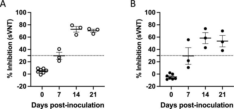
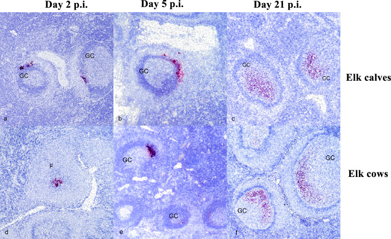
White-tailed deer (Odocoileus virginianus) have emerged as a reservoir host for SARS-CoV-2 given their susceptibility to infection and demonstrated high rates of seroprevalence and infection across the United States. As SARS-CoV-2 circulates within free-ranging white-tailed deer populations, there is the risk of transmission to other wildlife species and even back to the human population. The goal of this study was to determine the susceptibility, shedding, and immune response of North American elk (Cervus elaphus canadensis) to experimental infection with SARS-CoV-2, to determine if another wide-ranging cervid species could potentially serve as a reservoir host for the virus. Here we demonstrate that while North American elk do not develop clinical signs of disease, they do develop a neutralizing antibody response to infection, suggesting the virus is capable of replicating in this mammalian host. Additionally, we demonstrate SARS-CoV-2 RNA presence in the medial retropharyngeal lymph nodes of infected elk three weeks after experimental infection. Consistent with previous observations in humans, these data may highlight a mechanism of viral persistence for SARS-CoV-2 in elk.


Conflict of interest statement

The authors declare no competing interests.

Figures

Figure 1
Figure 1
Presence of SARS-CoV-2 neutralizing antibodies in serum from calves and adult elk. Serum samples were collected at various timepoints following intranasal infection with SARS-CoV-2 and assessed via sVNT for antibodies against the virus. Shown are percent inhibition results for (A) elk calves and (B) adult elk. Bars indicate mean percent inhibition values, and error bars indicate ± SD. Dotted line indicates assay cut off for positive results.
Figure 2
Figure 2
SARS-CoV-2 RNA detected via in situ hybridization (ISH) in the germinal centers of medial retropharyngeal lymph nodes of infected elk calves and cows. Medial retropharyngeal lymph nodes (mRPLN) from elk calves (ac) and elk cows (df) experimentally infected with SARS-CoV-2. In both elk calves and cows, labeling was observed on 2 (a,d), 5 (b,e), and 21 (c,f) days post inoculation (p.i). Red labeling indicates presence of viral RNA within germinal centers (GC) or follicles (F). At days 2 and 5 p.i., this labeling is observed primarily within the marginal zone, while at day 21, labeling for SARS-CoV-2 RNA is seen within the GC.
Figure 3
Figure 3
SARS-CoV-2 Spike protein detected via immunohistochemistry (IHC) in the lymphoid follicles of the medial retropharyngeal lymph nodes of infected elk. Medial retropharyngeal lymph nodes (mRPLN) from elk calves (ac) and elk cows (df) experimentally infected with SARS-CoV-2 at 2 (a,d), 5 (b,e), and 21 (c,f) days post inoculation (p.i). In calves, immunolabeling was present (dark brown color at the point of arrowheads) at day 2 (a) and 5 p.i. (b), but not at day 21 p.i. (c). In the elk cows, no immunolabeling for SARS-CoV-2 Spike protein was observed in the lymph nodes at day 2 p.i. (d) or day 21 p.i. (f) but was present at 5 days p.i. (e) (arrowhead). In all cases, labeling was observed within lymphoid follicles, specifically, within the marginal zone (MZ) of follicles and not the germinal center (GC).

References

    1. Martins M, et al. From Deer-to-Deer: SARS-CoV-2 is efficiently transmitted and presents broad tissue tropism and replication sites in white-tailed deer. PLoS Pathog. 2022;18:e1010197. doi: 10.1371/journal.ppat.1010197. - DOI - PMC - PubMed
    1. Palmer MV, et al. Susceptibility of white-tailed deer (Odocoileus virginianus) to SARS-CoV-2. J. Virol. 2021 doi: 10.1128/JVI.00083-21. - DOI - PMC - PubMed
    1. Chandler JC, et al. SARS-CoV-2 exposure in wild white-tailed deer (Odocoileus virginianus) Proc. Natl. Acad. Sci. USA. 2021 doi: 10.1073/pnas.2114828118. - DOI - PMC - PubMed
    1. Hale VL, et al. SARS-CoV-2 infection in free-ranging white-tailed deer. Nature. 2022;602:481–486. doi: 10.1038/s41586-021-04353-x. - DOI - PMC - PubMed
    1. Kuchipudi SV, et al. Multiple spillovers from humans and onward transmission of SARS-CoV-2 in white-tailed deer. Proc. Natl. Acad. Sci. USA. 2022 doi: 10.1073/pnas.2121644119. - DOI - PMC - PubMed