Clinical Trial
. 2024 May 15;14(1):11103.
doi: 10.1038/s41598-024-61791-z.

Transcriptomic signature, bioactivity and safety of a non-hepatotoxic analgesic generating AM404 in the midbrain PAG region

Affiliations
Clinical Trial

Transcriptomic signature, bioactivity and safety of a non-hepatotoxic analgesic generating AM404 in the midbrain PAG region

Hernan A Bazan et al. Sci Rep. .

Abstract

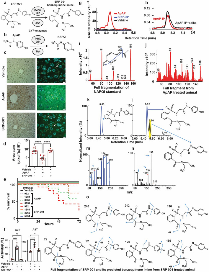
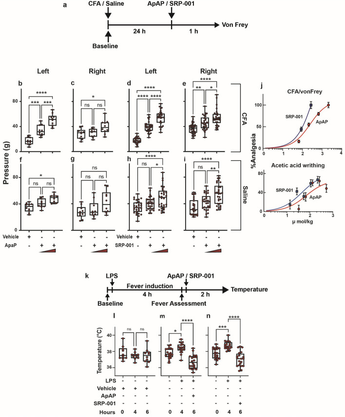
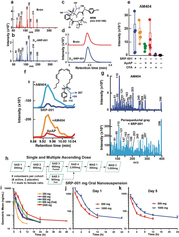
Safe and effective pain management is a critical healthcare and societal need. The potential for acute liver injury from paracetamol (ApAP) overdose; nephrotoxicity and gastrointestinal damage from chronic non-steroidal anti-inflammatory drug (NSAID) use; and opioids' addiction are unresolved challenges. We developed SRP-001, a non-opioid and non-hepatotoxic small molecule that, unlike ApAP, does not produce the hepatotoxic metabolite N-acetyl-p-benzoquinone-imine (NAPQI) and preserves hepatic tight junction integrity at high doses. CD-1 mice exposed to SRP-001 showed no mortality, unlike a 70% mortality observed with increasing equimolar doses of ApAP within 72 h. SRP-001 and ApAP have comparable antinociceptive effects, including the complete Freund's adjuvant-induced inflammatory von Frey model. Both induce analgesia via N-arachidonoylphenolamine (AM404) formation in the midbrain periaqueductal grey (PAG) nociception region, with SRP-001 generating higher amounts of AM404 than ApAP. Single-cell transcriptomics of PAG uncovered that SRP-001 and ApAP also share modulation of pain-related gene expression and cell signaling pathways/networks, including endocannabinoid signaling, genes pertaining to mechanical nociception, and fatty acid amide hydrolase (FAAH). Both regulate the expression of key genes encoding FAAH, 2-arachidonoylglycerol (2-AG), cannabinoid receptor 1 (CNR1), CNR2, transient receptor potential vanilloid type 4 (TRPV4), and voltage-gated Ca2+ channel. Phase 1 trial (NCT05484414) (02/08/2022) demonstrates SRP-001's safety, tolerability, and favorable pharmacokinetics, including a half-life from 4.9 to 9.8 h. Given its non-hepatotoxicity and clinically validated analgesic mechanisms, SRP-001 offers a promising alternative to ApAP, NSAIDs, and opioids for safer pain treatment.

Keywords: CNS; Liver toxicity; Non-opioid; Phase 1 trial; scRNA-seq.


Conflict of interest statement

H.A.B., J.A.-B., and N.G.B. are named on a patent assigned to the Board of Supervisors of Louisiana State University and Agricultural and Mechanical College and University Alcalá de Henares describing the synthesis and characterization of the novel non-hepatotoxic acetaminophen analogs, patent application PCT/US2018/022029, international filing data 12.03.2018; publication date 28.02.2019; which has been nationalized in numerous jurisdictions. H.A.B. and N.G.B. are co-founders of South Rampart Pharma, which has a license and commercial interest in the above patent applications, including a financial interest in the commercial success of SRP-001. They stand to potentially receive financial payments if SRP-001 is commercially successful.

Figures

Figure 1
Figure 1
SRP-001’s absent hepatotoxicity is due to lack of NAPQI formation and maintenance of hepatic tight junction integrity. (a) Possible oxidation of SRP-001 to the corresponding N-acyl-p-benzoquinone imine by CYP enzymes (CYP P450 2E1 and 3A4). (b) ApAP is metabolized by oxidation into NAPQI by CYP P450 2E1 and 3A4. (c) Doses known to be hepatotoxic for ApAP (600 mg/kg) but not SRP-001 (600 mg/kg) demonstrates centrilobular hepatic necrosis in nitrotyrosine-labeled hepatic sections from CD1 mice (100x, (c) first column), [(n = 5) mice per treatment group]. Hepatic tight junctions (ZO-1 label [3D-stacking, 1000x; scale bar = 5 µm] (c) second column) present between hepatocytes reveal a clear “chicken wire” configuration (vehicle) and are disrupted with toxic doses of ApAP but not SRP-001. (d) Quantification of the hepatic tight junctions by ZO-1 staining. There is a marked decrease in ZO-1 in ApAP-treated liver sections compared to vehicle and SRP-001, wherein ZO-1 staining is preserved. (e) Kaplan–Meier survival curves comparing equimolar doses of ApAP (150 to 900 mg/kg) and SRP-001 (402 to 2414 mg/kg) demonstrate a dose-dependent increase in mortality by 72 h for ApAP-treated groups (1/10 at 1984 mM/kg, 3/10 at 3969 mM/kg, and 7/10 at 5954 mM/kg), with no mortality observed in the SRP-001 and placebo cohorts; n = 10 per group (CD-1 male mice). (f) ApAP exposure but not SRP-001 led to an increase in the liver transaminases and 75% (15 out of 20) of ApAP treated animals were dead in 48 h and none in SRP-001 treated animals (data not shown). (g) ApAP-treated (red) but not SRP-001-treated (blue) mice produce the hepatotoxic metabolite NAPQI. Serum NAPQI peak is shown at 5.1 min on the chromatogram. NAPQI peak appears after IP-injection with ApAP but not SRP-001 (600 mg/kg). (h) ApAP-IP sample (red) with a standard NAPQI spike (black) demonstrates the same chromatographic retention time. (i) Full fragmentation pattern of the NAPQI standard and its likely fragments. (j) The full fragmentation spectrum from IP-ApAP sample’s NAPQI peak. Note: The spectrum matches well the standard in (i). (kn) LC–MS/MS retention time peak at 5.63 demonstrates SRP-001 (k) as it matches the full fragmentation pattern of SRP-001 from an ip-injected SRP-001 animal showing its fragmentation peaks (m); similarly, retention time 5.76 demonstrates the benzoquinoimine produced by SRP-001 (l); and (n) the resulting fragmentation pattern of the N-acyl-p-benzoquinone imine of SRP-001. (o) Structures matching full fragmentation pattern peaks of SRP-001 and its predicted benzoquinoimine.
Figure 2
Figure 2
Both ApAP and SRP-001 induce analgesia in the von Frey hyperalgesia in vivo assay and are antipyretic. (a) Timeline outlining the experimental design for the von Frey with electronic detection hyperalgesia/allodynia in vivo assay. (bi) Two separate doses of ApAP and SRP-001-32 and 100 mg/kg body weight—were tested using young male rats in von Frey. The threshold for paw withdrawal increased from 18 to 40 g and subsequently to 55 g in SRP-001-treated animals; it is more efficacious than ApAP at similar doses. (n = 10) rats for ApAP, and (n = 20) rats for SRP-001. Note: The left hind paw is treated with either CFA or saline, and the right hind paw is an internal control and is not injected. (j) SRP-001 has a more potent antinociceptive effect compared to ApAP at equimolar doses (µmol/kg). ED50 curves for SRP-001 are shifted to the left compared to ApAP in the hyperalgesia/allodynia (von Frey) and in a visceral pain model (acetic acid writing assay); see Supplementary Figs. 3 and 4 for detailed cohort data and Supplementary Fig. 5 for equimolar ED50 curves for each antinociceptive assay. (k) Timeline showing experimental design for antipyresis using LPS from Escherichia coli (100 μg/kg, 0111:B4). (l) there are no significant changes in body temperature of mice injected with 0.9% saline (vehicle) throughout the course of the experiment (n = 10) (m,n) SRP-001 and ApAP have comparable antipyresis. (n = 20) mice.
Figure 3
Figure 3
Antinociception/Analgesia is mediated via AM404 production in the periaqueductal gray (PAG) region of the brain and human single and multiple ascending doses (SAD and MAD) Phase 1 clinical trial reveals a favorable pharmacokinetic profile. (a) Detection of deuterated (D10-SRP-001) in the rat brain 30 min after IP injection. The full fragmentation spectrum of D10-SRP-001 detected in the brain (red) matches (b) the standard SRP-001 compound spectrum (blue). (c) The structure of D10-SRP-001 shows fragmentation patterns of D10-SRP-001 that match the spectra shown in the brain (red) and with the deuterated standard (blue). (d) The MRM (m/z 416 → 186) for D10-SRP-001 shows the elution time at ~ 2.2 min. (e) Quantification of AM404 production in the CNS periaqueductal gray region. (f) AM404 is expressed in the periaqueductal gray area of the brain following SRP-001 IP injection. LC–MS/MS detection of AM404 in the rat periaqueductal gray area after SRP-001 (top panel) and ApAP (bottom panel) IP injections. The AM404 structure and major fragments. (g) The peaks are confirmed to be AM404 by co-spiking AM404 standard to the samples (red and orange, respectively). The major peaks detected by LC–MS/MS demonstrate that the peak 9.9 min from LC is the AM404 compound. These peaks align to the (e) AM404 fragments. (h) Design of the first-in-human Phase 1 clinical trial for SRP-001. A randomized, double-blind, placebo-controlled study to assess the safety, tolerability, and pharmacokinetics of single and multiple ascending oral doses of SRP-001 and to characterize the effect of food on the pharmacokinetics of SRP-001 in healthy male and female subjects. Single ascending dose (SAD) escalation from 300 to 2000 mg (fasted state) and 900 mg (fed state). (i) and (j) Geometric mean SRP-001 plasma concentration–time profiles following oral administration (logarithmic plot) show a proportional or super-proportional increase in Cmax (peak time to concentration at 1 h post-dose) with a mono- or bi-phasic concentration decline in the SAD (i) and the MAD cohorts with an arithmetic mean T1/2 of 5.57 h and a geometric mean T1/2 of 4.92 h.
Figure 4
Figure 4
Single-cell transcriptomics of midbrain periaqueductal gray (PAG) cells in CFA-induced chronic inflammatory pain model. (a) Overview of single-cell RNA sequencing (scRNA-seq) experimental design to define the mechanism of action (MOA) of ApAP and SRP-001. The periaqueductal gray (PAG) midbrain region was dissected to isolate cell nuclei for sequencing library generation using the 10 × Genomics Chromium platform. Single cell data was embedded after dimensionality reduction using uniform manifold approximation and projection (UMAP). Cell clusters were annotated, followed by differential gene expression analysis. (b) UMAP plots showing the distribution of annotated cell clusters color-coded by each cluster – astrocytes, excitatory neurons, inhibitory interneurons, oligodendrocytes, oligodendrocyte precursor cells (OPC), and microglia, from the aggregated clusters of all the 4 samples. (c) UMAP plots showing the distribution of annotated cell clusters generated by the workflow of Seurat across 4 different samples – Vehicle, CFA_Veh, CFA_ApAP, and CFA_SRP-001, respectively. (di) Marker gene feature plots showing selected distinctive marker genes used for cell annotation of single cell clusters into 6 different cellular subpopulations. (d,e) Oligodendrocytes (f), Astrocytes (g) OPC (h) Glutamatergic neurons i GABAergic neurons. (j–k) GO enrichment analysis results for top 50 differentially expressed genes between CFA_Vehicle and CFA_ApAP, and between CFA_Vehicle and CFA_SRP-001 – gene concept network showing the linkage between the DE genes and GO terms, and Barplot of the enriched GO terms from the selected genes.
Figure 5
Figure 5
Cell annotation, marker gene feature plots, and endocannabinoid signaling pathway from neurons of PAG region cells in CFA-induced chronic inflammatory pain model with ApAP and SRP-001 treatments. (a) Heatmap of top 40 canonical pathways for neuronal clusters across samples – CFA_Veh, CFA_ApAP, and CFA_SRP-001 with the comparisons between them as follows – Vehicle vs CFA_Vehicle, CFA_Vehicle vs CFA_ApAP, and CFA_ApAP vs CFA_SRP-001. The heatmap shows that when CFA_Vehicle is compared to Vehicle, there is upregulation of several genes that are predicted to affect cellular signaling related to these pathways highlighted in the heatmap, and when CFA_ApAP and CFA_SRP-001 are compared to CFA_Vehicle, there is downregulation of those upregulated pathways in both cases, with more downregulation for CFA_SRP-001 than CFA_ApAP. (b,c) CellChat computed cell–cell interaction network for CFA_ApAP and CFA_SRP-001 group datasets showing number and strength of interactions describing similarities between the two groups. (d,e) Outgoing communication patterns in individual secreting cell types for CFA_ApAP and CFA_SRP-001. (f) Heatmap of DESeq2-normalized expression values from scRNA-seq data for genes involved in endocannabinoid signaling genes. (g) Endocannabinoid signaling pathways generated by Qiagen Ingenuity Pathway Analysis (IPA) with an overlay based on DESeq2-normalized expression values of differentially expressed genes (DEGs) in neuronal clusters CFA_Vehicle vs CFA_SRP-001 treatments, respectively.

Update of

References

    1. Zimmer Z, Fraser K, Grol-Prokopczyk H, Zajacova A. A global study of pain prevalence across 52 countries: examining the role of country-level contextual factors. Pain. 2022;163:1740–1750. doi: 10.1097/j.pain.0000000000002557. - DOI - PMC - PubMed
    1. Henschke N, Kamper SJ, Maher CG. The epidemiology and economic consequences of pain. Mayo Clin. Proc. 2015;90:139–147. doi: 10.1016/j.mayocp.2014.09.010. - DOI - PubMed
    1. Chang AK, Bijur PE, Esses D, Barnaby DP, Baer J. Effect of a single dose of oral opioid and nonopioid analgesics on acute extremity pain in the emergency department: A randomized clinical trial. JAMA J. Am. Med. Assoc. 2017;318:1661–1667. doi: 10.1001/jama.2017.16190. - DOI - PMC - PubMed
    1. Moore PA, Hersh EV. Combining ibuprofen and acetaminophen for acute pain management after third-molar extractions: Translating clinical research to dental practice. J. Am. Dent. Assoc. 2013;144:898–908. doi: 10.14219/jada.archive.2013.0207. - DOI - PubMed
    1. Wininger SJ, et al. A randomized, double-blind, placebo-controlled, multicenter, repeat-dose study of two intravenous acetaminophen dosing regimens for the treatment of pain after abdominal laparoscopic surgery. Clin. Ther. 2010;32:2348–2369. doi: 10.1016/j.clinthera.2010.12.011. - DOI - PubMed

Publication types