. 2024;18(3):101356.
doi: 10.1016/j.jcmgh.2024.05.004. Epub 2024 May 14.

NF-κB Inducing Kinase Attenuates Colorectal Cancer by Regulating Noncanonical NF-κB Mediated Colonic Epithelial Cell Regeneration

Affiliations

NF-κB Inducing Kinase Attenuates Colorectal Cancer by Regulating Noncanonical NF-κB Mediated Colonic Epithelial Cell Regeneration

Holly A Morrison et al. Cell Mol Gastroenterol Hepatol. 2024.

Abstract

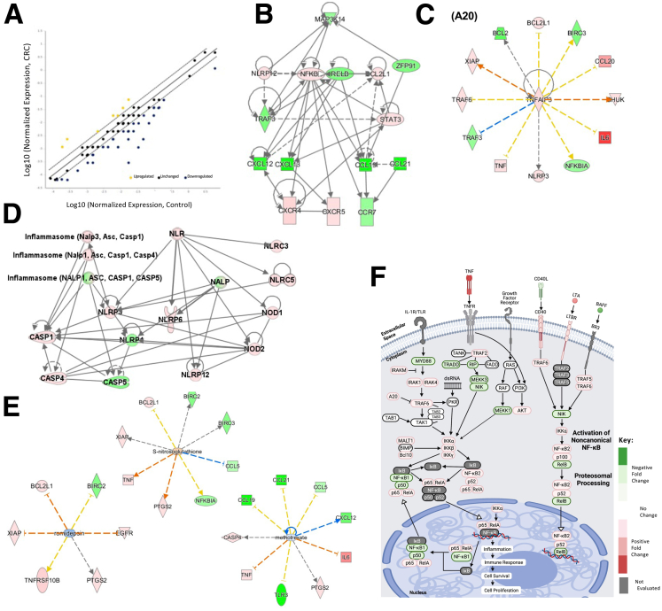
Background & aims: Dysregulated colonic epithelial cell (CEC) proliferation is a critical feature in the development of colorectal cancer. We show that NF-κB-inducing kinase (NIK) attenuates colorectal cancer through coordinating CEC regeneration/differentiation via noncanonical NF-κB signaling that is unique from canonical NF-kB signaling.

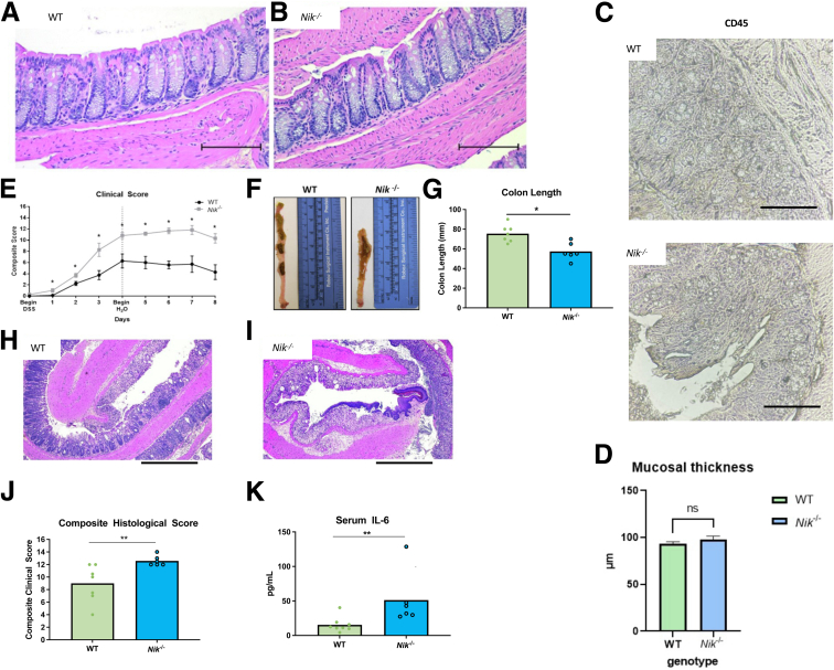
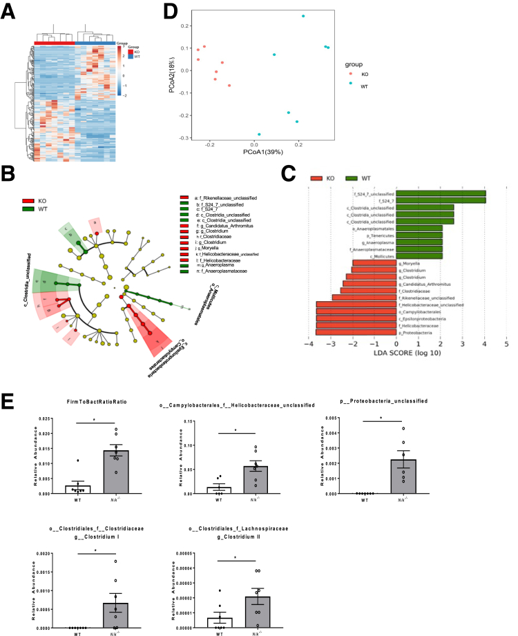
Methods: Initial studies evaluated crypt morphology/functionality, organoid generation, transcriptome profiles, and the microbiome. Inflammation and inflammation-induced tumorigenesis were initiated in whole-body NIK knockout mice (Nik-/-) and conditional-knockout mice following administration of azoxymethane and dextran sulfate sodium.

Results: Human transcriptomic data revealed dysregulated noncanonical NF-kB signaling. In vitro studies evaluating Nik-/- crypts and organoids derived from mature, nondividing CECs, and colonic stem cells exhibited increased accumulation and stunted growth, respectively. Transcriptomic analysis of Nik-/- cells revealed gene expression signatures associated with altered differentiation-regeneration. When assessed in vivo, Nik-/- mice exhibited more severe colitis with dextran sulfate sodium administration and an altered microbiome characterized by increased colitogenic microbiota. In the inflammation-induced tumorigenesis model, we observed both increased tumor burdens and inflammation in mice where NIK is knocked out in CECs (NikΔCEC). Interestingly, this was not recapitulated when NIK was conditionally knocked out in myeloid cells (NikΔMYE). Surprisingly, conditional knockout of the canonical pathway in myeloid cells (RelAΔMYE) revealed decreased tumor burden and inflammation and no significant changes when conditionally knocked out in CECs (RelAΔCEC).

Conclusions: Dysregulated noncanonical NF-κB signaling is associated with the development of colorectal cancer in a tissue-dependent manner and defines a critical role for NIK in regulating gastrointestinal inflammation and regeneration associated with colorectal cancer.

Keywords: Colitis-Associated Tumorigenesis; Differentiation; Epithelial Cells; Organoids.


Figures

None
Graphical abstract
Figure 1
Figure 1
Noncanonical NF-κB signaling is attenuated in human colorectal cancer patients. (A–H) Data from the TCGA data sets were analyzed, and the expression of key genes associated with noncanonical NF-κB signaling was compared between normal healthy controls (n = 22) and CRC patients (n = 215). Significant changes were observed in (A) BAFF, (B) BAFFR, (C) CD40, (D) CD40L, (E) LTB, (F) LTBR, and (G) MAP3K14 (NIK). (H) Using TCGA data sets, the gene expression levels of 4 effector chemokines produced by noncanonical NF-κB signaling (CCL19, CCL21, CXCL12, and CXCL13) were analyzed across different types of CRC. Data sets were only included if they contained all 4 chemokines. Significance was defined as ±2-fold change in expression. (I) RNA from human colon biopsies (n = 6 control, 6 CRC) were pooled and analyzed using a custom Superarray containing the 80 genes evaluated in the metadata analysis. (J–N) Data from the retrospective studies were confirmed by reverse transcriptase-polymerase chain reaction from colon biopsies from CRC patients (n = 6 control, 6 CRC). ∗∗P < .01; ∗∗∗P < 0.005; ∗∗∗∗P < .001.
Figure 2
Figure 2
Noncanonical NF-κB signaling is significantly down-regulated in biopsies from CRC patients. (A) RNA was extracted from human colon biopsies (n = 6 control; n = 6 CRC), and gene expression was evaluated using custom NF-κB signaling Superarray. Compared with control tissue, tissue from CRC patients showed a general down-regulation of genes related to noncanonical signaling (A; yellow, up-regulation; blue, down-regulation). Solid line represents no change from control, and each dotted line marks the borders of likely physiological significance (ie, change in fold regulation of >2). (B–E) Gene expression data from the GEO data set was evaluated using IPA. (B) IPA confirmed that noncanonical NF-κB signaling was significantly down-regulated and identified NIK (MAP3K14) as a critical driver of this dysregulation. (C) A20 and (D) NOD-like receptor signaling were identified as significant regulatory hubs for the altered NF-κB signaling identified by IPA. (B–D) Red correlates to up-regulation, and green correlates to down-regulation. (C–E) Solid lines indicate a direct interaction, dotted lines indicate an indirect interaction, arrowheads on the end of the line indicate activation, and flat lines on the end indicate inhibition. For colors, orange lines indicate a straightforward predicted activation, and blue lines indicate a straightforward predicted inhibition. Yellow lines indicate a more complex relationship with some inconsistencies in relationships that might indicate additional players. Grey lines indicate an effect is not predicted. (E) Based on the gene expression changes identified by IPA, romidepsin, s-nitrosoglutathione, and methotrexate were predicted by IPA to potentially impact disease progression. (F) Schematic of key aspects of noncanonical NF-κB signaling that were significantly altered in our CRC patient population. n = 6 control; n = 6 CRC human patients used in Superarray. Significant values ≥2 (yellow/red) and ≤2 (blue/green) reported as fold-change.
Figure 3
Figure 3
Colonic crypts from Nik-/-mice display decreased levels of stem cell proliferation and mature colonic epithelial cell death. (A and B) Immunocytochemistry of wild-type and Nik-/- crypts revealed decreased Ki-67 expression in the crypt base. (C) Lgr5 gene expression was significantly attenuated in the Nik-/- crypts. (D) Crypts from the Nik-/- animals were significantly elongated compared with those from wild-type mice. (E) Krt20 gene expression was significantly increased in the Nik-/- crypts. (F) Western blot analysis of the crypt fractions revealed decreased mature and cleaved poly-ADP ribose (PARP) levels in the Nik-/- crypts compared with wild-type. (G) Organoids from Apcmin mice displayed typical overzealous proliferation, whereas wild-type organoid growth was steady and highly uniform. Nik-/- organoids remained small compared with both wild-type and Apcmin. Colonic crypts were isolated and reduced to a single-cell suspension from wild-type, Nik-/-, and Apcmin mice and grown in culture for 6 days, without passage. (H) Blinded diameter measurement of randomly chosen organoids from wild-type and Nik-/- mice. (I) Organoids were manually disassociated from Matrigel and stained cytologically with Diff-Quik to evaluate morphology (wild-type/left; Nik-/-/right) despite the difference in size. Data represent the average of 3 independent trials. Measurements were taken from 30 randomly chosen organoids spread over a total of 12 wells. ∗P ≤ .05.
Figure 4
Figure 4
Nik-/-mice are sensitive to DSS-induced experimental colitis. (A) Wild-type (WT) mice and (B) Nik-/- mice display almost identical architecture, with healthy crypts and lack of inflammation under unstimulated conditions at the time points evaluated in the AOM+DSS study. Scale bars represent 100 μm. (C) Wild-type and Nik-/- mice exhibit minimal leukocyte infiltration into the crypts and villi at baseline demonstrated by CD45 staining. Original magnification, ×40; scale bars represent 100 μm. (D) Nik-/- mice and WT mice exhibit no significant differences between mucosal thickness, confirming no significant morphologic or pathologic differences at baseline. N = 8–9 mice/group. Each histologic sample from each mouse had the mucosal depth measured from the crypt up to the villus tip at 5 randomly chosen histologic areas. (E) Nik-/- mice exposed to 5% DSS in the acute experimental colitis model demonstrated enhanced clinical features of disease progression (weight loss, blood in stool, stool consistency, body condition, and behavior) compared with wild-type mice. (F and G) Colon length, a typical gross marker of inflammation and damage, was significantly decreased in the Nik-/- mice. (H and I) Histopathology assessments revealed increased inflammation in the Nik-/- colons, including increased damage to the epithelial cell barrier, compared with wild-type. (J) Semiquantitative scoring of pathology, assessing inflammation, epithelial cell defects, dysplasia, hyperplasia, which was translated into a composite score as previously described. (K) Serum cytokine levels were evaluated by enzyme-linked immunosorbent assay, with significant increases in IL-6 observed in the Nik-/- mice compared with the wild-type animals. n = 6; wild-type, n = 7. ∗P ≤ .05; ∗∗P ≤ .01.
Figure 5
Figure 5
Nik-/-mice display an altered microbiome with differential expression of species important to gut health. (A) The operational taxonomic unit heatmap shows hierarchical clustering associated with altered expression patterns of different bacterial taxa in the colonic contents of Nik-/- mice versus wild-type littermates. Red represents KO samples, and blue represents WT samples (KO = Nik-/-, WT = wild-type). (B) The phylogenic tree reveals the relationships between different orders, families, and genera that were changed in the Nik-/- and wild-type microbiomes. (C)The LDA score is a measure of bacterial species abundance. An LDA score of more than 2-fold change is considered significant. Nik-/- and wild-type mice exhibited different patterns of bacteria abundance, with particular emphasis on Clostridia, Helicobacter, and Camplylobacter. (D) PCA graph shows distinct clustering based on genotype. (E) Increased Firmicutes to Bacteroidetes ratio and unclassified bacteria in Nik-/- mice. n = 7 mice per genotype. Significance was defined as P ≤ .05.
Figure 6
Figure 6
Intestinal epithelial cell specific NIK knockout (NikΔCEC) mice display increased susceptibility to colorectal tumorigenesis. (A) Schematic illustrating the key components of the targeting construct used to insert loxP sites flanking key exons in the kinase domain of the Map3k14 gene that encodes NIK, ultimately generating Nikfl/fl mice. Mice carrying the floxed alleles appear to be phenotypically normal before crossing with Cre expressing animals, with no detrimental effects noted to date. The mice were then subjected to the AOM+DSS model for inflammation-driven tumorigenesis. (B) Composite clinical scores were assessed throughout the AOM+DSS model for NikΔMYE and Nikflox/flox (littermate control) mice. (C) Histopathology scoring of NikΔMYE and control tissues at completion of the model reflecting experimental colitis (IBD Index) and tumorigenesis (CRC Index). (D and E) Gross assessments of macroscopic polyps from NikΔMYE and control mice measuring (D) diameter and (E) number of polyps. (F) Composite clinical scores from the NikΔCEC and Nikflox/flox (littermate control) mice. (G) Representative histopathology of colonic tissue with immunohistochemistry staining of CD45+ cells (left) and Cd11b+cells (right). For the CD45+ image, the lymphoid follicle at the bottom of the image serves as an internal positive control. 10× objective; scale bar represents 100 μm. Upper portion of the Cd11b+ image represents positively staining leukocytes in the sero-cellular crust overlying the mucosa. (H) Subsequent histopathology scoring of NikΔCEC and control tissues at completion of the model. (I and J) Gross assessments of macroscopic polyps from NikΔCEC and control mice measuring (I) diameter and (J) number of polyps. (K) Gross examination of the colon further emphasized the increased tumor burden in the NikΔCEC mice. Grossly, polyps were large, raised, smooth to slightly cauliflower-like projections from the mucosa (arrowheads) that were typically confined to the distal colon in the Nikflox/flox littermates but were found up to the level of the transverse colon in the NikΔCEC mice (asterisks). (L) Histologically, all polyps were determined to represent well-differentiated adenocarcinomas. Histology scale bar = 500 μm. n = 5–7 mice per group. (M) Representative images of immunocytochemistry staining for Ki-67 in Nikflox/flox (left) and NikΔCEC (right) colons. Increased staining extending toward top of crypt observed in NikΔCEC colons. ∗P ≤ .05.
Figure 7
Figure 7
Myeloid cell-specific RelA knockout (RelAΔMYE) mice display attenuated colorectal tumorigenesis. The mice were subjected to the AOM+DSS model of inflammation-driven tumorigenesis. (A) Composite clinical scores composed of weight loss, fecal consistency, rectal bleeding, and behavior evaluations were assessed throughout the AOM+DSS model for RelAΔMYE and RelAflox/flox (littermate control) mice. (B and C) Histopathology scoring of RelAΔMYE and control tissues at completion of the model reflecting experimental colitis (IBD Index) and tumorigenesis (CRC Index). (D and E) Gross assessments of macroscopic polyps from RelAΔMYE and control mice measuring (D) diameter and (E) number of polyps. (F) Gross examination of the colon further emphasized the decrease in tumor burden in the RelAΔMYE mice. Grossly, polyps from the RelAΔMYE mice were seldom observed. All polyps in both genotypes were confined to the distal colon (asterisks). (G) Histologically, all polyps were determined to represent well-differentiated adenocarcinomas. Hematoxylin-eosin, 10×; scale bar = 250 μm. (H) Composite clinical scores from the RelAΔCEC and RelAflox/flox (littermate control) mice. (I) Histopathology scoring of RelAΔCEC and control tissues at completion of the model reflecting IBD Index and CRC Index. (J and K) Gross assessments of macroscopic polyps from RelAΔCEC and control mice measuring (J) diameter and (K) number of polyps. Histology scale bar = 500 μm. n = 5–7 mice per group. ∗P ≤ .05; ∗∗P ≤ .01.
Figure 8
Figure 8
Gene expression signature after loss of NIK reveals altered differentiation-regeneration profile. Gene expression profiles for (A) NF-κB signaling, (B) top-25 up-regulated genes, and (C) top 25 down-regulated genes. (D and E) Altered biological functions predicted by Gene Ontology (GO) given unique transcriptome profiles. (D) NikΔCEC lesions are predicted to have increased defense responses to bacterial infection. (E) NikΔCEC lesion compared with Nikfl/fl lesion tissue predicted to have altered regulation of epithelial cell proliferation. Gene expression profiles for (F) stem cell functions, (G) epithelial cell differentiation, (H) epithelial cell development, (I) cancer pathways, (J) apoptosis, and (K) wound healing. n = 3 representative mice/group selected from AOM+DSS study performed in Figure 6F–M or crypt organoid study in Figure 3 for transcriptome analysis. Significant values ≥2 (red) and ≤2 (green) reported as fold-change. Values that did not reach significance (white) in heatmap.

References

    1. McDaniel D.K., Eden K., Ringel V.M., et al. Emerging Roles for Noncanonical NF-kappaB Signaling in the Modulation of Inflammatory Bowel Disease Pathobiology. Inflamm Bowel Dis. 2016;22(9):2265–2279. - PMC - PubMed
    1. Nguyen V.Q., Eden K., Morrison H.A., et al. Noncanonical NF-kappaB Signaling Upregulation in Inflammatory Bowel Disease Patients is Associated With Loss of Response to Anti-TNF Agents. Front Pharmacol. 2021;12 - PMC - PubMed
    1. Eden K., Rothschild D.E., McDaniel D.K., et al. Noncanonical NF-kappaB signaling and the essential kinase NIK modulate crucial features associated with eosinophilic esophagitis pathogenesis. Dis Model Mech. 2017;10(12):1517–1527. - PMC - PubMed
    1. Häcker H., Chi L., Rehg J.E., et al. NIK prevents the development of hypereosinophilic syndrome-like disease in mice independent of IKKalpha activation. J Immunol. 2012;188(9):4602–4610. - PMC - PubMed
    1. Allen I.C., Wilson J.E., Schneider M., et al. NLRP12 suppresses colon inflammation and tumorigenesis through the negative regulation of noncanonical NF-kappaB signaling. Immunity. 2012;36(5):742–754. - PMC - PubMed

MeSH terms