

Reporting breast density on chest computed tomography
- PMID: 38751487
- PMCID: PMC11093103
- DOI: 10.21037/tbcr-23-36
Reporting breast density on chest computed tomography
Abstract
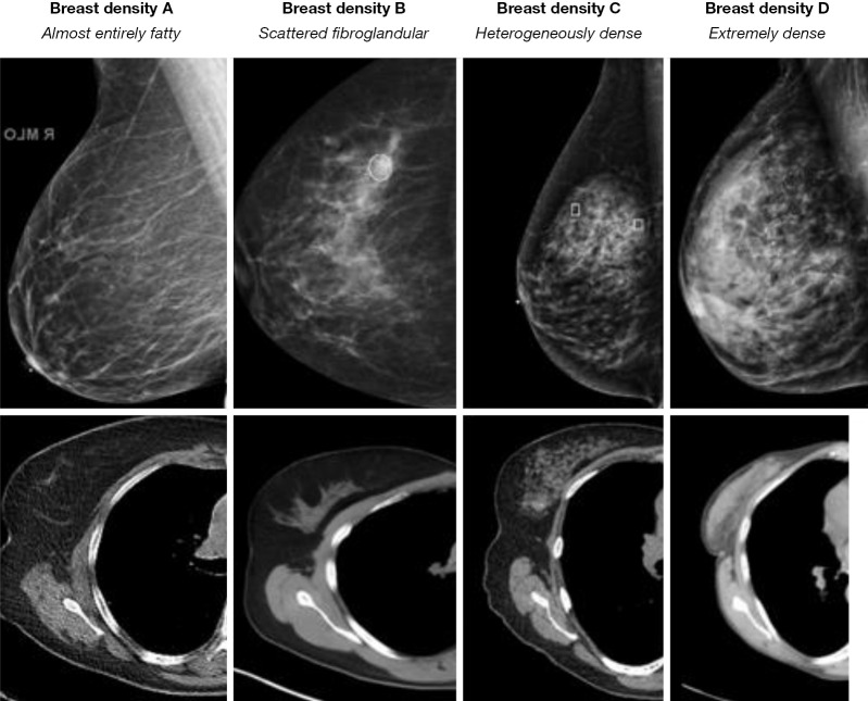
Women are encouraged to have a yearly mammogram and in addition to screening for breast cancer, the radiologist reports the patient's breast density. High breast density increases a woman's risk of developing breast cancer. The number of chest computed tomography (CT) scans performed each year is increasing. Chest CT scans for lung cancer screening in high-risk patients are the standard of care. Important additional findings can be identified on these exams including coronary artery calcifications, thyroid nodules, and breast density. Our previous research has shown that breast density can be reliably graded on chest CT and is comparable to mammographic grading. However, the inter-reader agreement was higher for chest CT. It is important that thoracic radiologists include the grading of breast density in their chest CT reports. According to mammography literature, this information has proven to be helpful for early detection of breast cancer. Federal legislation recommends notifying both providers and patients about breast density on mammography and so it follows that if we see the same information on chest CT, we should report it so that at the very least the clinician can encourage their patient to have a routine mammogram.
Keywords: Breast density; breast cancer; chest computed tomography (chest CT).
2023 Translational Breast Cancer Research. All rights reserved.
Conflict of interest statement
Conflicts of Interest: All authors have completed the ICMJE uniform disclosure form (available at https://tbcr.amegroups.com/article/view/10.21037/tbcr-23-36/coif). MMS reports grant funding from Boehringer Ingelheim and Genentech, research with Bioclinica, LungLifeAI, AbbVie, and speaking with Peer View and France Foundation. KMC served as an advisor for Cardinal Health Oncology Summits. The other authors have no conflicts of interest to declare.
Figures
Similar articles
-
Breast density: comparison of chest CT with mammography.Radiology. 2014 Jan;270(1):67-73. doi: 10.1148/radiol.13130733. Epub 2013 Oct 28. Radiology. 2014. PMID: 24126363
-
Chest CT for Breast Cancer Diagnosis.Life (Basel). 2022 Oct 26;12(11):1699. doi: 10.3390/life12111699. Life (Basel). 2022. PMID: 36362854 Free PMC article.
-
The chest radiologist's role in invasive breast cancer detection.Clin Imaging. 2018 Jul-Aug;50:13-19. doi: 10.1016/j.clinimag.2017.12.002. Epub 2017 Dec 7. Clin Imaging. 2018. PMID: 29245142
-
Mammographic density and breast cancer screening.Climacteric. 2020 Oct;23(5):460-465. doi: 10.1080/13697137.2020.1785418. Epub 2020 Jul 8. Climacteric. 2020. PMID: 32643449 Review.
-
Opportunistic screening at chest computed tomography: literature review of cardiovascular significance of incidental findings.Cardiovasc Diagn Ther. 2023 Aug 31;13(4):743-761. doi: 10.21037/cdt-23-79. Epub 2023 Jul 21. Cardiovasc Diagn Ther. 2023. PMID: 37675086 Free PMC article. Review.
References
Publication types
LinkOut - more resources
Full Text Sources