

Case Reports
doi: 10.1016/j.jaccas.2024.102357.
eCollection 2024 Jun 19.
Hydropneumopericardium Due to a Traumatic Esophageal-Pericardial Fistula
Affiliations
- PMID: 38751806
- PMCID: PMC11090896
- DOI: 10.1016/j.jaccas.2024.102357
Item in Clipboard
Case Reports
Hydropneumopericardium Due to a Traumatic Esophageal-Pericardial Fistula
JACC Case Rep.
.
Abstract
Esophago-pericardial fistula is a rare, life-threatening condition, usually arising as a complication of benign esophageal disorders or iatrogenic causes. Prompt diagnosis via multimodality imaging is crucial, with computed tomography being the most sensitive. Management varies based on severity, with a growing trend toward early endoscopic interventions, which result in improved outcomes.
Keywords: esophageal perforation; esophago-pericardial fistula; hydropneumopericardium.
© 2024 The Authors.
Conflict of interest statement
The authors have reported that they have no relationships relevant to the contents of this paper to disclose.
Figures
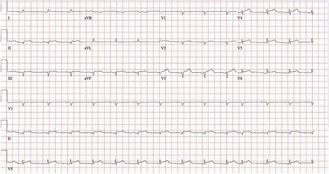
12-Lead Electrocardiogram Sinus rhythm, low QRS voltage, and ST-segment elevations in lead II, III, aVF, and V3-V6 suggestive of pericarditis.
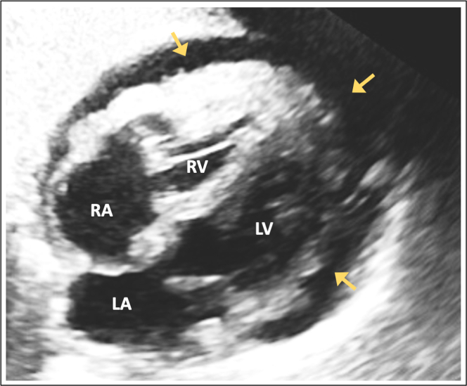
Transthoracic Echocardiography: Subcostal 4-Chamber View A moderate pericardial effusion (echolucency) is present (arrows).
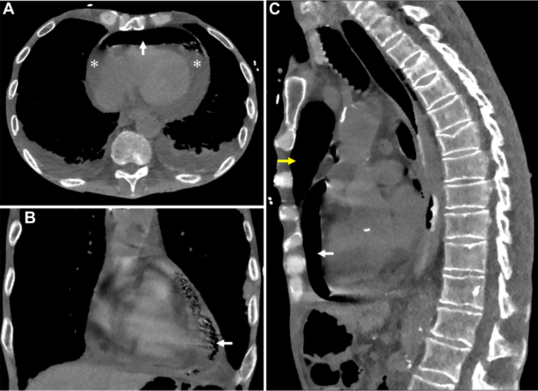
Computed Tomography of the Chest Without Contrast (A) Axial, (B) coronal, and (C) sagittal views, showing pneumopericardium (white arrow), a complex pericardial effusion (asterisk), and pneumomediastinum (yellow arrow).
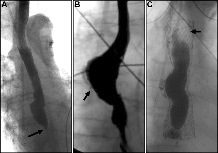
Series of Contrast Esophagrams: All in Anteroposterior Projection (A) Three weeks before hospitalization: proximal esophageal dilatation and distal narrowing (arrow) due to esophageal stricture, with no evidence of extraluminal contrast to suggest perforation. (B) Day 2 of hospitalization: esophageal mucosal irregularity with extravasation of contrast (arrow) suggestive of perforation. (C) Day 3 of hospitalization: esophageal stent in place (arrow) without evidence of contrast extravasation.
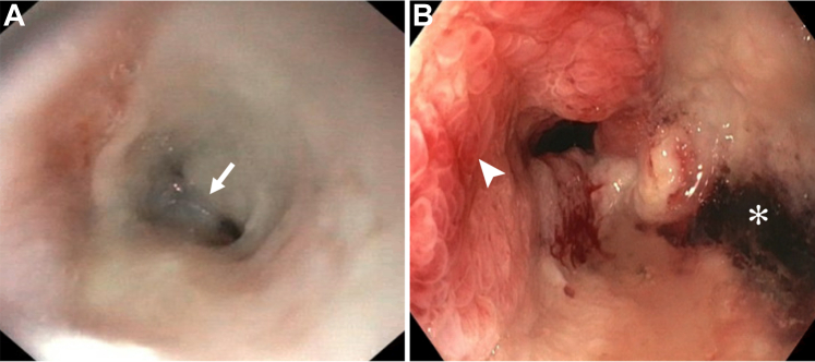
Series of Esophagogastroduodenoscopy Findings (Both Taken at the Same Level in the Lower Third of the Esophagus) (A) Three weeks before hospitalization showing a distal esophageal stricture (arrow). (B) Day 2 of hospitalization revealing severe esophagitis (arrowhead) and ulceration (asterisk) in the distal esophagus, likely the site of perforation.
References
-
- Salehpoor A., Shiehmorteza M., Terrazas M., Thompson W. Imaging in the evaluation of esophageal trauma including surgery. Contemp Diagn Radiol. 2022;45:1–7.
Publication types
LinkOut - more resources
Full Text Sources
