. 2024 May 1:14:1397940.
doi: 10.3389/fcimb.2024.1397940. eCollection 2024.

Modeling airway persistent infection of Moraxella catarrhalis and nontypeable Haemophilus influenzae by using human in vitro models

Affiliations

Modeling airway persistent infection of Moraxella catarrhalis and nontypeable Haemophilus influenzae by using human in vitro models

Andrea Ariolli et al. Front Cell Infect Microbiol. .

Abstract

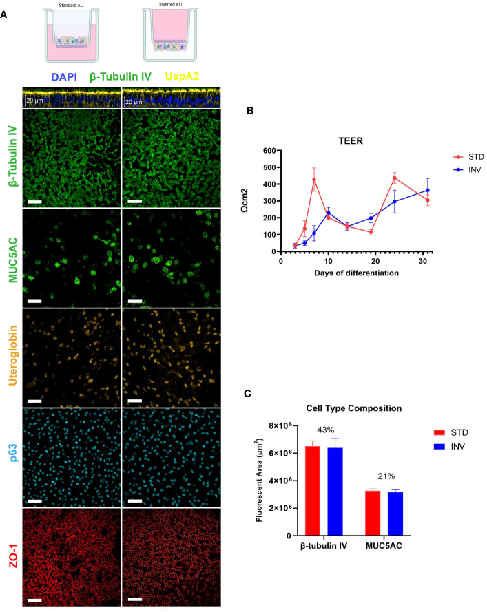
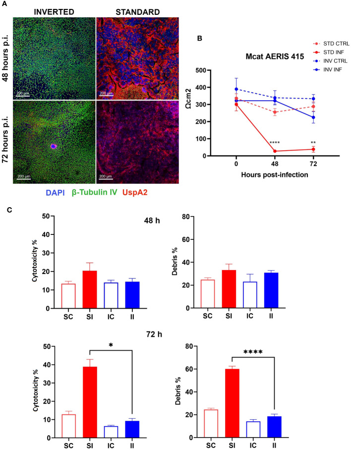
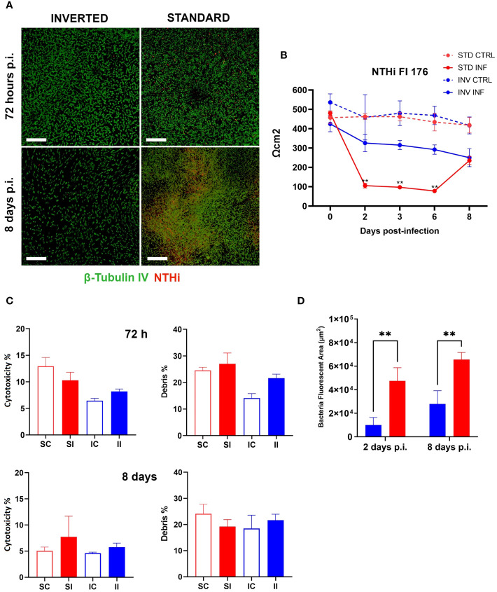
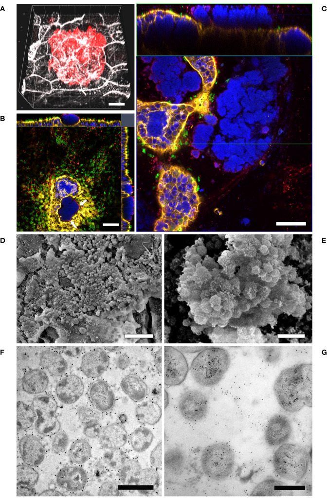
Non-typeable Haemophilus influenzae (NTHi) and Moraxella catarrhalis (Mcat) are two common respiratory tract pathogens often associated with acute exacerbations in Chronic Obstructive Pulmonary Disease (COPD) as well as with otitis media (OM) in children. Although there is evidence that these pathogens can adopt persistence mechanisms such as biofilm formation, the precise means through which they contribute to disease severity and chronicity remains incompletely understood, posing challenges for their effective eradication. The identification of potential vaccine candidates frequently entails the characterization of the host-pathogen interplay in vitro even though this approach is limited by the fact that conventional models do not permit long term bacterial infections. In the present work, by using air-liquid-interface (ALI) human airway in vitro models, we aimed to recreate COPD-related persistent bacterial infections. In particular, we explored an alternative use of the ALI system consisting in the assembly of an inverted epithelium grown on the basal part of a transwell membrane with the aim to enable the functionality of natural defense mechanisms such as mucociliary clearance and cellular extrusion that are usually hampered during conventional ALI infection experiments. The inversion of the epithelium did not affect tissue differentiation and considerably delayed NTHi or Mcat infection progression, allowing one to monitor host-pathogen interactions for up to three weeks. Notably, the use of these models, coupled with confocal and transmission electron microscopy, revealed unique features associated with NTHi and Mcat infection, highlighting persistence strategies including the formation of intracellular bacterial communities (IBCs) and surface-associated biofilm-like structures. Overall, this study demonstrates the possibility to perform long term host-pathogen investigations in vitro with the aim to define persistence mechanisms adopted by respiratory pathogens and individuate potential new vaccine targets.

Keywords: AECOPD; CIVM; airways; in vitro models; microscopy; replacement.


Conflict of interest statement

At the time of the study AA was recipient of a GSK fellowship from the PhD program of the University of Tuscia. MC was a PhD student recipient of a PhD fellowship funded by the MIUR at the University of Naples Federico II. AT is an employee of the University of Tuscia. MD, ST, KB, ID, SP and AP are employees of the GSK group of companies. The remaining authors declare that the research was conducted in the absence of any commercial or financial relationships that could be construed as a potential conflict of interest.

Figures

Figure 1
Figure 1
Characterization of ALI respiratory models. (A) LSCM analysis of epithelial differentiation: cilia (β-tubulin IV), mucus (MUC5AC), club cells (Uteroglobin), basal cells (p63) and Zonula Occludens 1 (ZO-1) were present in both standard and inverted models (white bars = 50µm). (B) Trans-epithelial electrical resistance measurement during epithelial differentiation. Results are expressed as mean values plus standard deviation calculated on 25 replicates for each configuration. (C) Evaluation of ciliated and goblet cells relative proportion based on the quantification of the fluorescent area related to β-tubulin IV and MUC5AC, respectively. Bars represent mean values plus standard deviation calculated on three replicates from three independent experiments (144 fields were analyzed for each sample).
Figure 2
Figure 2
Inverted respiratory models better tolerate Mcat infection. (A) LSCM analysis of standard and inverted models 48 and 72 hours after Mcat AERIS 415 inoculum. Unlike inverted models, standard models are completely covered by bacteria (Green are cilia, red is Mcat, Blue is DNA; white bars = 200 µm). (B) TEER measurement during 72 hours of Mcat infection. Results are expressed as mean values (plus standard deviation) of four replicates for each condition. (C) Cytotoxicity and debris analysis of standard and inverted models 48 and 72 hours after Mcat challenge. Bars represent mean value (plus standard deviation) calculated on three replicates for each condition (SC, Standard Control; IC, Inverted Control; SI, Standard Infected; II, Inverted Infected); data is representative of three independent experiments. Data were analyzed according to Welch and Brown-Forsythe ANOVA followed by a Dunnet’s T3 post-hoc test. Statistical significance for the difference between the means of SI and II is reported (*p < 0.05; **p < 0.005; ****p < 0.0001).
Figure 3
Figure 3
NTHi infection is better controlled on inverted models. (A) LSCM analysis of standard and inverted models 72 hours and 8 days after NTHi infection (Green are cilia, red is NTHi; white bars = 200 µm). (B) TEER measurement during 8 days of NTHi Fi176 infection. Results are expressed as mean values [plus standard deviation] of four replicates for each condition. (C) Cytotoxicity and debris analysis of standard and inverted models 72 hours and 8 days after NTHi challenge. Bars represent mean value (plus standard deviation) calculated on three replicates for each condition (SC, Standard Control; IC, Inverted Control; SI, Standard Infected; II, Inverted Infected); data is representative of three independent experiments. (D) Quantification of bacteria colonizing standard or inverted models 2 and 8 days after infection. The total area associated with bacterial fluorescence from 25 random fields was quantified with the Harmony software. Bars represent mean values (plus standard deviation) calculated on three replicates. Data were analyzed according to Welch and Brown-Forsythe ANOVA followed by a Dunnet’s T3 post-hoc test. Statistical significance for the difference between the means is reported (**p < 0.005).
Figure 4
Figure 4
LSCM analysis of airway models infected with Mcat AERIS 415. (A) Mcat infection expands intraepithelially after 48 hours from the challenge (red is Mcat; grey is cell membrane; white bar = 5 µm). (B) Mcat colonies emerge apically from the epithelium. Only bacteria residing on the external perimeter of the aggregate (white arrows) can be efficiently immuno-stained with anti-UspA2 antibodies; white bar = 50 µm. (C) At seven days post infection, Mcat macroaggregates pervade the epithelial surface and most cells contain IBCs; the upper panel represent a Z/Y section of the epithelium while the lower panel is a X/Y view of the same stack (blue is DNA; yellow is F-actin; green are cilia; red is Mcat; white bar = 50 µm). (D, E) Scanning Electron Microscopy analysis of inverted airway models 14 days after Mcat inoculum shows the presence of an extra-polymeric substance enveloping bacterial clusters (white bar is 10 µm and 2 µm for (D, E) respectively). (F) Immunogold labeling assay conducted on Mcat biofilm ultrathin sections shows that UspA2 is expressed by bacterial cells residing inside the aggregate; white bar = 1 µm (G) dsDNA immunogold staining of a biofilm section; white bar = 0.5 µm.
Figure 5
Figure 5
LSCM and TEM analysis of airway models infected with NTHi Fi176. (A) After 72 hours of infection with strain Fi176 bacteria can be found adhering to the epithelial surface, entering or inside cells; white bar = 50 µm. (B) NTHi forms IBCs after 1 week of infection (red arrows). Bacteria residing in the internal part of IBCs are negative for an anti-Fi176 antibody unlike those located in the margin (stained in red and indicated by white arrowheads); white bar = 10 µm. (C) TEM analysis of IBCs residing below the apical surface of the epithelium (black arrows). Bacteria localized paracellularly are indicated by black arrowheads. Cell-cell junctions appear loose (black asterisks); white bar = 5 µm. (D) Higher magnification of an IBC formed by NTHi; white bar = 2 µm.

Similar articles

References

    1. Ahearn C. P., Gallo M. C., Murphy T. F. (2017). Insights on persistent airway infection by non-typeable Haemophilus influenzae in chronic obstructive pulmonary disease. Pathog. Dis. 75. doi: 10.1093/femspd/ftx042 - DOI - PMC - PubMed
    1. Anderson G. G., Martin S. M., Hultgren S. J. (2004). Host subversion by formation of intracellular bacterial communities in the urinary tract. Microbes Infection 6, 1094–1101. doi: 10.1016/j.micinf.2004.05.023 - DOI - PubMed
    1. Armbruster C. E., Hong W., Pang B., Weimer K. E., Juneau R. A., Turner J., et al. . (2010). Indirect pathogenicity of Haemophilus influenzae and Moraxella catarrhalis in polymicrobial otitis media occurs via interspecies quorum signaling. mBio 1 (3). doi: 10.1128/mBio.00102-10 - DOI - PMC - PubMed
    1. Bair K. L., Campagnari A. A. (2019). Moraxella catarrhalis promotes stable polymicrobial biofilms with the major otopathogens. Front. Microbiol. 10, 3006. doi: 10.3389/fmicb.2019.03006 - DOI - PMC - PubMed
    1. Bakaletz L. O., Novotny L. A. (2018). Nontypeable haemophilus influenzae (NTHi). Trends Microbiol. 26, 727–728. doi: 10.1016/j.tim.2018.05.001 - DOI - PubMed

MeSH terms

LinkOut - more resources