

[Application of mixed reality technology in free fibular flap transplantation for repairing mandibular defects]
- PMID: 38752246
- PMCID: PMC11096888
- DOI: 10.7507/1002-1892.202402027
[Application of mixed reality technology in free fibular flap transplantation for repairing mandibular defects]
Abstract
Objective: To explore the feasibility and effectiveness of mixed reality technology for localizing perforator vessels in the repair of mandibular defects using free fibular flap.
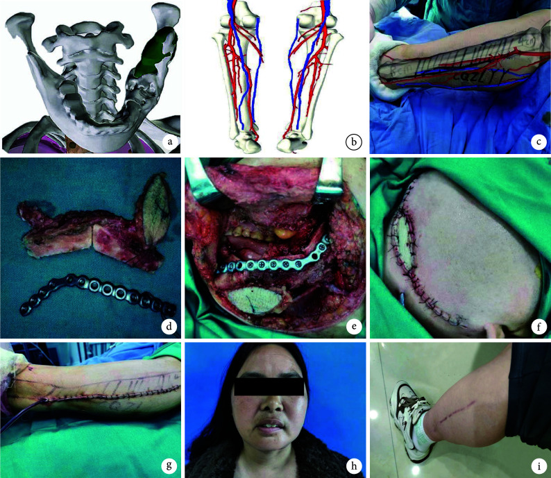
Methods: Between June 2020 and June 2023, 12 patients with mandibular defects were repaired with free fibular flap. There were 8 males and 4 females, with an average age of 61 years (range, 35-78 years). There were 9 cases of ameloblastomas and 3 cases of squamous cell carcinomas involving the mandible. The disease duration ranged from 15 days to 2 years (median, 14.2 months). The length of mandibular defects ranged from 5 to 14 cm (mean, 8.5 cm). The area of soft tissue defects ranged from 5 cm×4 cm to 8 cm×6 cm. Preoperative enhanced CT scans of the maxillofacial region and CT angiography of the lower limbs were performed, and the data was used to create three-dimensional models of the mandible and lower limb perforator vessels. During operation, the mixed reality technology was used to overlay the three-dimensional model of perforator vessels onto the body surface for harvesting the free fibular flap. The length of the fibula harvested ranged from 6 to 15 cm, with a mean of 9.5 cm; the size of the flap ranged from 6 cm×5 cm to 10 cm×8 cm. The donor sites were sutured directly in 7 cases and repaired with free skin grafting in 5 cases.
Results: Thirty perforator vessels were located by mixed reality technology before operation, with an average of 2.5 vessels per case; the distance between the exit point of the perforator vessels located before operation and the actual exit point ranged from 1 to 4 mm, with a mean of 2.8 mm. All fibular flaps survived; 1 case had necrosis at the distal end of flap, which healed after dressing changes. One donor site had infection, which healed after anti-inflammatory dressing changes; the remaining incisions healed by first intention, and the grafts survived smoothly. All patients were followed up 8-36 months (median, 21 months). The repaired facial appearance was satisfactory, with no flap swelling. Among the patients underwent postoperative radiotherapy, 2 patients had normal bone healing and 1 had delayed healing at 6 months.
Conclusion: In free fibular flap reconstruction of mandibular defects, the use of mixed reality technology for perforator vessel localization can achieve three-dimensional visualization, simplify surgical procedures, and reduce errors.
目的: 探讨在游离腓骨皮瓣修复下颌骨缺损中,使用混合现实技术定位穿支血管的可行性和效果。.
方法: 2020年6月—2023年6月,采用游离腓骨皮瓣修复12例下颌骨缺损患者。男8例,女4例;年龄35~78岁,平均61岁。下颌骨成釉细胞瘤9例,侵犯下颌骨口腔鳞癌3例。病程15 d~2年,中位病程14.2个月。下颌骨缺损长度5~14 cm,平均8.5 cm;软组织缺损范围为5 cm×4 cm~8 cm×6 cm。术前均行颌面部增强CT和下肢CT血管造影检查,将所得数据制成下颌骨、下肢穿支血管三维模型。术中使用混合现实技术将穿支血管三维模型重叠于患者体表,制取游离腓骨皮瓣修复缺损。腓骨切取长度为6~15 cm,平均9.5 cm;皮瓣切取范围6 cm×5 cm~10 cm×8 cm,供区拉拢缝合(7例)或游离植皮修复(5例)。.
结果: 12例患者术前使用混合现实技术定位穿支血管30支,每例平均2.5支;术中测量术前定位穿支血管穿出点与实际穿出点距离为1~4 mm,平均2.8 mm。术后12例腓骨瓣顺利成活;1例皮瓣远端边缘坏死,换药后延期愈合。供区发生感染1例,经抗炎换药治疗后愈合;其余患者切口Ⅰ期愈合,植皮顺利成活。患者均获随访,随访时间8~36个月,中位时间21个月。修复面部外形好,皮瓣无臃肿。3例术后接受放化疗患者中,随访6个月时2例正常骨愈合、1例延迟愈合。.
结论: 在游离腓骨皮瓣修复下颌骨缺损手术中使用混合现实技术,可以实现穿支血管三维可视化,手术操作简便,误差较小。.
Keywords: Mixed reality technology; free fibular flap; mandibular defect; perforator vessel; repair and reconstruction.
Conflict of interest statement
利益冲突 在课题研究和文章撰写过程中不存在利益冲突;经费支持没有影响文章观点和对研究数据客观结果的统计分析及其报道
References
Publication types
MeSH terms
LinkOut - more resources
Full Text Sources