Clinical Trial
. 2024 Oct 1;119(10):2010-2018.
doi: 10.14309/ajg.0000000000002871. Epub 2024 May 16.

Comparison of Effective Imaging Modalities for Detecting Gastric Neoplasms: A Randomized 3-Arm Phase II Trial

Affiliations
Clinical Trial

Comparison of Effective Imaging Modalities for Detecting Gastric Neoplasms: A Randomized 3-Arm Phase II Trial

Tomohiro Kadota et al. Am J Gastroenterol. .

Abstract

Introduction: The early detection of gastric neoplasms (GNs) leads to favorable treatment outcomes. The latest endoscopic system, EVIS X1, includes third-generation narrow-band imaging (3G-NBI), texture and color enhancement imaging (TXI), and high-definition white-light imaging (WLI). Therefore, this randomized phase II trial aimed to identify the most promising imaging modality for GN detection using 3G-NBI and TXI.

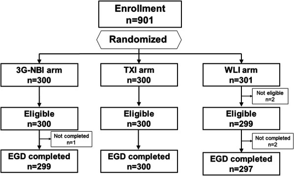
Methods: Patients with scheduled surveillance endoscopy after a history of esophageal cancer or GN or preoperative endoscopy for known esophageal cancer or GN were randomly assigned to the 3G-NBI, TXI, or WLI groups. Endoscopic observations were performed to detect new GN lesions, and all suspected lesions were biopsied. The primary endpoint was the GN detection rate during primary observation. Secondary endpoints were the rate of missed GNs, early gastric cancer detection rate, and positive predictive value for a GN diagnosis. The decision rule had a higher GN detection rate between 3G-NBI and TXI, outperforming WLI by >1.0%.

Results: Finally, 901 patients were enrolled and assigned to the 3G-NBI, TXI, and WLI groups (300, 300, and 301 patients, respectively). GN detection rates in the 3G-NBI, TXI, and WLI groups were 7.3, 5.0, and 5.6%, respectively. The rates of missed GNs were 1.0, 0.7, and 1.0%, the detection rates of early gastric cancer were 5.7, 4.0, and 5.6%, and the positive predictive values for the diagnosis of GN were 36.5, 21.3, and 36.8% in the 3G-NBI, TXI, and WLI groups, respectively.

Discussion: Compared with TXI and WLI, 3G-NBI is a more promising modality for GN detection.


Conflict of interest statement

Guarantor of the article: Tomonori Yano, MD, PhD.

Specific author contributions: T.K., S.A., N.U., H.D., Y.F., M.M., N.Y., C.K., T.I., M.W., and T.Y.: designed the study. T.K., S.A., N.U., H.D., Y.F., M.M., S.N., H.T., T.M., K.N., Y.T., Y.O., A.K., N.Y., A.W., C.K., M.T., A.Y., and T.Y.: collected the data. T.I. and M.W.: analyzed the data. H.F.: managed and coordinated the study. T.K. and T.Y.: wrote the manuscript. All authors reviewed and approved the final version of the manuscript.

Financial support: This work was supported by research funding and free device loans provided by Olympus Corporation. This company was not involved in this study.

Potential competing interests: T.Y. received a research grant from Olympus as a principal investigator. S.A., N.U., H.D., Y.F., and M.M. received research grants from Olympus as facility study managers. Seiichiro Abe received personal fees from Boston Scientific, personal fees and research funding from FUJIFILM, and personal fees and research funding from Olympus. N.U. received honoraria for lectures from Olympus Co., Ltd., FUJIFILM Co., Ltd., Boston Scientific, Japan, and AI Medical Service Co., Ltd. H.D. received honoraria for lectures at Olympus. S.N. has received honoraria for lectures at Olympus, Japan. T.Y. received honoraria for lectures and research grants from Olympus and research grants from HOYA PENTAX and FUJIFILM. T.K., H.T., T.M., K.N., Y.T., Y.O., A.K., N.Y., A.W., C.K., M.T., A.Y., H.F., T.I., and M.W. have no conflict of interest.

Clinical trial registration: Japan Registry of Clinical Trials, Number: jRCT1032210213 (https://jrct.niph.go.jp/en-latest-detail/jRCT1032210213).

Figures

Figure 1.
Figure 1.
Representative images of target lesions. A depressed lesion in the middle third of the stomach is shown (arrowheads). The final histopathological diagnosis was well-differentiated adenocarcinoma confined to the mucosa. (a and b) On white-light imaging (WLI), the lesion appears as a reddish area with irregular margins and surfaces. (c and d) On texture and color enhancement imaging, the lesion appears as a reddish area with irregular margin surfaces, with a greater color difference than that in WLI. (e and f) Third-generation narrow-band imaging shows the lesion as a brownish area with irregular margins and surfaces.
Figure 2.
Figure 2.
Representative images of target lesions. A slightly elevated lesion in the middle third of the stomach is observed (arrowheads). The final histopathological diagnosis was well-differentiated adenocarcinoma confined to the mucosa. (a) On white-light imaging, the lesion appears as a same-colored area with irregular margins and surfaces. (b) On texture and color enhancement imaging, the lesion appears as a same-colored area with irregular margins and surfaces. (c and d) Third-generation narrow-band imaging: The lesion appears as a brownish area with irregular margins and surfaces.
Figure 3.
Figure 3.
Patient flow diagram. 3G-NBI, third-generation narrow-band imaging; EGD, esophagogastroduodenoscopy; TXI, texture and color enhancement imaging; WLI, white-light imaging.

References

    1. Hamashima C, Ogoshi K, Okamoto M, et al. . A community-based, case-control study evaluating mortality reduction from gastric cancer by endoscopic screening in Japan. PLoS One 2013;8(11):e79088. - PMC - PubMed
    1. Choi KS, Jun JK, Suh M, et al. . Effect of endoscopy screening on stage at gastric cancer diagnosis: Results of the national cancer screening programme in Korea. Br J Cancer 2015;112(3):608–12. - PMC - PubMed
    1. Maekawa A, Kato M, Nakamura T, et al. . Incidence of gastric adenocarcinoma among lesions diagnosed as low-grade adenoma/dysplasia on endoscopic biopsy: A multicenter, prospective, observational study. Dig Endosc 2018;30(2):228–35. - PubMed
    1. Kinami S, Yamada S, Takamura H. Confusion and prospects for carcinogenesis of gastric adenoma and dysplasia: What is the correct answer currently? World J Gastroenterol 2022;28(48):6900–8. - PMC - PubMed
    1. Hamashima C. Benefits and harms of endoscopic screening for gastric cancer. World J Gastroenterol 2016;22(28):6385–92. - PMC - PubMed

Publication types