

Nano-organization of synaptic calcium signaling
- PMID: 38752834
- PMCID: PMC11346461
- DOI: 10.1042/BST20231385
Nano-organization of synaptic calcium signaling
Abstract
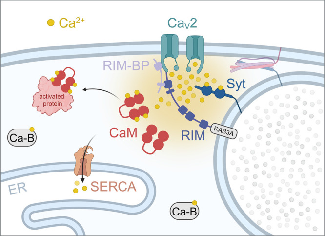
Recent studies suggest an exquisite structural nano-organization within single synapses, where sites of evoked fusion - marked by clustering of synaptic vesicles, active zone proteins and voltage-gated calcium channels - are directly juxtaposed to postsynaptic receptor clusters within nanocolumns. This direct nanometer scale alignment between presynaptic fusion apparatus and postsynaptic receptors is thought to ensure the fidelity of synaptic signaling and possibly allow multiple distinct signals to occur without interference from each other within a single active zone. The functional specificity of this organization is made possible by the inherent nano-organization of calcium signals, where all the different calcium sources such as voltage-gated calcium channels, intracellular stores and store-operated calcium entry have dedicated local targets within their nanodomain to ensure precision of action. Here, we discuss synaptic nano-organization from the perspective of calcium signals, where some of the principal findings from early work in the 1980s continue to inspire current studies that exploit new genetic tools and super-resolution imaging technologies.
Keywords: calcium signaling; synapse; synaptic plasticity.
© 2024 The Author(s).
Conflict of interest statement
The authors declare that there are no competing interests associated with the manuscript.
Figures

Similar articles
-
Synapse and Active Zone Assembly in the Absence of Presynaptic Ca2+ Channels and Ca2+ Entry.Neuron. 2020 Aug 19;107(4):667-683.e9. doi: 10.1016/j.neuron.2020.05.032. Epub 2020 Jun 16. Neuron. 2020. PMID: 32616470 Free PMC article.
-
Transsynaptic Assemblies Link Domains of Presynaptic and Postsynaptic Intracellular Structures across the Synaptic Cleft.J Neurosci. 2023 Aug 16;43(33):5883-5892. doi: 10.1523/JNEUROSCI.2195-22.2023. Epub 2023 Jun 27. J Neurosci. 2023. PMID: 37369583 Free PMC article.
-
Spatially non-overlapping Ca2+ signals drive distinct forms of neurotransmission.Cell Rep. 2023 Oct 31;42(10):113201. doi: 10.1016/j.celrep.2023.113201. Epub 2023 Sep 30. Cell Rep. 2023. PMID: 37777959 Free PMC article.
-
Millisecond Ca2+ dynamics activate multiple protein cascades for synaptic vesicle control.Proc Jpn Acad Ser B Phys Biol Sci. 2017;93(10):802-820. doi: 10.2183/pjab.93.050. Proc Jpn Acad Ser B Phys Biol Sci. 2017. PMID: 29225307 Free PMC article. Review.
-
Molecular mechanism of active zone organization at vertebrate neuromuscular junctions.Mol Neurobiol. 2012 Feb;45(1):1-16. doi: 10.1007/s12035-011-8216-y. Epub 2011 Dec 2. Mol Neurobiol. 2012. PMID: 22135013 Free PMC article. Review.
Cited by
-
High frequency stimulation activates hot spots of spontaneous synaptic transmission.Front Synaptic Neurosci. 2025 Apr 14;17:1539868. doi: 10.3389/fnsyn.2025.1539868. eCollection 2025. Front Synaptic Neurosci. 2025. PMID: 40297638 Free PMC article.
-
Resolving synaptic events using subsynaptically targeted GCaMP8 variants.bioRxiv [Preprint]. 2025 Jun 19:2025.06.19.660577. doi: 10.1101/2025.06.19.660577. bioRxiv. 2025. PMID: 40611906 Free PMC article. Preprint.
References
Publication types
MeSH terms
Substances
Grants and funding
LinkOut - more resources
Full Text Sources