. 2024 May 15;16(10):8645-8656.
doi: 10.18632/aging.205830. Epub 2024 May 15.

Mangiferin attenuates lipopolysaccharide-induced neuronal injuries in primary cultured hippocampal neurons

Affiliations

Mangiferin attenuates lipopolysaccharide-induced neuronal injuries in primary cultured hippocampal neurons

Hongling Tan et al. Aging (Albany NY). .

Abstract

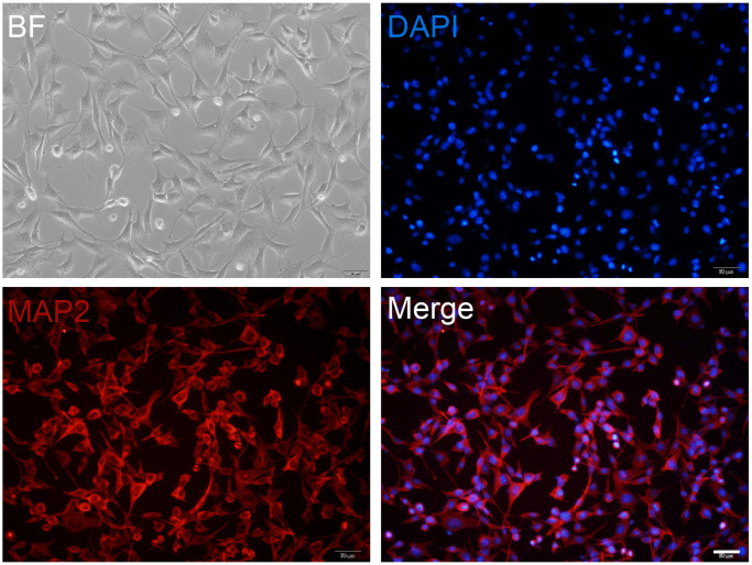
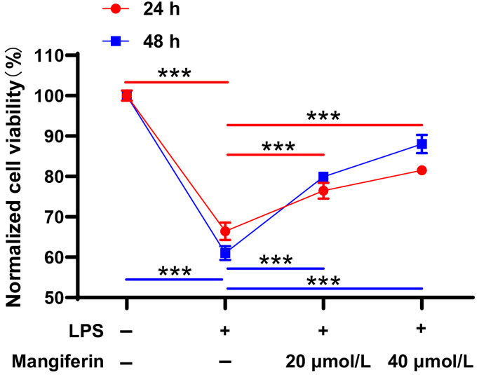
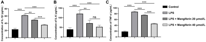
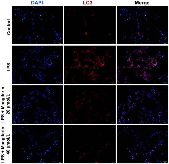
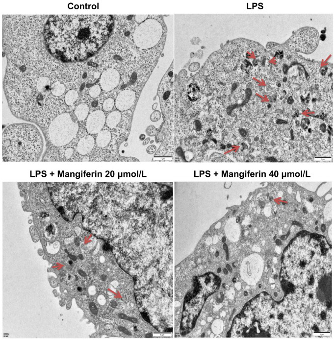
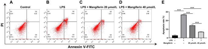
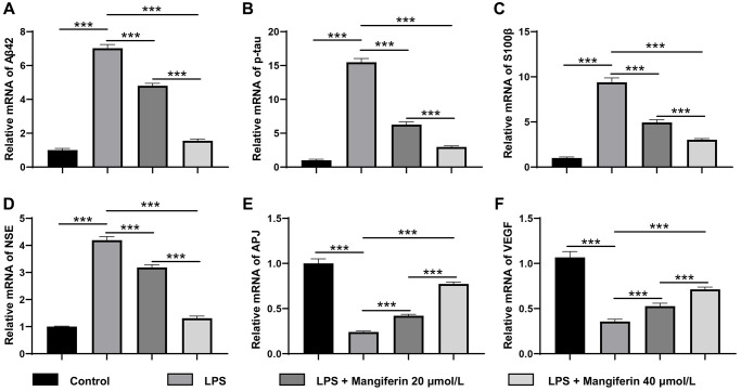
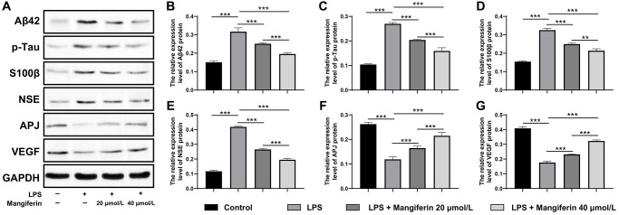
Mangiferin, a naturally occurring potent glucosylxanthone, is mainly isolated from the Mangifera indica plant and shows potential pharmacological properties, including anti-bacterial, anti-inflammation, and antioxidant in sepsis-induced lung and kidney injury. However, there was a puzzle as to whether mangiferin had a protective effect on sepsis-associated encephalopathy. To answer this question, we established an in vitro cell model of sepsis-associated encephalopathy and investigated the neuroprotective effects of mangiferin in primary cultured hippocampal neurons challenged with lipopolysaccharide (LPS). Neurons treated with 20 μmol/L or 40 μmol/L mangiferin for 48 h can significantly reverse cell injuries induced by LPS treatment, including improved cell viability, decreased inflammatory cytokines secretion, relief of microtubule-associated light chain 3 expression levels and several autophagosomes, as well as attenuated cell apoptosis. Furthermore, mangiferin eliminated pathogenic proteins and elevated neuroprotective factors at both the mRNA and protein levels, showing strong neuroprotective effects of mangiferin, including anti-inflammatory, anti-autophagy, and anti-apoptotic effects on neurons in vitro.

Keywords: lipopolysaccharide; mangiferin; neuron; sepsis; sepsis-associated encephalopathy.


Conflict of interest statement

CONFLICTS OF INTEREST: The authors declare no conflicts of interest related to this study.

Figures

Figure 1
Figure 1
Primary cultured neuron preparation. Primary cultured hippocampal neurons are maintained at DIV12 and are observed in a bright field (BF) or stained with MAP2 (red). DAPI (blue) is used as a nuclear marker. Scale bar, 50 μm.
Figure 2
Figure 2
Neuron viability with different concentrations of mangiferin treatment. DIV12 neurons are incubated with different doses of mangiferin for 24 h and absorption is measured at 450 nm using a microplate reader. Data are collected from three independent experiments and presented as mean ± SD. One-way ANOVA is used for statistical analysis; *P < 0.05, **P < 0.01, ***P < 0.001.
Figure 3
Figure 3
Mangiferin protects neurons from LPS injury. LPS-exposed neurons are incubated with 20 μmol/L or 40 μmol/L of mangiferin for 24 h or 48 h and then analyzed with the CCK8 assay. LPS treatment significantly affected cell viability (66.41% in 24 h, 61.07% in 48 h, ***P < 0.001 compared to the PBS control group), while 20 μmol/L (76.50% in 24 h, 79.91% in 48 h, ***P < 0.001) and 40 μmol/L (81.54% in 24 h, 88.02% in 48 h, ***P < 0.001) of mangiferin countered the LPS-induced phenotype. Data are presented as mean ± SD. One-way ANOVA is used for statistical analysis; ***P < 0.001.
Figure 4
Figure 4
Anti-inflammatory effect of mangiferin on LPS-exposed cells. The concentrations of IL-1β, IL-6, and TNF-a in mangiferin-treated culture medium are measured by ELISA assay. Data are presented as mean ± SD. One-way ANOVA is used for statistical analysis. (A) Concentrations of IL-1β in control, LPS, 20 μmol/L mangiferin with LPS, and 40 μmol/L mangiferin with LPS are 23.23 pg/mL, 62.11 pg/mL, 49.37 pg/mL, and 35.64 pg/mL, respectively, all ***P < 0.001. (B) Concentrations of IL-6 in control, LPS, 20 μmol/L mangiferin with LPS, 40 μmol/L mangiferin with LPS are 39.52 pg/ml, 120.70 pg/mL, 71.41 pg/mL, and 52.57 pg/mL, respectively. **P = 0.0066 for LPS vs. 20 μmol/L mangiferin with LPS, P = 0.23 for 20 μmol/L mangiferin with LPS vs. 40 μmol/L mangiferin with LPS, no significance is observed. All other comparisons are presented as ***P < 0.001. (C) Concentrations of TNF-a in control, LPS, 20 μmol/L mangiferin with LPS, and 40 μmol/L mangiferin with LPS are 18.61 pg/mL, 86.98 pg/mL, 75.60 pg/mL, and 45.80 pg/mL, respectively, all ***P < 0.001.
Figure 5
Figure 5
Mangiferin treatment inhibits the autophagy process induced by LPS. Neurons are stained with LC3 (red) to label the autophagosomes and DAPI (blue) to mark the nucleus. Scale bars, 50 μm.
Figure 6
Figure 6
Mangiferin treatment alleviates the number of autophagosomes induced by LPS by TEM. Different neuronal treatments were performed for transmission electron microscopy (TEM). Red arrowheads indicate autophagosomes. Scale bars, 1 μm.
Figure 7
Figure 7
Mangiferin mediates an anti-apoptotic effect in LPS-exposed cells. (AD) The plots show the flow cytometry of different treatments, and the apoptosis rates are analyzed (E). In the plots, quadrant Q4 shows surviving cells, Q2 and Q3 show dead cells, and Q1 shows cell debris; Q2 and Q3 are used to assess apoptosis. The apoptosis rates in the control, LPS, 20 μmol/L mangiferin with LPS, 40 μmol/L mangiferin with LPS are 2.87%, 26.8%, 14.39%, and 7.02% respectively, all ***P < 0.001.
Figure 8
Figure 8
Mangiferin eliminates pathogenic proteins and increases neuroprotective factors in LPS-challenged cells by RT-PCR. (A) The relative mRNA levels of Aβ42 in LPS, 20 μmol/L mangiferin with LPS, and 40 μmol/L mangiferin with LPS compared to control are 7.02, 4.80, and 1.56 times, respectively, all ***P < 0.001. One-way ANOVA, F = 1199, ***P < 0.001. (B) Relative mRNA levels of p-tau in LPS, 20 μmol/L mangiferin with LPS, and 40 μmol/L mangiferin with LPS compared to control are 15.49, 6.27, and 3.00 times, respectively, all ***P < 0.001. One-way ANOVA, F = 1013, ***P < 0.001. (C) Relative mRNA levels of S100β in LPS, 20 μmol/L mangiferin with LPS, and 40 μM mangiferin with LPS compared to the control are 9.39, 4.95, and 3.03, respectively, all ***P < 0.001. One-way ANOVA, F = 446, ***P < 0.001. (D) Relative mRNA levels of NSE in LPS, 20 μmol/L mangiferin with LPS, 40 μmol/L mangiferin with LPS compared to control were 4.19, 3.19, and 1.31 times, respectively, all ***P < 0.001. One-way ANOVA, F = 834, ***P < 0.001. (E) Relative mRNA levels of APJ in LPS, 20 μmol/L mangiferin with LPS, and 40 μmol/L mangiferin with LPS compared to control are 0.24, 0.42, and 0.77 times, respectively, all ***P < 0.001. One-way ANOVA, F = 1688, ***P < 0.001. (F) Relative mRNA levels of VEGF in LPS, 20 μmol/L mangiferin with LPS, and 40 μmol/L mangiferin with LPS compared to control are 0.36, 0.53, and 0.71 times, respectively, all ***P < 0.001. One-way ANOVA, F = 396, ***P < 0.001.
Figure 9
Figure 9
Mangiferin eliminates pathogenic proteins and elevates neuroprotective factors in LPS-challenged cells by Western blot. (A) Western blot showed the protein level of Aβ42, p-tau, S100β, NSE, APJ, and VEGF change with different treatments. (BG) Quantitative analysis of A, protein levels of Aβ42 in LPS, 20 μmol/L mangiferin with LPS, 40 μmol/L mangiferin with LPS compared to control were 2.10, 1.67, and 1.30 times, respectively, all ***P < 0.001. One-way ANOVA, F = 111.7, ***P < 0.001. Protein levels of p-tau in LPS, 20 μmol/L mangiferin with LPS, 40 μmol/L mangiferin with LPS compared to the control are 2.60, 1.94, and 1.53 times, respectively, all ***P < 0.001. One-way ANOVA, F = 285.7, ***P < 0.001. Protein levels of S100β in LPS, 20 μmol/L mangiferin with LPS, and 40 μmol/L mangiferin with LPS compared to the control are 2.11, 1.61, and 1.38 times, respectively, **P = 0.0026 for 20 μmol/L mangiferin with LPS vs. 40 μmol/L mangiferin with LPS. One-way ANOVA, F = 241.0, ***P < 0.001. Protein levels of NSE in LPS, 20 μmol/L mangiferin with LPS, 40 μM mangiferin with LPS compared to the control are 3.57, 2.27, and 1.67 times, respectively, all ***P < 0.001. One-way ANOVA, F = 990.9, ***P < 0.001. Protein levels of APJ in LPS, 20 μmol/L mangiferin with LPS, 40 μmol/L mangiferin with LPS compared to control are 0.45, 0.63, and 0.82 in fold, respectively, **P = 0.0028 for LPS vs. 20 μmol/L mangiferin with LPS, **P = 0.0015 for 20 μmol/L mangiferin with LPS vs. 40 μmol/L mangiferin with LPS. One-way ANOVA, F = 108.3, ***P < 0.001. Protein levels of VEGF in LPS, 20 μmol/L mangiferin with LPS, 40 μM mangiferin with LPS compared to control were 0.43, 0.57, and 0.79 times, respectively, all ***P < 0.001. One-way ANOVA, F = 467.7, ***P < 0.001.

Similar articles

References

    1. Ji MH, Gao YZ, Shi CN, Wu XM, Yang JJ. Acute and long-term cognitive impairment following sepsis: mechanism and prevention. Expert Rev Neurother. 2023; 23:931–43. 10.1080/14737175.2023.2250917 - DOI - PubMed
    1. Jarczak D, Kluge S, Nierhaus A. Sepsis-Pathophysiology and Therapeutic Concepts. Front Med (Lausanne). 2021; 8:628302. 10.3389/fmed.2021.628302 - DOI - PMC - PubMed
    1. Sekino N, Selim M, Shehadah A. Sepsis-associated brain injury: underlying mechanisms and potential therapeutic strategies for acute and long-term cognitive impairments. J Neuroinflammation. 2022; 19:101. 10.1186/s12974-022-02464-4 - DOI - PMC - PubMed
    1. Pan S, Lv Z, Wang R, Shu H, Yuan S, Yu Y, Shang Y. Sepsis-Induced Brain Dysfunction: Pathogenesis, Diagnosis, and Treatment. Oxid Med Cell Longev. 2022; 2022:1328729. 10.1155/2022/1328729 - DOI - PMC - PubMed
    1. Jacobi J. The pathophysiology of sepsis-2021 update: Part 1, immunology and coagulopathy leading to endothelial injury. Am J Health Syst Pharm. 2022; 79:329–37. 10.1093/ajhp/zxab380 - DOI - PMC - PubMed

Publication types

LinkOut - more resources