. 2024 May 16;14(3):228-242.
doi: 10.1556/1886.2024.00045. Print 2024 Sep 11.

Preclinical assessment of a recombinant RBD-Fc fusion protein as SARS-CoV-2 candidate vaccine

Affiliations

Preclinical assessment of a recombinant RBD-Fc fusion protein as SARS-CoV-2 candidate vaccine

Navid Dashti et al. Eur J Microbiol Immunol (Bp). .

Abstract

Background: Waning immunity and emergence of new variants of severe acute respiratory syndrome coronavirus 2 (SARS-CoV-2), highlight the need for further research in vaccine development.

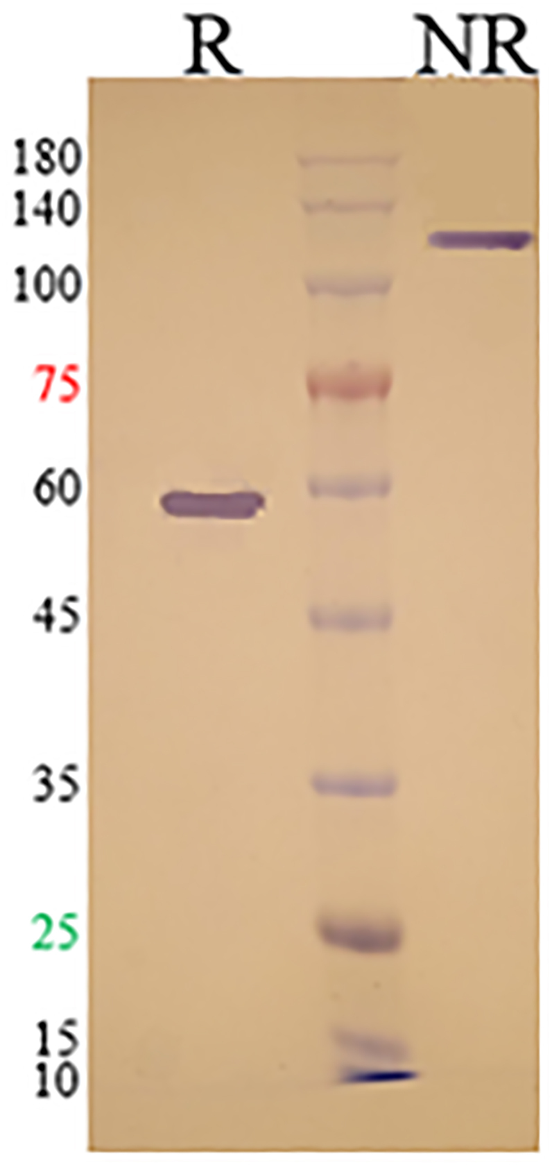
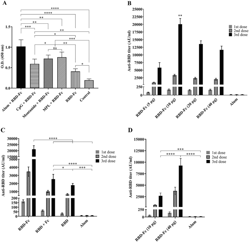
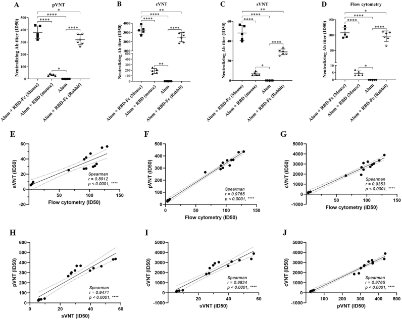
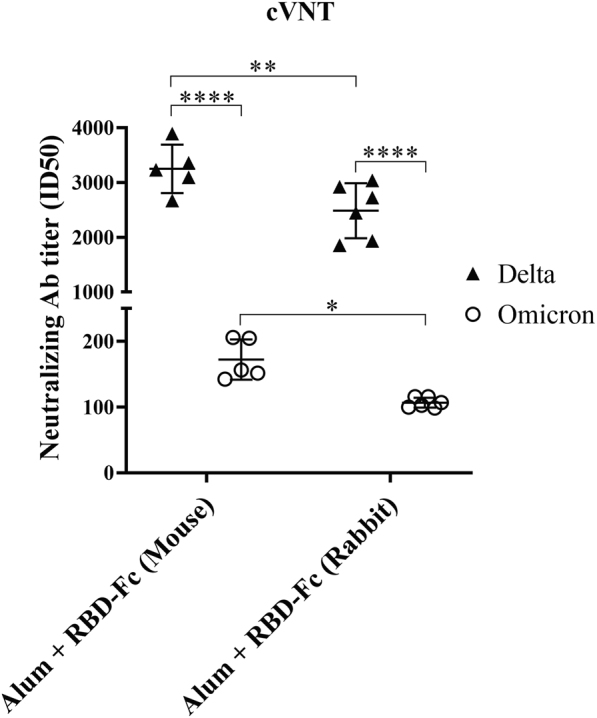
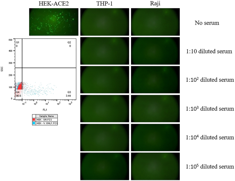
Methods: A recombinant fusion protein containing the receptor-binding domain (RBD) fused to the human IgG1 Fc (RBD-Fc) was produced in CHO-K1 cells. RBD-Fc was emulsified with four adjuvants to evaluate its immunogenicity. The RBD-specific humoral and cellular immune responses were assessed by ELISA. The virus neutralizing potency of the vaccine was investigated using four neutralization methods. Safety was studied in mice and rabbits, and Antibody-Dependent Enhancement (ADE) effects were investigated by flow cytometry.

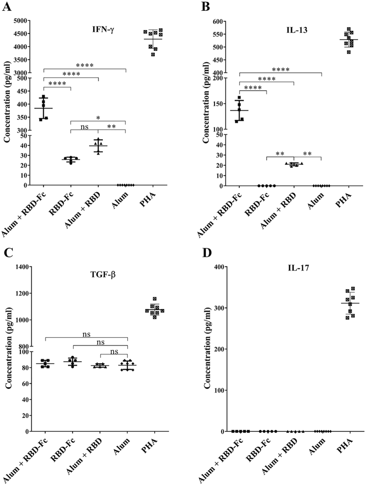
Results: RBD-Fc emulsified in Alum induced a high titer of anti-RBD antibodies with remarkable efficacy in neutralizing both pseudotyped and live SARS-CoV-2 Delta variant. The neutralization potency dropped significantly in response to the Omicron variant. RBD-Fc induced both TH2 and particularly TH1 immune responses. Histopathologic examinations demonstrated no substantial pathologic changes in different organs. No changes in serum biochemical and hematologic parameters were observed. ADE effect was not observed following immunization with RBD-Fc.

Conclusion: RBD-Fc elicits highly robust neutralizing antibodies and cellular immune responses, with no adverse effects. Therefore, it could be considered a promising and safe subunit vaccine against SARS-CoV-2.

Keywords: RBD-Fc; SARS-CoV-2; immunogenicity; neutralization; safety; vaccine.


Conflict of interest statement

Funding sources: This work was partially supported by Tehran University of Medical Sciences (TUMS) under Grant number 1401-1-99-57094.

Authors' contributions: Navid Dashti: Investigation, Formal analysis, Writing – original draft. Forough Golsaz-Shirazi, Amir-Hassan Zarnani and Mahmood Jeddi-Tehrani: Data curation, Validation, Writing – review & editing. Haleh Soltanghoraee: Investigation, Validation. Mehdi Mohammadi and Danyal Imani: Methodology. Mohammad Mehdi Amiri: Conceptualization, Methodology, Software, Supervision, Writing – review & editing, Data curation. Fazel Shokri: Conceptualization, Data curation, Funding acquisition, Project administration, Supervision, Validation, Writing – review & editing.

All authors had full access to all data in the study and take responsibility for the integrity of the data and the accuracy of the data analysis.

Conflicts of interest: The authors declare no conflict of interest.

Figures

Fig. 1.
Fig. 1.
SDS-PAGE analysis of RBD-Fc. Five µg of purified RBD-Fc was loaded on 10% gel at reduced (R) and non-reduced (NR) conditions. A band at approximately 120 kDa in the absence of 2 ME (NR) and approximately 60 kDa in the presence of 2 ME was detected
Fig. 2.
Fig. 2.
RBD-Fc candidate vaccine elicited robust antibody response against RBD in mice and rabbits. (A) Adjuvant selection study: RBD-Fc (5 µg) was formulated in different adjuvants. Two doses of each vaccine formulation were administered to animals at 21 days intervals. (B) Dose escalation study in mice: Four doses of RBD-Fc (5, 10, 20, and 40 µg) were emulsified with Alum and administered three times at 21 days intervals. (C) Fc immunogenicity study: Different compositions, including RBD-Fc, RBD, and RBD plus human Fc fragment were emulsified with Alum, and each mice group was administered with one formulation three times. (D) Dose escalation study in rabbits: Two doses of RBD-Fc (10 and 40 µg) with Alum were administered three times. Alum was injected in control group. Data are presented as the mean ± SD. P-values are determined by one-way ANOVA
Fig. 3.
Fig. 3.
RBD-Fc candidate vaccine is capable of eliciting both TH1 and TH2 responses. Mice immunized with RBD-Fc, RBD, or PBS were sacrificed and their splenocytes were isolated. After stimulation with 5 μg mL−1 RBD, the supernatants were collected after 72 h and the level of (A) IFN-γ, (B) IL-13, (C) TGF-β, and (D) IL-17 were detected by ELISA. Splenocytes cultured without RBD protein were considered as negative control, whereas phytohaemagglutinin (PHA) was used to stimulate cytokine response as a positive control. Data are presented as the mean±SD. P-values are determined by one-way ANOVA
Fig. 4.
Fig. 4.
RBD-Fc induced strong neutralizing antibody responses in mice and rabbits. The neutralizing potency of immunized sera was determined by different viral neutralization assays. (A) pseudotyped Virus Neutralization Test (pVNT): eGFP-pseudotyped lentivirus containing SARS-CoV-2 Delta variant spike protein was mixed with serial dilutions of sera. The mixture was added to HEK293T cells expressing ACE2. After 48 h, the pseudovirus-infected eGFP-positive cells were imaged and detected using fluorescence microscopy. (B) conventional Virus Neutralization Test (cVNT): Serum samples were heat-inactivated to destroy complement. The, 100 Tissue Culture Infectious Dose 50 (TCID50) of live Delta variant SARS-CoV-2 was mixed with serial dilutions of sera. The virus/serum mixtures were then added to Vero-E6 cells. After 1 h incubation, the supernatant was washed away, and the infected cells incubated in DMEM. The virus-specific CPE of each well was recorded under microscopes 72 h post-infection, and the ID50 was determined as the inhibitory dilution. (C) surrogate Virus Neutralization Test (sVNT): The ability of RBD-specific neutralizing antibodies to inhibit binding of RBD to ACE2 was investigated in competitive ELISA. After coating RBD in ELISA plate, serially diluted serum samples or standard solutions and HRP-conjugated ACE2 were added to individual wells. Following addition of chromogen solution and stop solution, the OD of the reactions were measured. (D) Flow cytometry: Serially diluted serum samples were mixed with an equal volume of RBD-Fc (0.25 μg mL−1). Serum/RBD-Fc mixture was then added to HEK293T cells that were expressing ACE2. Human immunoglobulin was used as an isotype control. Then, FITC-labeled sheep anti-human antibody was added at a final dilution of 1:100. Finally, data were acquired on a flow cytometer device and analyzed using FlowJo V10 software. Data are presented as the mean±SD. P-values are determined by one-way ANOVA. (E–J) The correlation of obtained ID50 values between flow cytometry, sVNT, pVNT, and cVNT was calculated by Spearman analysis
Fig. 5.
Fig. 5.
Comparison of RBD-Fc induced neutralizing antibody responses against Omicron (BA.1) and Delta variants of live SARS-CoV-2 virus (cVNT) in mice and rabbits. Serum samples were heat-inactivated to destroy complement. The, 100 Tissue Culture Infectious Dose 50 (TCID50) of live Delta or Omicron (BA.1) variant SARS-CoV-2 was mixed with serial dilutions of sera. The virus/serum mixtures were then added to Vero-E6 cells. After 1 h incubation, the supernatant was washed away, and the infected cells incubated in DMEM. The virus-specific CPE of each well was recorded under microscopes 72 h post-infection, and the ID50 was determined as the inhibitory dilution
Fig. 6.
Fig. 6.
Serum from immunized mice and rabbits did not induce ADE of SARS-CoV-2 in both THP-1 and Raji cell lines. Representative fluorescence images of cells infected with eGFP-pseudotyped lentiviruses were obtained in presence or absence of various dilutions of immunized sera. The entry of SARS-CoV-2 pseudovirus into FcγR-expressing cell lines (THP-1 and Raji) was investigated using serially diluted immunized or non-immunized (control) sera, ranging from 1:10 to 1:105. Treatment with the same quantity of SARS-CoV-2 pseudovirus resulted in an infection rate of approximately 4% in HEK-ACE2 cells

Similar articles

Cited by

References

    1. Li C, Ye Z, Zhang AJX, Chan JFW, Song W, Liu F, et al. . Severe acute respiratory syndrome coronavirus 2 (SARS-CoV-2) infection by intranasal or intratesticular route induces testicular damage. Clin Infect Dis. 2022;75(1):e974–e90. - PMC - PubMed
    1. World Health Organization . COVID-19 weekly epidemiological update. edition 164. 16 February 2024.
    1. Silk BJ, Scobie HM, Duck WM, Palmer T, Ahmad FB, Binder AM, et al. . COVID-19 surveillance after expiration of the public health emergency declaration―United States, may 11, 2023. Morbidity Mortality Weekly Rep. 2023;72(19):523. - PMC - PubMed
    1. Rubin R. COVID-19 vaccines vs variants—determining how much immunity is enough. Jama. 2021;325(13):1241–3. - PubMed
    1. Bok K, Sitar S, Graham BS, Mascola JR. Accelerated COVID-19 vaccine development: milestones, lessons, and prospects. Immunity. 2021;54(8):1636–51. - PMC - PubMed

LinkOut - more resources