. 2024 Jun;17(6):e012734.
doi: 10.1161/CIRCEP.124.012734. Epub 2024 May 16.

Preclinical Study of Pulsed Field Ablation of Difficult Ventricular Targets: Intracavitary Mobile Structures, Interventricular Septum, and Left Ventricular Free Wall

Affiliations

Preclinical Study of Pulsed Field Ablation of Difficult Ventricular Targets: Intracavitary Mobile Structures, Interventricular Septum, and Left Ventricular Free Wall

Moritz Nies et al. Circ Arrhythm Electrophysiol. 2024 Jun.

Abstract

Background: Endocardial catheter-based pulsed field ablation (PFA) of the ventricular myocardium is promising. However, little is known about PFA's ability to target intracavitary structures, epicardium, and ways to achieve transmural lesions across thick ventricular tissue.

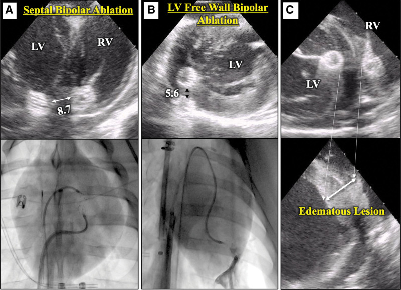
Methods: A lattice-tip catheter was used to deliver biphasic monopolar PFA to swine ventricles under general anesthesia, with electroanatomical mapping, fluoroscopy and intracardiac echocardiography guidance. We conducted experiments to assess the feasibility and safety of repetitive monopolar PFA applications to ablate (1) intracavitary papillary muscles and moderator bands, (2) epicardial targets, and (3) bipolar PFA for midmyocardial targets in the interventricular septum and left ventricular free wall.

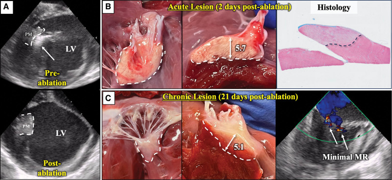
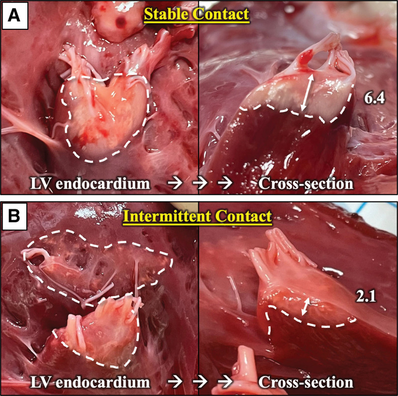
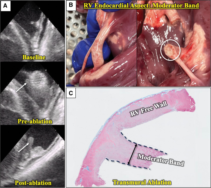
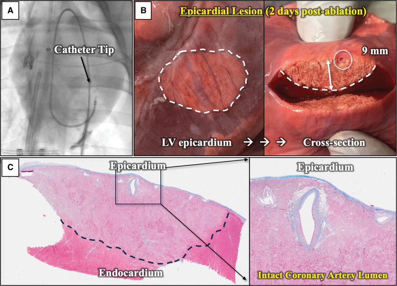
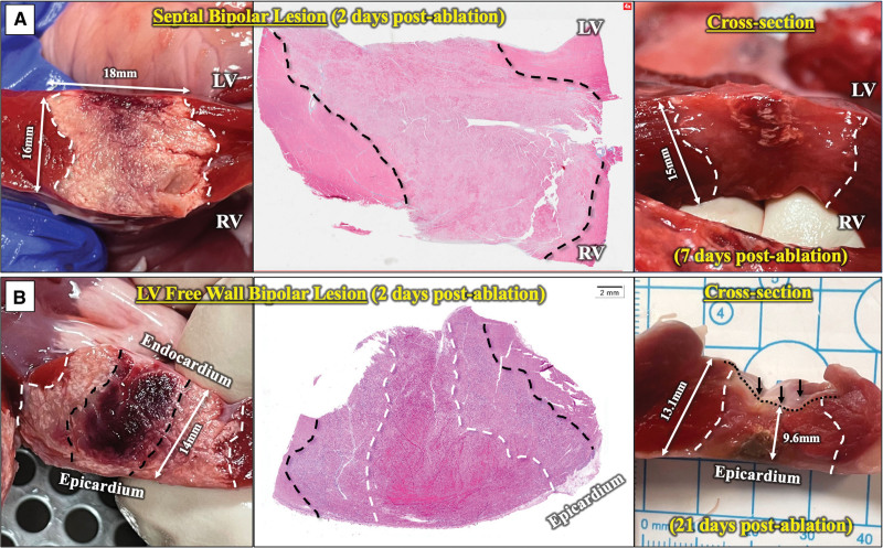
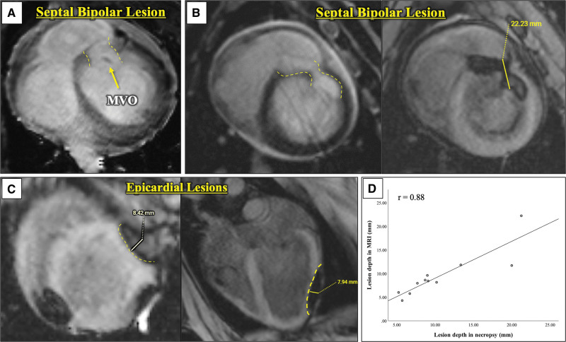
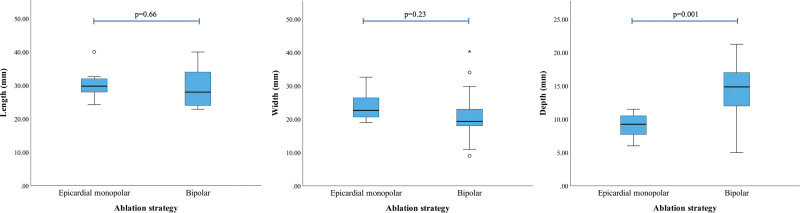
Results: (1) Papillary muscles (n=13) were successfully ablated and then evaluated at 2, 7, and 21 days. Nine lesions with stable contact measured 18.3±2.4 mm long, 15.3±1.5 mm wide, and 5.8±1.0 mm deep at 2 days. Chronic lesions demonstrated preserved chordae without mitral regurgitation. Two targeted moderator bands were transmurally ablated without structural disruption. (2) Transatrial saline/carbon dioxide assisted epicardial access was obtained successfully and epicardial monopolar lesions had a mean length, width, and depth of 30.4±4.2, 23.5±4.1, and 9.1±1.9 mm, respectively. (3) Bipolar PFA lesions were delivered across the septum (n=11) and the left ventricular free wall (n=7). Twelve completed bipolar lesions had a mean length, width, and depth of 29.6±5.5, 21.0±7.3, and 14.3±4.7 mm, respectively. Chronically, these lesions demonstrated uniform fibrotic changes without tissue disruption. Bipolar lesions were significantly deeper than the monopolar epicardial lesions.

Conclusions: This in vivo evaluation demonstrates that PFA can successfully ablate intracavitary structures and create deep epicardial lesions and transmural left ventricular lesions.

Keywords: catheter ablation; electroporation; heart ventricles; papillary muscles; swine; tachycardia, ventricular.


Conflict of interest statement

Disclosures Drs Koruth and Reddy have served as a consultant to and received grant support and equity from Affera-Medtronic. A comprehensive list of all financial disclosures (unrelated to this article) is included in the Supplemental Material. Dr Nies has received a scholarship from the German Research Foundation (Deutsche Forschungsgemeinschaft). The other authors report no conflicts.

Figures

Figure 1.
Figure 1.
Papillary muscle (PM) ablation. A, Intracardiac echocardiography (ICE) images. Top, Catheter tip (arrow) in contact with the left ventricular (LV) posterior PM before ablation. Bottom, ICE image 90 minutes post-ablation with echogenic changes. B, Necropsy and histology findings 2 days post-ablation. Lesion confined to the PM. A small area of hemorrhage is seen. Maximum lesion depth was 5.7 mm. Histology (Masson Trichrome) reveals homogeneous necrosis (dotted line). C, After 21 days of survival, the PMs retain their integrity and overall structure. Chordae were intact (left) with a maximum lesion depth of 5.1 mm. There was no significant mitral valve regurgitation noted on ICE. MR indicates mitral regurgitation.
Figure 2.
Figure 2.
Papillary muscle (PM) ablation, effect of tissue contact. A, PM lesion (dotted line) 2 days post-ablation with good, stable contact. The lesion is confined to the PM and achieved a maximum depth of 6.4 mm. B, PM ablation with intermittent contact with wider but shallower (2.1 mm depth) lesion, extending to neighboring myocardium. LV indicates left ventricle.
Figure 3.
Figure 3.
Moderator band ablation. A, Intracardiac echocardiography images demonstrate the moderator band (MB) at baseline, preablation with the catheter in position, and postablation changes 10 minutes after. The white arrow points to the MB branch in contact with the catheter tip. B, Necropsy findings 2 days after ablation show transmural ablation of the MB as well as ablation of insertion points. C, Histology confirms transmural ablation of the MB (dotted lines) and the neighboring right ventricular (RV) wall. Masson Trichrome staining.
Figure 4.
Figure 4.
Epicardial monopolar ablation. A, Fluoroscopic image of the catheter tip (black arrow) on the epicardial surface. Small region of contrast staining seen related to subxiphoid needle epicardial access. B, Necropsy findings 2 days after ablation. A wide, homogenous lesion is seen on the epicardial surface (left). Cross section (right) reveals a lesion depth of 9 mm. Note the large epicardial coronary artery branch within the ablation lesion (white circle). C, Histology (Masson Trichrome) demonstrating a homogenous lesion (left). The coronary artery lumen remains patent as shown in the zoomed inset (right). Note part of the arterial medial wall is ablated as well. LV indicates left ventricle.
Figure 5.
Figure 5.
Bipolar ablation—intracardiac echocardiography (ICE) and fluoroscopic views. A, Septal bipolar ablation. Catheter tips are placed on opposing sites of the interventricular septum and positioned with optimized contact with 8.7-mm separation on ICE (top). The corresponding fluoroscopy image is shown in the bottom (AP-view). B, Bipolar ablation of the left ventricular (LV) free wall: One catheter tip was introduced retrogradely into the LV, and the other is in the epicardial space. Top, ICE view with the catheter tips 5.6 mm separated. The corresponding fluoroscopy image is shown in the bottom. C, ICE images during (top) and after septal bipolar ablation (bottom), showing increased echogenicity and edematous swelling (double arrow) of the ablated tissue. RV indicates right ventricle.
Figure 6.
Figure 6.
Bipolar ablation—pathology. A, Left, Gross appearance 2 days post-ablation. Septal bipolar lesion (white dotted line) shows central hemorrhagic changes extending to the center on the left ventricular (LV) aspect. This lesion measures 18 mm wide and 16 mm deep. Middle, Histology (H&E-stain) demonstrates the septal lesion (black dotted line). Right, Gross appearance 7 days post-ablation: a transmural septal lesion demonstrates mild hemorrhage and surrounding calcification limited to the LV aspect, with the rest of the lesion appearing pale typical of chronic PFA lesions. There were no signs of tissue disruption. B, Left, Gross appearance 2 days post-ablation. A bipolar LV free wall lesion with a central dark area (black dotted line) spanned through the entire thickness that corresponds to transmural hemorrhagic changes in histology (mid, H&E-stain). Right, Gross appearance 21 days post-ablation. A transmural LV wall bipolar lesion (white dotted line) is seen with a fibrotic appearance and without signs of hemorrhage. Compared with the neighboring unablated LV free wall (13.1 mm), the lesion center demonstrates reduced wall thickness (9.6 mm) related to wall thinning from fibrotic remodeling. RV indicates right ventricle.
Figure 7.
Figure 7.
Magnetic resonance imaging. A, Bright-blood late gadolinium enhancement (LGE) sequence of a septal bipolar lesion. The ablation lesion (yellow dotted line) is well demarcated by LGE, with a small region of microvascular obstruction (MVO, arrow). B, Septal bipolar lesion (yellow dotted line) in bright- and dark-blood LGE-sequences. C, Epicardial lesions (yellow dotted lines). D, Strong correlation (r=0.88) of lesion depth measured in magnetic resonance imaging (MRI) to that measured on necropsy.
Figure 8.
Figure 8.
Lesion dimensions with different ablation strategies. Box plots illustrate the differences in lesion size. Bipolar lesions were significantly deeper than epicardial monopolar lesions, but there were no significant differences in lesion length or width.

Similar articles

Cited by

References

    1. Della Bella P, Baratto F, Tsiachris D, Trevisi N, Vergara P, Bisceglia C, Petracca F, Carbucicchio C, Benussi S, Maisano F, et al. . Management of ventricular tachycardia in the setting of a dedicated unit for the treatment of complex ventricular arrhythmias: long-term outcome after ablation. Circulation. 2013;127:1359–1368. doi: 10.1161/CIRCULATIONAHA.112.000872 - PubMed
    1. Wasmer K, Reinecke H, Heitmann M, Dechering DG, Reinke F, Lange PS, Frommeyer G, Kochhauser S, Leitz P, Eckardt L, et al. . Clinical, procedural and long-term outcome of ischemic VT ablation in patients with previous anterior versus inferior myocardial infarction. Clin Res Cardiol. 2020;109:1282–1291. doi: 10.1007/s00392-020-01622-z - PMC - PubMed
    1. Yamashita S, Cochet H, Sacher F, Mahida S, Berte B, Hooks D, Sellal JM, Al Jefairi N, Frontera A, Komatsu Y, et al. . Impact of new technologies and approaches for post-myocardial infarction ventricular tachycardia ablation during long-term follow-up. Circ Arrhythm Electrophysiol. 2016;9:e003901. doi: 10.1161/CIRCEP.116.003901 - PubMed
    1. Barkagan M, Leshem E, Shapira-Daniels A, Sroubek J, Buxton AE, Saffitz JE, Anter E. Histopathological characterization of radiofrequency ablation in ventricular scar Tissue. JACC Clin Electrophysiol. 2019;5:920–931. doi: 10.1016/j.jacep.2019.05.011 - PubMed
    1. Schwartzman D, Chang I, Michele JJ, Mirotznik MS, Foster KR. Electrical impedance properties of normal and chronically infarcted left ventricular myocardium. J Interv Card Electrophysiol. 1999;3:213–224. doi: 10.1023/a:1009887306055 - PubMed

MeSH terms