. 2024 May 16;20(5):e1012010.
doi: 10.1371/journal.ppat.1012010. eCollection 2024 May.

Exploiting bacterial effector proteins to uncover evolutionarily conserved antiviral host machinery

Affiliations

Exploiting bacterial effector proteins to uncover evolutionarily conserved antiviral host machinery

Aaron Embry et al. PLoS Pathog. .

Abstract

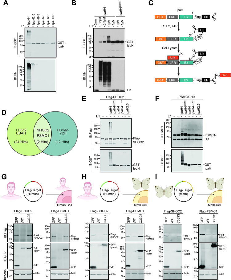
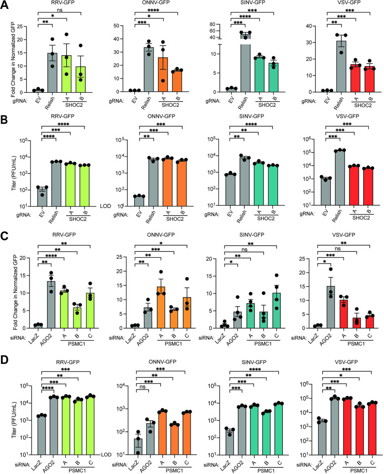
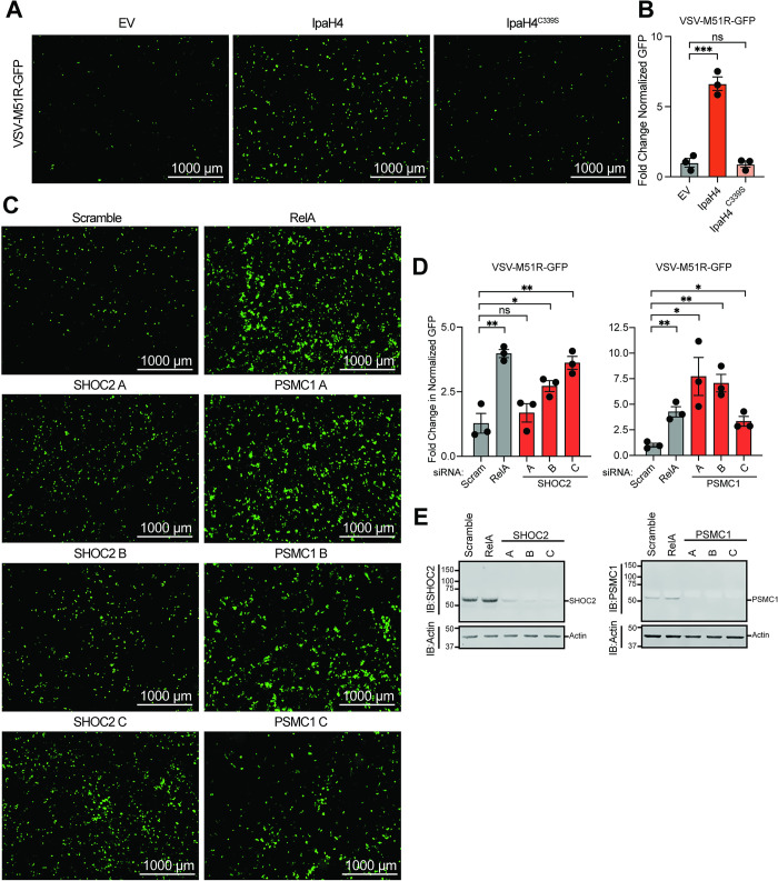
Arboviruses are a diverse group of insect-transmitted pathogens that pose global public health challenges. Identifying evolutionarily conserved host factors that combat arbovirus replication in disparate eukaryotic hosts is important as they may tip the balance between productive and abortive viral replication, and thus determine virus host range. Here, we exploit naturally abortive arbovirus infections that we identified in lepidopteran cells and use bacterial effector proteins to uncover host factors restricting arbovirus replication. Bacterial effectors are proteins secreted by pathogenic bacteria into eukaryotic hosts cells that can inhibit antimicrobial defenses. Since bacteria and viruses can encounter common host defenses, we hypothesized that some bacterial effectors may inhibit host factors that restrict arbovirus replication in lepidopteran cells. Thus, we used bacterial effectors as molecular tools to identify host factors that restrict four distinct arboviruses in lepidopteran cells. By screening 210 effectors encoded by seven different bacterial pathogens, we identify several effectors that individually rescue the replication of all four arboviruses. We show that these effectors encode diverse enzymatic activities that are required to break arbovirus restriction. We further characterize Shigella flexneri-encoded IpaH4 as an E3 ubiquitin ligase that directly ubiquitinates two evolutionarily conserved proteins, SHOC2 and PSMC1, promoting their degradation in insect and human cells. We show that depletion of either SHOC2 or PSMC1 in insect or human cells promotes arbovirus replication, indicating that these are ancient virus restriction factors conserved across invertebrate and vertebrate hosts. Collectively, our study reveals a novel pathogen-guided approach to identify conserved antimicrobial machinery, new effector functions, and conserved roles for SHOC2 and PSMC1 in virus restriction.


Conflict of interest statement

The authors declare that there are no competing interests.

Figures

Fig 1
Fig 1. Abortive arbovirus replication in LD652 cells can be relieved with ActD treatment.
A. Representative fluorescence microscopy images (GFP channel) of LD652 cells treated with DMSO (vehicle) or 0.05 μg/mL ActD and infected with the indicated GFP reporter strains for 72 h. B. Fold-change in normalized GFP signals in ActD-treated cultures relative to DMSO treatments. Cells were stained 72 hpi with CellTracker Orange dye (not shown) and imaged in GFP and RFP channels to calculate fold-change in GFP signal after normalization of cell number using CellTracker (RFP) channel signals. C. Fold-change in titer of supernatants from LD652 cell cultures treated as in A-B 72 hpi relative to input inoculum (dotted line). Data in B-C are means ± SD; n = 3. Statistical significance was determined with unpaired student’s t-test; ns = P>0.1234, * = P<0.0332, ** = P<0.0021, *** = P<0.0002, **** = P<0.0001.
Fig 2
Fig 2. Specific Bacterial Effectors Relieve Arbovirus Restriction in LD652 Cells.
A. Schematic outlining screen for bacterial effectors that rescue arbovirus restriction in LD652 cells. Cells were transfected with expression plasmids from a library consisting of 210 different effector proteins. After 48 h, cells were infected with either GFP or luciferase reporter strains. At 72 hpi, viral replication was quantified using fluorescence microscopy (RRV-GFP and ONNV-GFP) or luciferase assays (VSV-LUC and SINV-LUC). Image was created with BioRender.com. B-E. Fold-change in reporter readout, normalized to empty vector controls for all four screens. The cutoff for fold-change in GFP-based assays was set to >2.5, while the cutoff for luciferase reporters was set to >4-fold (represented by dotted horizontal lines). Data points are means. RLU = relative light units. F. Summary of bacterial effector proteins that rescued at least one virus. Green blocks indicate the effector rescued the virus indicated in the column header. The bacterium encoding each effector is noted to the right: Shigella flexneri (S. flexneri), Pseudomonas syringae (P. syringae), Salmonella enterica (S. enterica), Legionella pneumophila (L. pneumo.) Enterohemorrhagic Escherichia coli 0157:H7 (EHEC). Additional effector proteins from Yersinia pseudotuberculosis and Bartonella henselae were also screened but did not rescue arbovirus replication. The complete list of effectors screened and the raw results of the screens can be found in S1 Table.
Fig 3
Fig 3. Validation and characterization of top hits from bacterial effector screens.
A. AlphaFold predicted structures, and known or predicted enzymatic functions for indicated effector proteins identified as hits in arbovirus screens. Effector catalytic residues where substitution mutations were made are highlighted in red in AlphaFold structures. B. Representative immunoblots of Flag-tagged bacterial effector expression in LD652 cells 48 h post transfection. C-F. Fold-change in normalized viral GFP signal relative to empty vector (EV) controls 72 hpi with after transfection with indicated Flag-tagged effector constructs. Wild-type (WT) effectors are compared to their mutants (E/A or C/S). Cells were stained 72 hpi with CellTracker dye and imaged to calculate fold-change in normalized GFP signal over signals in EV treatments. Data in C-F are means ± SD; n = 3. Statistical significance was determined with unpaired student’s t-test; ns = P>0.1234, * = P<0.0332, ** = P<0.0021, *** = P<0.0002, **** = P<0.0001.
Fig 4
Fig 4. Structural analysis of Legionella pneumophila effector Ceg10.
A. Six structural homologs of Ceg10, shown in the same orientation with the putative active site near the top of the figure. Top row, from left: Ceg10 (this study), L. pneumophila RavJ, L. pneumophila LapG. Bottom row, from left: S. enterica SseI, S. flexneri OspI, P. savastanoi AvrPphB. Table shows these structural homologs as determined via the Dali Lite server and their PBD ID. B. The putative active site of Ceg10 with residues shown in stick representation and hydrogen bonds are shown as dotted yellow lines. The catalytic Cys (C159), Asp (D204) and His (H192) are labeled as well as residues Asp110 and Trp 206 which are hydrogen bonded to each other in both structures. In the S-nitrosylated structure, Asp110 also hydrogen bonds to the nitrosylated-C159 and van der Waals interactions occur between the aromatic ring of Trp206 and the nitrosylation moiety. C. Electron density for C159 in the native (left) and S-nitrosylated (right) Ceg10 structures. The final refined 2Fo-mDFc electron density map, contoured at the 1σ level, is shown superimposed on each residue, as well as a 180° rotation of this region. C. Superposition of native (blue) and S-nitrosylated (brown) Ceg10 structures. D. Electrostatic surface potential of both Ceg10 structures and RavJ. All structures are orientated with the putative catalytic cysteine residue in approximately the center of the surface, and the orientations between Ceg10 and RavJ correspond to protein alignments. The displayed surface is colored by electrostatic potential from -10 kT (red) to + 10 kT (blue), as calculated by the APBS plugin in PyMOL.
Fig 5
Fig 5. Identification of host SHOC2 and PSMC1 as conserved targets of IpaH4.
A. Representative immunoblot of in vitro ubiquitination assay performed with indicated GST-IpaH proteins in the absence of substrates. B. Representative immunoblot of in vitro ubiquitination assay performed with indicated concentrations of wild-type GST-IpaH4 or GST-IpaH4C339S mutant proteins. C. Schematic outlining UBAIT protocol [53,56]. D. Venn diagram showing conserved putative substrates (overlapping region) of IpaH4 across UBAIT experiments (n = 3) in LD652 cell lysates and Y2H screens (n = 2) against a human prey library. E. Representative immunoblot of in vitro ubiquitination assay showing IpaH4-mediated ubiquitination of human Flag-SHOC2 proteins. F. Representative immunoblot of in vitro ubiquitination assay showing IpaH4-mediated ubiquitination of human PSMC1-His proteins. G. Representative immunoblot of degradation assays using indicated Flag-tagged human proteins in transfected HEK293T cells co-expressing GFP, IpaH4 (WT) or catalytic mutant GFP-IpaH4C339S (C339S). H. Representative immunoblot of degradation assays using indicated Flag-tagged human proteins in transfected LD652 cells co-expressing GFP, IpaH4 (WT) or catalytic mutant GFP-IpaH4C339S (C339S). I. Representative immunoblot of degradation assays of Flag-tagged moth (L. dispar) protein in LD652 cells expressing GFP, IpaH4 (WT) or catalytic mutant GFP-IpaH4C339S (C339S). Images in G-I were created with BioRender.com.
Fig 6
Fig 6. Depletion of IpaH4 substrates SHOC2 and PSMC1 enhances arbovirus replication in LD652 cells.
A. Fold-change in normalized viral GFP signals in cells expressing gRNA targeting Relish or SHOC2 relative to empty vector controls 72 hpi. Cells were stained with CellTracker dye 72 hpi and imaged to calculate fold-change in normalized GFP signal over empty vector (control) treatments. Data are means ± SD; n = 3. Statistical significance was determined with unpaired student’s t-test. B. Titer of supernatants from LD652 cell cultures treated as described in A. C. Fold-change in normalized viral GFP signals relative to LacZ siRNA (control) treatments. Cells were stained with CellTracker dye 72 hpi and imaged to calculate fold-change in normalized GFP signal over LacZ (control) siRNA treatments. D. Titer of supernatants from LD652 cell cultures treated as described in C. Data are means ± SD; n = 3. Statistical significance was determined with unpaired student’s t-test; ns = P>0.1234, * = P<0.0332, ** = P<0.0021, *** = P<0.0002, **** = P<0.0001.
Fig 7
Fig 7. Bacterial effector expression or depletion of effector targets enhances oncolytic virus replication in human cancer cells.
A. Representative fluorescence microscopy images (GFP channel) of human 786–0 renal adenocarcinoma cell line infected with VSV-M51R-GFP at 16 hpi. B. Fold-change in normalized viral GFP signal relative to empty vector (EV) control from experiments as in A. Cells were stained with CellTracker Orange dye 16 hpi and imaged to calculate fold-change in normalized GFP signal over EV treatments. Data are means ± SD; n = 3. Statistical significance was determined with unpaired student’s t-test. C. Representative fluorescence microscopy images (GFP channel) of 786–0 cells infected with VSV-M51R-GFP at 16 hpi after transfection with indicated siRNAs. D. Fold-change in normalized viral GFP signal relative to scrambled (control) treatments as in C. Cells were stained with CellTracker dye 16 hpi and imaged to calculate fold-change in normalized GFP signal over control treatments. E. Representative immunoblot of 786–0 whole cell lysate 72 h post-transfection with indicated siRNAs. For B and D: ns = P>0.1234, * = P<0.0332, ** = P<0.0021, *** = P<0.0002, **** = P<0.0001.

Update of

Similar articles

Cited by

References

    1. Velazquez-Salinas L, Pauszek SJ, Stenfeldt C, O’Hearn ES, Pacheco JM, Borca MV, et al.. Increased Virulence of an Epidemic Strain of Vesicular Stomatitis Virus Is Associated With Interference of the Innate Response in Pigs. Front Microbiol. 2018;9:1891. Epub 2018/08/31. doi: 10.3389/fmicb.2018.01891 ; PubMed Central PMCID: PMC6104175. - DOI - PMC - PubMed
    1. Madewell ZJ. Arboviruses and Their Vectors. South Med J. 2020;113(10):520–3. Epub 2020/10/03. doi: 10.14423/SMJ.0000000000001152 ; PubMed Central PMCID: PMC8055094. - DOI - PMC - PubMed
    1. Balakrishnan VS. WHO launches global initiative for arboviral diseases. Lancet Microbe. 2022;3(6):e407. Epub 2022/06/07. doi: 10.1016/S2666-5247(22)00130-6 ; PubMed Central PMCID: PMC9159734. - DOI - PMC - PubMed
    1. Guerrero-Arguero I, Tellez-Freitas CM, Weber KS, Berges BK, Robison RA, Pickett BE. Alphaviruses: Host pathogenesis, immune response, and vaccine & treatment updates. J Gen Virol. 2021;102(8). Epub 2021/08/27. doi: 10.1099/jgv.0.001644 . - DOI - PubMed
    1. Abdelnabi R, Delang L. Antiviral Strategies against Arthritogenic Alphaviruses. Microorganisms. 2020;8(9). Epub 2020/09/11. doi: 10.3390/microorganisms8091365 ; PubMed Central PMCID: PMC7563460. - DOI - PMC - PubMed